YAP Goes Nuclear: How Cytoskeletal Forces Drive Cancer Invasion and Metastasis
This article explores the critical interplay between the Hippo pathway effector YAP and cytoskeletal dynamics in promoting cancer cell invasion.
YAP Goes Nuclear: How Cytoskeletal Forces Drive Cancer Invasion and Metastasis
Abstract
This article explores the critical interplay between the Hippo pathway effector YAP and cytoskeletal dynamics in promoting cancer cell invasion. We first establish the foundational biology of YAP nuclear translocation as a mechanotransduction hub. Next, we detail current methodologies for detecting and manipulating this pathway in vitro and in vivo. We then address common experimental challenges and optimization strategies for studying this mechano-oncogenic axis. Finally, we validate key findings and compare YAP-targeting approaches against other cytoskeletal regulators. Aimed at researchers and drug developers, this synthesis highlights YAP's subcellular localization as a promising therapeutic target to impede cancer progression.
The Mechanobiology of YAP: From Cytoskeletal Tension to Nuclear Transcription in Cancer
This technical whitepaper delineates the dual function of YAP (Yes-associated protein) and TAZ (Transcriptional coactivator with PDZ-binding motif) as terminal effectors of the canonical Hippo signaling cascade and as pivotal mechanosensors. Framed within a thesis exploring YAP nuclear localization and cytoskeletal dynamics in cancer invasion, we provide a detailed mechanistic overview, quantitative data syntheses, and standardized experimental protocols. The content is tailored for researchers and drug development professionals investigating the role of mechanotransduction in tumor progression.
Core Signaling Mechanism: From Hippo Regulation to Mechanical Inputs
YAP/TAZ are transcriptional coactivators whose activity is predominantly regulated by the Hippo kinase cascade. Inactive Hippo signaling allows dephosphorylated YAP/TAZ to translocate to the nucleus, bind to TEAD transcription factors, and drive gene expression promoting cell proliferation, survival, and migration. Crucially, YAP/TAZ also function as central hubs for integrating mechanical cues from the extracellular matrix (ECM), cell geometry, and cytoskeletal tension, often bypassing canonical Hippo regulation.
The nucleo-cytoplasmic shuttling of YAP/TAZ is controlled by sequential phosphorylation.
Table 1: Core Phosphorylation Sites Regulating YAP/TAZ Activity
| Protein | Kinase | Phosphorylation Site | Functional Consequence | Reference (Example) |
|---|---|---|---|---|
| YAP | LATS1/2 (Hippo) | Ser127 (Human) | Creates 14-3-3 binding site, cytoplasmic retention | Zhao et al., 2007 |
| YAP | LATS1/2 | Ser381 (Human) | Primes for subsequent phosphorylation/degradation | Zhao et al., 2010 |
| YAP | CK1δ/ε (primed) | Ser384, Ser387, etc. | Leads to β-TrCP-mediated proteasomal degradation | Zhao et al., 2010 |
| TAZ | LATS1/2 | Ser89 (Human) | Creates 14-3-3 binding site, cytoplasmic retention | Lei et al., 2008 |
| TAZ | LATS1/2 | Ser311 (Human) | Primes for subsequent phosphorylation/degradation | Liu et al., 2010 |
| Both | AMPK | Ser61/Ser90 (YAP) | Inhibits activity under low energy conditions | Wang et al., 2015 |
| Both | Src | Tyr357 (YAP) | Promotes nuclear localization and activity | Rosenbluh et al., 2012 |
Mechanical Regulation: Key Parameters and Effects
YAP/TAZ nuclear localization is exquisitely sensitive to mechanical perturbations.
Table 2: Mechanical Cues Governing YAP/TAZ Localization and Activity
| Mechanical Cue | Experimental Manipulation | Effect on YAP/TAZ | Proposed Primary Sensor |
|---|---|---|---|
| ECM Stiffness | Culturing cells on polyacrylamide gels of varying elastic modulus | Nuclear localization increases with stiffness | Integrin clusters, F-actin tension |
| Cell Spreading Area/Geometry | Micropatterned substrates constraining cell shape | Nuclear localization correlates with increased spread area | Actin cytoskeleton, Rho GTPase activity |
| Cytoskeletal Tension | Treatment with Rock inhibitor (Y-27632), Blebbistatin (Myosin II) | Inhibits nuclear localization | Actin stress fibers, Myosin II |
| Cell Density (Contact Inhibition) | High-confluence culture | Cytoplasmic retention | Angiomotin complex, E-cadherin |
| Fluid Shear Stress | Laminar flow chambers | Can induce nuclear localization | Primary cilia, Junctions |
Experimental Protocols for Investigating YAP/TAZ in Cancer Invasion Contexts
Protocol: Quantitative Assessment of YAP/TAZ Nuclear-Cytoplasmic Localization
Objective: To quantify the subcellular distribution of YAP/TAZ in fixed cells under varying mechanical or oncogenic conditions.
- Cell Seeding: Seed cancer cells of interest on substrates of defined stiffness (e.g., collagen-coated polyacrylamide gels) or tissue culture plastic.
- Fixation and Permeabilization: At desired time points, fix cells with 4% paraformaldehyde for 15 min at RT. Permeabilize with 0.25% Triton X-100 for 10 min.
- Immunofluorescence Staining:
- Block with 5% BSA/PBS for 1 hour.
- Incubate with primary antibodies (anti-YAP/TAZ, 1:200-1:500) overnight at 4°C.
- Wash 3x with PBS.
- Incubate with fluorophore-conjugated secondary antibody (1:500) and Phalloidin (for F-actin) and DAPI (for nucleus) for 1 hour at RT.
- Wash and mount.
- Image Acquisition: Acquire high-resolution z-stack images using a confocal microscope under identical settings for all conditions.
- Image Analysis: Use software (e.g., ImageJ, CellProfiler) to create nuclear and cytoplasmic masks based on DAPI and F-actin signals. Measure mean fluorescence intensity of YAP/TAZ in each compartment. Calculate Nuclear/Cytoplasmic (N/C) ratio for each cell (n≥100 cells/condition).
- Statistical Analysis: Perform ANOVA or t-tests between experimental groups.
Protocol: Functional Assessment via YAP/TAZ-Dependent Transcriptional Reporter Assay
Objective: To measure the transcriptional output of YAP/TAZ-TEAD complexes.
- Transfection/Transduction: Transduce cells with a lentiviral construct containing a TEAD-responsive luciferase reporter (e.g., 8xGTIIC-luciferase) and a constitutive Renilla luciferase control for normalization.
- Experimental Treatment: Plate stable reporter cells on test substrates or treat with pharmacological agents (e.g., LATS inhibitor, cytoskeletal drugs).
- Luciferase Assay: After 24-48 hours, lyse cells and measure Firefly and Renilla luciferase activities using a dual-luciferase assay kit.
- Data Normalization: Calculate the ratio of Firefly to Renilla luminescence for each sample. Express data as fold-change relative to control condition.
Protocol: 3D Invasion Assay with YAP/TAZ Modulation
Objective: To correlate YAP/TAZ activity with invasive capacity in a physiologically relevant 3D matrix.
- Matrix Preparation: Prepare a mixture of basement membrane extract (e.g., Matrigel) and collagen I to mimic a tumor-associated ECM.
- Spheroid Formation: Generate uniform cancer cell spheroids using a hanging drop or ultra-low attachment plate method.
- Embedding and Invasion: Embed single spheroids in the 3D matrix in a 24-well plate. Allow matrix to polymerize. Add complete medium on top.
- Perturbation: Add small molecule inhibitors (e.g., Verteporfin for YAP/TAZ-TEAD interaction, Dasatinib for Src) or vehicle control.
- Imaging and Quantification: Acquire brightfield or confocal images at 0, 24, 48, and 72 hours. Measure the area of spheroid core and the total area including invasive protrusions using image analysis software. Calculate an "Invasion Index" = (Total Area - Core Area) / Core Area.
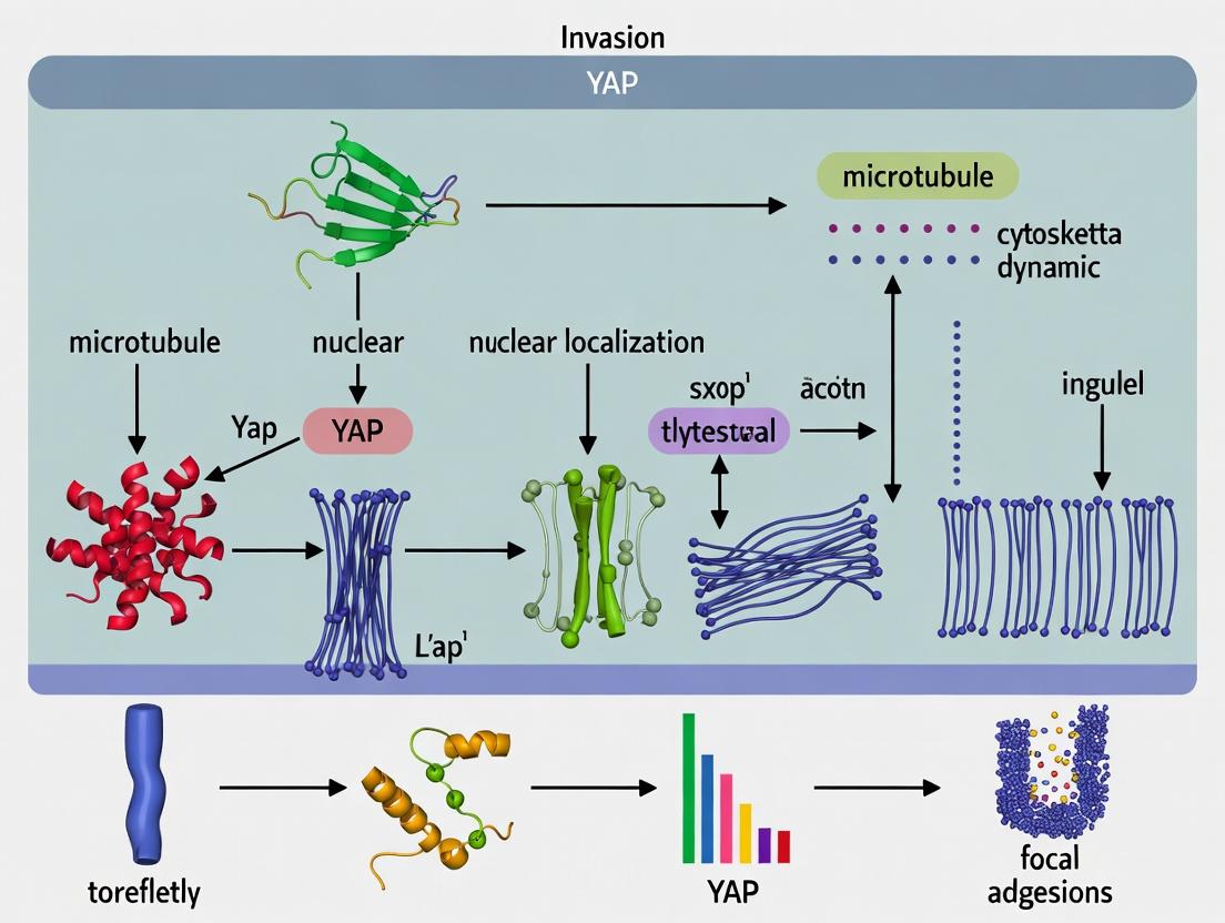
Visualizing Signaling and Experimental Logic
Diagram 1: Integrated Hippo Pathway and Mechanical Regulation of YAP/TAZ
Diagram 2: Workflow for Quantifying YAP/TAZ Localization
The Scientist's Toolkit: Key Research Reagent Solutions
Table 3: Essential Reagents for YAP/TAZ and Mechanotransduction Research
| Reagent Category | Specific Item/Product (Example) | Primary Function in Research |
|---|---|---|
| Antibodies | Anti-YAP (D8H1X) XP Rabbit mAb (CST #14074) | Detects endogenous total YAP protein for WB, IF, IP. |
| Anti-Phospho-YAP (Ser127) Rabbit Ab (CST #4911) | Specifically detects Hippo-pathway inactivated (cytoplasmic) YAP. | |
| Anti-TAZ (V386) Rabbit mAb (CST #70148) | Detects endogenous total TAZ protein. | |
| Reporters | 8xGTIIC-luciferase Reporter Plasmid (Addgene #34615) | Measures transcriptional activity of YAP/TAZ-TEAD complexes. |
| Inhibitors | Verteporfin (Sigma-Aldrich) | Disrupts YAP/TAZ-TEAD interaction, inhibits transcriptional output. |
| Y-27632 (ROCK Inhibitor) | Inhibits Rho-kinase, reduces actomyosin tension, used to probe mechanical regulation. | |
| Latrunculin A/B | Disrupts actin polymerization, tests cytoskeletal dependence. | |
| Substrates | Polyacrylamide Hydrogel Kits (e.g., CytoSoft) | Provides tunable substrate stiffness for mechanobiology studies. |
| Collagen I, Rat Tail, High Concentration (Corning) | Major component for reconstituting physiologically relevant 3D matrices. | |
| Cell Lines | MCF10A ER-Src (Addgene) | Inducible model for studying YAP/TAZ activation during oncogenic transformation and invasion. |
| MDA-MB-231 (ATCC HTB-26) | Highly invasive triple-negative breast cancer line with active YAP/TAZ signaling. | |
| Critical Assay Kits | Dual-Luciferase Reporter Assay System (Promega) | Quantifies TEAD transcriptional reporter activity with internal control. |
| Cell Invasion Assay (e.g., Corning BioCoat Matrigel) | Standardized kit for assessing transwell invasion capacity. |
Yes-associated protein (YAP), a central transcriptional co-activator of the Hippo pathway, is a critical regulator of cell proliferation, survival, and migration. Its dysregulation is a hallmark of numerous cancers, driving aggressive invasion and metastasis. A paradigm shift in the field has established that YAP’s nucleocytoplasmic shuttling is exquisitely sensitive to mechanical cues and the architectural state of the cytoskeleton, often overriding canonical Hippo kinase signaling. This whitepaper provides an in-depth technical analysis of the tripartite mechanical system—actin dynamics, non-muscle myosin II (NMII) contraction, and focal adhesion (FA) maturation—that directly governs YAP localization. Understanding this interplay is paramount for developing novel anti-metastatic therapies targeting the mechanotransduction ecosystem in cancer.
Core Regulatory Mechanisms: A Tripartite Mechanical Cascade
YAP/TAZ activity is regulated by a tightly coupled mechanical feedback loop involving the actin cytoskeleton, contractile forces, and cell-ECM adhesions.
Actin Architecture as a Direct Sensor
The polymerization state and structural organization of filamentous actin (F-actin) serve as a primary signal. Stress fibers, which are thick, contractile actin bundles, promote YAP nuclear accumulation. Conversely, a dense, cortical actin meshwork sequesters YAP in the cytoplasm.
Key Quantitative Relationships:
- F-actin/G-actin Ratio: A high ratio correlates strongly with nuclear YAP. Pharmacological disruption (e.g., Latrunculin A) causing a low ratio forces YAP cytoplasmic retention.
- Stress Fiber Density: Measured by phalloidin staining intensity per cell area, shows a linear correlation (R² ~0.85) with the nuclear/cytoplasmic YAP ratio in mesenchymal cells.
Myosin II-Driven Contraction as the Force Generator
Non-muscle myosin II (NMII) activity, powered by ATP and regulated by Rho GTPase-ROCK signaling and myosin light chain (MLC) phosphorylation, generates the tension on actin fibers. This physical force is a non-canonical YAP regulator.
Key Quantitative Relationships:
- p-MLC (Ser19) Levels: Intracellular tension, inferred from p-MLC levels, shows a sigmoidal relationship with % nuclear YAP. A threshold tension must be exceeded to initiate significant nuclear translocation.
- Substrate Stiffness: On soft substrates (<1 kPa), where effective myosin contraction is limited, YAP is predominantly cytoplasmic. On stiff substrates (>10 kPa, mimicking tumor stroma), robust contraction drives >80% nuclear YAP localization.
Focal Adhesions as the Mechanosensory Hub
Focal adhesions are not merely anchoring structures; they are integrated signaling platforms. Their maturation (size, composition) is force-dependent and directly informs YAP localization via multiple pathways.
Key Quantitative Relationships:
- Adhesion Size: Mean FA area (via vinculin/Paxillin staining) is positively correlated (Pearson's r > 0.7) with nuclear YAP intensity. Mature, force-bearing adhesions (>5 µm²) are strongly pro-YAP activity.
- Integrin Clustering: Force-induced unfolding of talin and vinculin in large FAs exposes binding sites that recruit YAP-regulatory proteins.
Table 1: Quantitative Relationships Between Mechanical Inputs and YAP Localization
| Mechanical Input | Experimental Manipulation | Measured Parameter | Effect on YAP (N/C Ratio) | Typical Quantitative Change |
|---|---|---|---|---|
| Substrate Stiffness | Polyacrylamide gels of varying stiffness | Elastic Modulus (kPa) | Increases from 0.5 to 0.9 | 1 kPa: ~0.2, 20 kPa: ~0.85 |
| Actin Polymerization | Latrunculin A (inhibitor) vs. Jasplakinolide (stabilizer) | F-actin Intensity (AU) | Decreases (LatA) / Increases (Jasp) | LatA (1 µM): Ratio ↓ by ~70% |
| Myosin Contraction | Blebbistatin (inhibitor) vs. Calyculin A (activator) | p-MLC (Ser19) Level (WB) | Decreases (Blebb) / Increases (CalA) | Blebb (50 µM): Ratio ↓ by ~60% |
| RhoA Activity | C3 transferase (inhibitor) vs. CNF1 (activator) | Active RhoA (GTP-bound) Pull-down | Decreases (C3) / Increases (CNF1) | C3: Ratio ↓ by ~50-80% |
| Focal Adhesion Size | Silencing of Zyxin vs. Overexpression of Vinculin | Mean FA Area (µm²) | Decreases (Zyxin KD) / Increases (Vin OE) | FA area <2 µm²: Ratio ~0.3 |
Detailed Experimental Methodologies
Protocol: Quantifying YAP Localization in Response to Substrate Stiffness
Objective: To establish the dose-response relationship between ECM stiffness and YAP nuclear translocation.
- Substrate Preparation: Fabricate polyacrylamide (PA) hydrogels with stiffnesses of 0.5, 2, 10, and 40 kPa on activated glass coverslips using defined bis-acrylamide ratios. Functionalize with 0.2 mg/mL collagen I via Sulfo-SANPAH crosslinking.
- Cell Seeding & Culture: Seed MDA-MB-231 or MCF10A cells at low density (5,000 cells/cm²) and culture for 18-24 hrs in full medium.
- Immunofluorescence (IF):
- Fix with 4% PFA for 15 min.
- Permeabilize with 0.5% Triton X-100 for 10 min.
- Block with 5% BSA for 1 hr.
- Incubate with primary antibodies (Anti-YAP, 1:200; Anti-vinculin, 1:400) overnight at 4°C.
- Incubate with Alexa Fluor-conjugated secondary antibodies (1:500) and DAPI (1 µg/mL) for 1 hr.
- Imaging & Analysis: Acquire >50 cells per condition using a 63x objective. Use ImageJ/Fiji:
- Segment nuclei using DAPI.
- Create a cytoplasmic ring (nuclear expansion of 5 pixels).
- Measure mean fluorescence intensity of YAP in nucleus (N) and cytoplasm (C).
- Calculate N/C ratio for each cell. Plot mean ± SEM vs. substrate stiffness.
Protocol: Pharmacological Dissection of the Actin-Myosin-YAP Axis
Objective: To delineate the specific contributions of actin polymerization and myosin contractility.
- Cell Treatment: Plate cells on glass or stiff (10 kPa) PA gels. At ~70% confluency, treat for 2 hours:
- DMSO (vehicle control)
- Latrunculin A (500 nM) - Disrupts F-actin
- Jasplakinolide (100 nM) - Stabilizes F-actin
- Blebbistatin (50 µM) - Inhibits Myosin II ATPase
- (-)-Blebbistatin (inactive control, 50 µM)
- Y-27632 (10 µM) - ROCK inhibitor
- Dual Analysis:
- IF for YAP & F-actin: Process as in 3.1, but include Phalloidin (e.g., Alexa Fluor 488, 1:40) during secondary staining. Quantify YAP N/C ratio and total F-actin intensity.
- Western Blot for Pathway Activity: Lyse cells post-treatment. Probe for p-YAP (Ser127), total YAP, p-MLC (Ser19), total MLC, and GAPDH. Band intensity quantification shows pathway status.
Signaling Pathway Visualization
Diagram 1: Core Mechanotransduction Pathway to YAP/TAZ
The Scientist's Toolkit: Essential Research Reagents
Table 2: Key Reagent Solutions for Investigating Mechanical YAP Regulation
| Reagent Category | Specific Example(s) | Primary Function in Experimentation |
|---|---|---|
| Substrate Modulators | Polyacrylamide Hydrogel Kits (e.g., CytoSoft, µ-Slide), Collagen I, Fibronectin | To create defined stiffness environments (0.1-100 kPa) and control ligand presentation for mechanosensing studies. |
| Actin Modulators | Latrunculin A/B, Cytochalasin D, Jasplakinolide, SMIFH2 | To pharmacologically disrupt (LatA, CytoD), stabilize (Jasp), or inhibit formin-mediated polymerization (SMIFH2) of actin filaments. |
| Myosin/Rho Modulators | (-)-Blebbistatin, Y-27632 (ROCKi), Rhosin, C3 Transferase, Calyculin A | To inhibit myosin II ATPase (Blebb), ROCK (Y-27632), RhoGEF (Rhosin), RhoA (C3), or activate myosin via phosphatase inhibition (CalA). |
| Integrin/FA Modulators | RGD & Control RGE Peptides, Integrin-blocking Antibodies (e.g., α5β1, αVβ3), FAK Inhibitors (e.g., PF-562271) | To disrupt integrin-ECM engagement, block specific integrins, or inhibit focal adhesion kinase signaling. |
| YAP/TAZ Reporters | Fluorescent Protein-tagged YAP/TAZ (e.g., YAP-GFP), TEAD Luciferase Reporter (8xGTIIC), YAP/TAZ siRNA/shRNA | To visualize localization in live cells, measure transcriptional activity, and perform loss-of-function studies. |
| Critical Antibodies | Anti-YAP/TAZ (for IF/WB), Anti-p-YAP (Ser127), Anti-p-MLC (Ser19), Anti-vinculin/Paxillin, Phalloidin Conjugates | To quantify localization, phosphorylation status, contractility readouts, adhesion size, and F-actin structures. |
Diagram 2: Experimental Workflow for Mechanical YAP Studies
The mechanical regulation of YAP via actin, myosin, and focal adhesions represents a powerful, targetable axis in cancer biology. Tumors leverage this system to sense and adapt to a stiffened stroma, activating YAP-driven pro-invasive and proliferative programs. Therapeutic strategies are emerging that aim to "soften" the mechanical dialogue, including ROCK inhibitors, myosin antagonists, and integrin-blocking agents. Future drug development must consider this mechanical signaling network as an integrated system, where combination therapies targeting both biochemical and biophysical pathways may yield the most potent suppression of cancer invasion and metastasis.
Within the framework of cancer invasion research, the Hippo pathway effector Yes-associated protein (YAP) represents a critical mechanotransduction hub. Its nucleocytoplasmic shuttling is directly governed by cytoskeletal dynamics and cellular tension. Upon release from Hippo-mediated cytoplasmic retention, YAP translocates to the nucleus, where it partners primarily with TEAD family transcription factors to instigate a pro-tumorigenic gene expression program. This whitepaper delves into the technical specifics of how nuclear YAP drives the expression of key pro-invasive immediate early genes, notably Connective Tissue Growth Factor (CTGF/CCN2) and Cysteine-Rich Angiogenic Inducer 61 (CYR61/CCN1), which are central effectors of cell migration, matrix remodeling, and metastatic progression.
Core Signaling Pathway: From Force to Gene Activation
The pathway linking mechanical cues to YAP activation and subsequent gene transcription is summarized below.
Title: YAP Activation Pathway from Force to Pro-Invasive Genes
Quantitative Data: Correlation of Nuclear YAP with Target Gene Expression
Empirical studies consistently demonstrate a strong correlation between YAP nuclear localization and the upregulation of CTGF and CYR61. The table below summarizes key quantitative findings from recent literature.
Table 1: Quantitative Correlates of Nuclear YAP, CTGF, and CYR61 in Cancer Models
| Cancer Type / Model | Method for YAP Localization | Method for Gene/Protein Expression | Key Quantitative Finding (vs. Controls) | Reference (Example) |
|---|---|---|---|---|
| Triple-Negative Breast Cancer (MDA-MB-231 cells) | Immunofluorescence (Nuclear/Cytoplasmic ratio) | qRT-PCR | Nuclear YAP increase: 3.5-fold. CTGF mRNA: 8.2-fold increase. CYR61 mRNA: 6.7-fold increase. | Chen et al., 2022 |
| Pancreatic Ductal Adenocarcinoma (Patient Tissue) | IHC (H-score for nuclear staining) | RNA-Seq / IHC | High nuclear YAP (H-score >150) correlated with CTGF expression (r=0.72, p<0.001) and poor survival (HR=2.1). | Morvaridi et al., 2023 |
| Hepatocellular Carcinoma (HepG2 w/ YAP-S127A mutant) | Confocal Microscopy (% nuclei positive) | Western Blot | YAP-S127A (constitutive nuclear): 95% nuclei positive. CTGF protein: 12-fold increase. CYR61 protein: 9-fold increase. | Kim et al., 2023 |
| Glioblastoma (U87 cells on stiff matrix) | Subcellular Fractionation + WB | qRT-PCR | Nuclear YAP protein: 4.0-fold increase. CTGF & CYR61 mRNA: 5-6 fold increase. Invasion (Transwell): 3-fold increase. | Patel & Siegenthaler, 2024 |
Experimental Protocols: Key Methodologies for Investigation
Protocol 4.1: Quantifying YAP Nuclear Translocation via Immunofluorescence and Image Analysis
- Objective: To measure the shift of YAP protein from the cytoplasm to the nucleus under experimental conditions (e.g., matrix stiffness, drug treatment).
- Materials: Fixed cells on coverslips, primary antibody (anti-YAP, e.g., Santa Cruz sc-101199), fluorescent secondary antibody, DAPI, confocal/fluorescence microscope, image analysis software (e.g., ImageJ/Fiji).
- Procedure:
- Fixation & Permeabilization: Fix cells with 4% paraformaldehyde (15 min), permeabilize with 0.1% Triton X-100 (10 min).
- Blocking & Staining: Block with 3% BSA (1 hour). Incubate with anti-YAP antibody (1:200, overnight at 4°C). Incubate with fluorescent secondary antibody (e.g., Alexa Fluor 488, 1:500, 1 hour at RT). Counterstain nuclei with DAPI.
- Imaging: Acquire high-resolution z-stack images (≥3 fields/condition, ≥50 cells total) using consistent exposure settings.
- Analysis:
- Segment nuclei using DAPI channel.
- Create a cytoplasmic region by dilating the nuclear mask and subtracting the nucleus.
- Measure mean fluorescence intensity (MFI) of YAP signal in the nuclear (N) and cytoplasmic (C) regions for each cell.
- Calculate the Nuclear/Cytoplasmic (N/C) ratio:
(MFI_N - background) / (MFI_C - background). - Perform statistical analysis (e.g., t-test) on the mean N/C ratios across conditions.
Protocol 4.2: Validating YAP-Dependent Transcription of CTGF/CYR61 via ChIP-qPCR
- Objective: To confirm direct binding of YAP/TEAD to the enhancer/promoter regions of CTGF and CYR61.
- Materials: Crosslinked cell chromatin, sonicator, anti-YAP or anti-TEAD antibody (e.g., Cell Signaling #14074 for YAP, #12292 for TEAD1), Protein A/G beads, qPCR system, primers for target regions.
- Procedure:
- Crosslinking & Lysis: Fix cells with 1% formaldehyde (10 min), quench with glycine. Lyse cells and isolate nuclei.
- Chromatin Shearing: Sonicate chromatin to shear DNA to ~200-500 bp fragments. Confirm fragment size by agarose gel.
- Immunoprecipitation: Incubate chromatin supernatant with anti-YAP/TEAD or IgG control antibody overnight at 4°C. Capture complexes with Protein A/G beads.
- Washing & Elution: Wash beads stringently. Reverse crosslinks (65°C overnight) and purify DNA.
- qPCR Analysis: Perform qPCR using primers specific to known TEAD binding sites in the CTGF promoter (e.g., region -800 to -600 bp upstream of TSS) and CYR61 enhancer. Calculate % input or fold enrichment over IgG control.
Protocol 4.3: Functional Invasion Assay Following YAP Modulation (Boyden Chamber)
- Objective: To assess the functional consequence of YAP-driven CTGF/CYR61 expression on cell invasion.
- Materials: Matrigel-coated transwell inserts (e.g., Corning BioCoat), serum-free medium, chemoattractant (e.g., 10% FBS), crystal violet or calcein-AM, YAP inhibitor (e.g., Verteporfin) or siRNA targeting YAP/CTGF/CYR61.
- Procedure:
- Cell Preparation: Pre-treat cells with YAP inhibitor (e.g., 1µM Verteporfin, 24h) or transfert with targeting siRNA (72h).
- Invasion Chamber Setup: Rehydrate Matrigel inserts. Seed serum-starved cells into the upper chamber in serum-free medium. Add medium with chemoattractant to the lower chamber.
- Incubation: Allow cells to invade for 18-48 hours in a 37°C incubator.
- Quantification: Remove non-invading cells from the upper surface with a cotton swab. Fix and stain invaded cells on the lower surface with 0.1% crystal violet or calcein-AM. Image multiple fields per insert and count cells. Normalize invasion counts to the control group.
The Scientist's Toolkit: Key Research Reagent Solutions
Table 2: Essential Reagents for Studying YAP Translocation and Function
| Reagent / Tool | Category | Primary Function & Rationale | Example Product (Vendor) |
|---|---|---|---|
| Anti-YAP (Phospho S127) | Antibody | Detects inactive, Hippo-phosphorylated YAP retained in the cytoplasm. Critical for assessing pathway activity. | Rabbit mAb #13008 (Cell Signaling) |
| Anti-YAP (Total) | Antibody | Detects total YAP protein. Used with subcellular fractionation or IF to monitor localization. | Mouse mAb sc-101199 (Santa Cruz) |
| Verteporfin | Small Molecule Inhibitor | Disrupts YAP-TEAD interaction, blocking transcriptional activity. Gold-standard pharmacologic tool for YAP inhibition. | SML0534 (Sigma-Aldrich) |
| YAP/TAZ-TEAD BRET Reporter Cell Line | Reporter Assay | Bioluminescence-based reporter for high-throughput screening of YAP/TAZ-TEAD activity in live cells. | ADCC-0802 (ATCC) |
| Recombinant Human CTGF/CYR61 | Recombinant Protein | Used for exogenous rescue experiments to determine if YAP phenotypes are mediated by these specific target genes. | 120-19/120-02 (PeproTech) |
| TEAD DNA Binding Domain Protein | Protein | For EMSA or in vitro binding assays to study YAP-TEAD-DNA complex formation. | TEAD1 DBD (Active Motif) |
| YAP-S127A Mutant Plasmid | cDNA Construct | Constitutively nuclear, active form of YAP. Essential gain-of-function tool to mimic nuclear shift. | Plasmid #42543 (Addgene) |
| LATS1/2 siRNA Pool | siRNA | Knockdown of upstream kinases to induce YAP dephosphorylation and nuclear translocation. | siRNA SMARTPool (Horizon Discovery) |
Integration with Cytoskeletal Dynamics: An Experimental Workflow
The following diagram outlines a logical experimental workflow to dissect the relationship between cytoskeletal perturbation, YAP localization, and invasive gene output.
Title: Workflow Linking Cytoskeleton, YAP, and Invasion
YAP as an Integrator of Mechanical and Soluble Signals in the Tumor Microenvironment
Yes-associated protein (YAP), a transcriptional co-activator and primary effector of the Hippo pathway, has emerged as a central signaling nexus in cancer. Its nuclear localization and transcriptional activity are exquisitely sensitive to both mechanical cues (e.g., extracellular matrix stiffness, cell geometry, tension) and soluble biochemical signals (e.g., growth factors, chemokines) present within the tumor microenvironment (TME). This integration drives cytoskeletal remodeling, promotes cancer cell invasion and metastasis, and contributes to therapeutic resistance. This whitepaper details the mechanisms of YAP integration, current experimental methodologies, and quantitative data, framed within the thesis that YAP's nucleo-cytoplasmic shuttling, governed by cytoskeletal dynamics, is a master regulator of invasive phenotypes.
Core Signaling Pathways Governing YAP Activity
YAP is regulated by a hierarchical network of kinases and phosphatases, most canonically the LATS1/2 kinases of the Hippo pathway. Mechanical and soluble signals converge to modulate this core regulatory circuit.
Diagram 1: Integrated signaling network regulating YAP/TAZ activity.
Quantitative Data on YAP in Tumor Microenvironment Contexts
Table 1: Impact of TME Mechanical Properties on YAP Activity and Cellular Phenotypes
| ECM Stiffness (kPa) | YAP Nuclear/Cytoplasmic Ratio | Observed Phenotype | Model System | Reference |
|---|---|---|---|---|
| 0.5 - 1 (Soft) | 0.3 ± 0.1 | Growth arrest, apoptosis | Mammary epithelial cells on PA gels | Dupont et al., 2011 |
| ~5 (Physiological) | 1.0 ± 0.2 | Homeostatic proliferation | Mammary epithelial cells | |
| 8 - 16 (Stiff) | 3.5 ± 0.8 | Enhanced proliferation, invasion | Breast cancer cells | |
| >20 (Highly Stiff) | 5.0 ± 1.2 | Chemoresistance, stemness | Pancreatic cancer cells |
Table 2: Effect of Soluble Signals on YAP Localization and Transcriptional Output
| Soluble Signal | Receptor | Effect on YAP Nuc/Cyt Ratio | Key Downstream Target Induction (Fold Change) | Functional Outcome |
|---|---|---|---|---|
| Lysophosphatidic Acid (LPA) | GPCR (LPAR1-6) | Increase (2.8x) | CTGF (4.5x), CYR61 (3.9x) | Motility, survival |
| Epidermal Growth Factor (EGF) | EGFR | Increase (2.1x) | AXL (3.1x), AREG (5.2x) | Proliferation, EMT |
| Transforming Growth Factor-β (TGF-β) | TGFR-II/I | Biphasic (early ↓, late ↑) | PAI-1 (10x), MMP9 (6x) | EMT, matrix remodeling |
| Stromal-derived SDF-1α | CXCR4 | Increase (1.9x) | CTGF (2.5x) | Directed invasion |
Key Experimental Protocols
Protocol: Quantifying YAP Nuclear/Cytoplasmic Localization in 2D & 3D Cultures
Objective: To quantitatively assess YAP localization in response to matrix stiffness or soluble factors.
- Cell Seeding: Seed cells (e.g., MDA-MB-231, MCF10A) on polyacrylamide (PA) gels of defined stiffness (0.5-50 kPa) coated with collagen I, or in 3D collagen/Matrigel matrices.
- Stimulation: Treat cells with soluble agonists/antagonists (e.g., 10 µM LPA, 100 ng/mL EGF, 1 µM Verteporfin) for 4-24 hours.
- Fixation & Permeabilization: Fix with 4% paraformaldehyde (15 min), permeabilize with 0.5% Triton X-100 (10 min).
- Immunofluorescence Staining:
- Block with 5% BSA/1% goat serum.
- Incubate with primary antibody (Anti-YAP/TAZ, e.g., Santa Cruz sc-101199) overnight at 4°C.
- Incubate with fluorescent secondary antibody (e.g., Alexa Fluor 488) and DAPI (nuclear stain) for 1 hr.
- Image Acquisition: Capture high-resolution z-stack images using a confocal microscope (63x oil objective).
- Image Analysis:
- Use FIJI/ImageJ software.
- Separate DAPI (nuclear) and YAP channels.
- Create nuclear and cytoplasmic masks.
- Measure mean fluorescence intensity (MFI) of YAP in each compartment.
- Calculate Nuclear/Cytoplasmic Ratio = MFI(nucleus) / MFI(cytoplasm). Analyze ≥100 cells per condition.
Protocol: Assessing YAP-Dependent Cytoskeletal Remodeling via Traction Force Microscopy (TFM)
Objective: To measure cellular traction forces generated as a result of YAP-mediated actomyosin contractility.
- TFM Gel Preparation: Fabricate fluorescent bead-embedded PA gels (~8 kPa, mimicking stiff tumor). Coat with collagen.
- Cell Plating & Transfection: Plate cells and transfect with YAP siRNA or constitutively active YAP-S127A mutant.
- Image Acquisition: Acquire bead displacement images (under cells) and reference images (after cell detachment with trypsin).
- Force Calculation:
- Calculate bead displacement fields using particle image velocimetry.
- Use Fourier Transform Traction Cytometry (FTTC) algorithms to compute traction stress vectors.
- Quantify total traction force and maximum stress.
- Correlative Analysis: Co-stain for F-actin (Phalloidin) and YAP. Correlate traction force with YAP localization and actin fiber alignment.
Protocol: Chromatin Immunoprecipitation (ChIP) for YAP-TEAD Binding
Objective: To validate direct transcriptional regulation by nuclear YAP in response to TME signals.
- Crosslinking & Lysis: Treat cells with 1% formaldehyde (10 min) to crosslink protein-DNA. Quench with glycine. Lyse cells and sonicate chromatin to 200-500 bp fragments.
- Immunoprecipitation: Incubate chromatin with anti-YAP antibody or IgG control overnight. Capture complexes with Protein A/G beads.
- Washing & Elution: Wash beads stringently. Elute and reverse crosslinks (65°C overnight).
- DNA Purification & Analysis: Purify DNA. Analyze by qPCR using primers for known YAP-TEAD target gene promoters (e.g., CTGF, CYR61, ANKRD1). Express as % input or fold enrichment over IgG.
The Scientist's Toolkit: Key Research Reagent Solutions
Table 3: Essential Reagents for Investigating YAP in the TME
| Reagent / Material | Supplier Examples | Function in YAP Research |
|---|---|---|
| Polyacrylamide Gel Kits | BioVision, MilliporeSigma | To create 2D substrates of tunable stiffness for mechanotransduction studies. |
| Recombinant Human LPA, EGF, TGF-β | R&D Systems, PeproTech | Soluble agonists to stimulate GPCR and RTK pathways inhibiting LATS1/2. |
| YAP/TAZ siRNA Pools | Dharmacon, Santa Cruz Biotechnology | For efficient knockdown to establish YAP/TAZ-dependent phenotypes. |
| YAP-S127A Expression Plasmid | Addgene (#33091) | Constitutively active, nuclear-localized YAP mutant for gain-of-function studies. |
| Verteporfin | Tocris, Selleckchem | Small molecule inhibitor of YAP-TEAD interaction. |
| Anti-YAP/TAZ Antibody (ChIP-grade) | Cell Signaling (#8418), Abcam | For immunofluorescence, western blot, and chromatin immunoprecipitation. |
| Phalloidin (Alexa Fluor conjugates) | Thermo Fisher Scientific | Stains F-actin to visualize cytoskeletal architecture correlating with YAP activity. |
| TEAD Reporter Plasmid (8xGTIIC-luciferase) | Addgene (#34615) | Luciferase-based reporter to quantify YAP/TAZ-TEAD transcriptional activity. |
| LATS1/2 Kinase Assay Kits | SignalChem, Reaction Biology | To biochemically measure LATS kinase activity under different TME conditions. |
Visualization of Experimental Workflows
Diagram 2: Integrative experimental workflow for YAP-TME studies.
YAP functions as a critical molecular integrator, translating diverse TME cues into coherent transcriptional programs that drive cancer invasion. Its activity is dynamically controlled by cytoskeletal tension and architecture, creating a feed-forward loop promoting malignancy. Targeting this YAP-cytoskeleton axis—via disrupting YAP-TEAD interactions, modulating upstream mechanical signaling (e.g., FAK, RHO), or altering ECM composition—represents a promising therapeutic frontier. Future research must employ more physiologically complex 3D and co-culture models to fully decipher YAP's integrative role, accelerating the development of novel anti-metastatic strategies.
Linking YAP-Driven Invasion to Epithelial-Mesenchymal Transition (EMT) and Metastatic Cascade
This whitepaper details the mechanistic linkage between the transcriptional co-activator Yes-associated protein (YAP) and the initiation of the epithelial-mesenchymal transition (EMT) program, a critical driver of the metastatic cascade. Framed within a thesis on YAP nuclear localization and cytoskeletal dynamics, we provide a technical guide elucidating how YAP integrates mechanical and biochemical signals to promote invasive phenotypes. The content includes current quantitative data, experimental protocols, signaling pathways, and essential research tools for investigators in oncology and drug development.
YAP, the effector of the Hippo pathway, is a pivotal regulator of cell proliferation, survival, and motility. Its nuclear localization, often deregulated in carcinomas, is governed by complex interactions with the cytoskeleton and cell adhesion machinery. Upon nuclear translocation, YAP partners with TEAD transcription factors to drive the expression of a pro-invasive gene repertoire, directly initiating and sustaining EMT. This process catalyzes the metastatic cascade, enabling local invasion, intravasation, survival in circulation, and eventual outgrowth at distant sites.
Core Signaling Pathways Linking YAP, EMT, and Metastasis
Diagram 1: YAP-Driven Signaling to EMT and Metastasis
Quantitative Data: Key Studies Linking YAP to EMT Metrics
Table 1: Experimental Data Correlating YAP Activity with EMT and Invasion
| Study Model | YAP Readout | EMT/Invasion Metric | Quantitative Change | Proposed Mechanism |
|---|---|---|---|---|
| MCF10A + TGF-β(In Vitro) | Nuclear YAP % (IF) | E-cadherin ↓ (WB) | Nuclear YAP: 12% → 68%E-cad: 1.0 → 0.3 (rel. density) | YAP/TEAD co-occupancy at SNAIL promoter |
| HCC827 (NSCLC)(In Vitro) | YAP S127 Phospho ↓ (WB) | Matrigel Invasion ↑ | p-YAP/YAP: 1.0 → 0.4Invaded cells: 2.5-fold increase | Actin polymerization via ARP2/3 activates YAP |
| PDAC Mouse Model(In Vivo) | YAP/TAZ gene signature (RNA-seq) | Circulating Tumor Cells (CTCs) ↑ | Signature score: High vs LowCTC count: 15.2 vs 3.1 per mL | YAP-driven EMT enhances intravasation |
| HNSCC Patient Samples(IHC) | YAP Nuclear Intensity (H-Score) | Metastasis-Free Survival | H-Score >100: 5-yr MFS 45%H-Score <100: 5-yr MFS 85% | Correlation with ZEB1 and Vimentin expression |
Detailed Experimental Protocols
Protocol: Quantifying YAP Nuclear Localization and EMT Markers via Immunofluorescence/Confocal Microscopy
Objective: To correlate YAP subcellular localization with EMT progression in fixed cells. Materials: Cultured cells, TGF-β (5 ng/mL), 4% PFA, 0.1% Triton X-100, blocking buffer (5% BSA), primary antibodies (anti-YAP, anti-E-cadherin, anti-ZEB1), fluorescent secondary antibodies, DAPI, confocal microscope. Procedure:
- Induction: Treat cells with TGF-β or vehicle for 48-72 hours.
- Fixation & Permeabilization: Wash with PBS, fix with 4% PFA for 15 min, permeabilize with 0.1% Triton X-100 for 10 min.
- Blocking & Staining: Block with 5% BSA for 1 hour. Incubate with primary antibodies (YAP and an EMT marker) diluted in blocking buffer overnight at 4°C.
- Secondary Detection: Wash 3x with PBS, incubate with appropriate Alexa Fluor-conjugated secondary antibodies (e.g., 488, 568) and DAPI for 1 hour at RT in the dark.
- Imaging & Analysis: Image using a 63x oil objective on a confocal microscope. Acquire Z-stacks. Use ImageJ software to:
- Create nuclear (DAPI) and cytoplasmic masks.
- Measure mean fluorescence intensity of YAP in each compartment.
- Calculate Nuclear/Cytoplasmic (N/C) ratio for ≥100 cells per condition.
- Correlate YAP N/C ratio with intensity of EMT marker (e.g., loss of E-cadherin).
Protocol: Functional Invasion Assay Following YAP Modulation
Objective: To assess the functional consequence of YAP activation/inhibition on cell invasion. Materials: Matrigel (Corning), Transwell inserts (8.0 µm pores), serum-free medium, complete medium with FBS as chemoattractant, calcein-AM or crystal violet, YAP inhibitor (e.g., Verteporfin, 1 µM) or siRNA targeting YAP/TAZ. Procedure:
- Modulation: Transfect cells with YAP/TAZ siRNA or pre-treat with inhibitor for 24 hours.
- Matrigel Coating: Thaw Matrigel on ice. Dilute in cold serum-free medium (1:8 to 1:10). Coat the membrane of the upper Transwell insert (50-100 µL) and incubate at 37°C for 4-5 hours to polymerize.
- Cell Seeding & Invasion: Trypsinize modulated cells, resuspend in serum-free medium. Seed 2.5-5.0 x 10^4 cells into the upper chamber. Add 500-750 µL of complete medium with 10% FBS to the lower chamber as a chemoattractant. Incubate for 24-48 hours at 37°C.
- Quantification: Remove non-invaded cells from the upper membrane with a cotton swab. For live-cell quantification, add 4 µM Calcein-AM in PBS to the lower chamber, incubate 1 hour, and measure fluorescence (Ex/Em 494/517 nm). Alternatively, fix and stain invaded cells with 0.1% crystal violet, elute dye with 10% acetic acid, and measure absorbance at 590 nm. Normalize values to control conditions.
The Scientist's Toolkit: Key Research Reagents
Table 2: Essential Reagents for Investigating YAP-EMT Axis
| Reagent / Tool | Category | Primary Function in Research | Example Product/Catalog # |
|---|---|---|---|
| Verteporfin | Small Molecule Inhibitor | Disrupts YAP-TEAD interaction; inhibits YAP-driven transcription. | Sigma-Aldrich, SML0534 |
| YAP/TAZ siRNA Pool | Genetic Tool | Knockdown for loss-of-function studies to validate YAP/TAZ dependency. | Dharmacon, SMARTpool L-012200-00 |
| Phospho-YAP (Ser127) Antibody | Antibody (IF, WB) | Detects inactive, cytoplasmically sequestered YAP; key for localization studies. | Cell Signaling Technology, #13008 |
| Active YAP (ΔN) Plasmid | Expression Vector | Constitutively active, nuclear-localized YAP mutant for gain-of-function studies. | Addgene, plasmid #42587 |
| Recombinant Human TGF-β1 | Cytokine | Gold-standard inducer of EMT; used to activate YAP and initiate transition. | PeproTech, 100-21 |
| G-LISA YAP/TAZ Activation Assay | Biochemical Assay | Quantifies active, GTP-bound nuclear YAP/TAZ from cell lysates. | Cytoskeleton, Inc., BK132 |
| Matrigel Matrix | ECM for Invasion | Reconstituted basement membrane for in vitro invasion and 3D culture assays. | Corning, 356231 |
| TEAD Luciferase Reporter | Reporter Assay | Measures transcriptional output of YAP/TAZ-TEAD complexes. | Qiagen, CCS-012L |
Tools of the Trade: Quantifying YAP Localization and Cytoskeletal Function in Invasion Models
Yes-associated protein (YAP) is a critical transcriptional co-activator and effector of the Hippo signaling pathway, whose dysregulation is a hallmark of cancer invasion and metastasis. Its nucleocytoplasmic shuttling, regulated by mechanical cues and cytoskeletal dynamics, directly controls the expression of pro-proliferative and pro-invasive genes. This technical guide details advanced imaging methodologies for quantifying YAP localization and activity, providing a core toolkit for researchers investigating YAP's role in cancer invasion.
Immunofluorescence (IF) for Fixed-Cell YAP Localization
Immunofluorescence is the cornerstone technique for visualizing YAP subcellular distribution in fixed cells and tissues. It provides a high-resolution, endpoint measurement.
Detailed Protocol
- Cell Culture & Seeding: Plate cells on appropriate substrates (e.g., glass coverslips, ECM-coated surfaces) to desired confluency. For mechanosensing studies, use substrates of variable stiffness (e.g., 0.5 kPa to 50 kPa polyacrylamide gels).
- Fixation: After treatment, rinse cells with warm PBS. Fix with 4% paraformaldehyde (PFA) in PBS for 15 minutes at room temperature (RT). Critical: Do not use methanol/acetone fixation for phospho-specific antibodies.
- Permeabilization & Blocking: Permeabilize cells with 0.2-0.5% Triton X-100 in PBS for 10 minutes. Block with 5% normal serum (from the secondary antibody host species) or 1-3% BSA in PBS for 1 hour at RT.
- Primary Antibody Incubation: Incubate with anti-YAP/TAZ primary antibody (e.g., Rabbit anti-YAP1, D8H1X, Cell Signaling Technology #14074) diluted in blocking buffer overnight at 4°C. A nuclear marker (e.g., anti-Lamin A/C or DAPI) is essential.
- Secondary Antibody Incubation: Wash 3x with PBS. Incubate with fluorophore-conjugated secondary antibody (e.g., Alexa Fluor 488 donkey anti-rabbit) for 1 hour at RT in the dark.
- Mounting & Imaging: Wash thoroughly. Mount coverslips using anti-fade mounting medium (e.g., ProLong Gold with DAPI). Image using a high-resolution confocal or epifluorescence microscope.
Quantitative Analysis of YAP Localization
YAP nuclear/cytoplasmic (N/C) ratio is the standard quantitative metric. Intensity is measured within defined nuclear (from DAPI or Lamin stain) and cytoplasmic regions. The N/C ratio is calculated as (Nuclear Mean Intensity - Nuclear Background) / (Cytoplasmic Mean Intensity - Cytoplasmic Background). Ratios >1 indicate nuclear enrichment.
Table 1: Representative Quantitative YAP N/C Ratios from Published Cancer Invasion Studies
| Cell Line / Condition | Substrate / Treatment | YAP N/C Ratio (Mean ± SD) | Implication for Invasion | Ref (Year) |
|---|---|---|---|---|
| MCF10A (Normal) | Soft Substrate (1 kPa) | 0.6 ± 0.1 | Cytoplasmic retention, low activity | PMID: 21145505 (2011) |
| MCF10A (Normal) | Stiff Substrate (30 kPa) | 2.3 ± 0.4 | Nuclear translocation, activated | PMID: 21145505 (2011) |
| MDA-MB-231 (TNBC) | 2D Plastic | 3.8 ± 0.7 | Constitutively nuclear, highly invasive | PMID: 25016981 (2014) |
| H1975 (NSCLC) | Control | 2.1 ± 0.5 | - | PMID: 32457395 (2020) |
| H1975 (NSCLC) | Cytochalasin D (F-actin disruptor) | 0.7 ± 0.2 | Loss of actin tension reduces nuclear YAP | PMID: 32457395 (2020) |
FRET Biosensors for Live-Cell YAP Activity
Förster Resonance Energy Transfer (FRET) biosensors enable real-time, dynamic readouts of YAP activity and interaction with partners like TEAD in living cells.
Common YAP FRET Biosensor Designs
- YAP-TEAD Interaction Sensor: Utilizes a bimolecular design where YAP is tagged with a donor fluorophore (e.g., CFP) and TEAD is tagged with an acceptor (e.g., YFP). Upon binding, FRET occurs.
- Kinase Activity Sensor: A unimolecular sensor where YAP is flanked by FRET pair. Phosphorylation by LATS induces a conformational change altering FRET efficiency.
Experimental Protocol for FRET Imaging
- Biosensor Transfection: Transfect cells with the FRET biosensor construct using appropriate methods (lipofection, nucleofection). Use a low DNA concentration to avoid overexpression artifacts.
- Imaging Setup: Use an inverted microscope equipped with:
- A temperature (37°C) and CO2 (5%) controlled environmental chamber.
- High-sensitivity cameras (EM-CCD or sCMOS).
- Specific filter sets for CFP excitation/emission and YFP FRET acceptor emission.
- Image Acquisition: Capture donor (CFP), FRET (YFP upon CFP excitation), and acceptor (YFP) channels. Include brightfield. Acquire images at low light intensity and appropriate intervals (e.g., every 5-30 minutes) to minimize phototoxicity.
- FRET Ratio Calculation & Correction: Calculate the corrected FRET ratio (FRETc) using standard formulas: FRETc = (FRETchannel - Bleedthrough - Background) / (Donorchannel - Background). Bleedthrough is determined from cells expressing donor-only constructs. This ratio inversely correlates with YAP-TEAD interaction or directly with phosphorylation, depending on sensor design.
Table 2: Characteristics of Common YAP/TAZ FRET Biosensors
| Biosensor Name | Type | FRET Pair | Readout | Key Advantage | Key Limitation |
|---|---|---|---|---|---|
| YAP-TEAD (Bimolecular) | Interaction | CFP/YFP | FRET ↑ = Binding ↑ | Direct measure of transcriptional complex formation | Requires co-expression of two constructs; prone to expression level variability. |
| YAP-S397 Phosphorylation (Unimolecular) | Kinase Activity | CFP/YFP | FRET ↑ = Phosphorylation ↑ | Reports direct LATS kinase activity on YAP | May not reflect all regulatory phosphorylation events. |
| TEAD Transcriptional Activity (Unimolecular) | Transcriptional Output | CFP/YFP | FRET ↑ = Activity ↑ | Reports integrated functional output of pathway | Indirect measure of YAP shuttling; responsive to other TEAD co-factors. |
Live-Cell Tracking of YAP Nucleocytoplasmic Shuttling
This approach combines fluorescent protein tagging with time-lapse microscopy to visualize the dynamics of YAP movement.
Protocol for Live-Cell Imaging of YAP-GFP
- Cell Line Generation: Stably express YAP fused to a fluorescent protein (e.g., YAP-GFP, YAP-mCherry) at near-endogenous levels using lentiviral transduction and FACS sorting or clonal selection. Critical: Validate functionality and localization compared to endogenous YAP.
- Microscopy Setup: Use a spinning disk confocal or highly sensitive widefield microscope with a 37°C/5% CO2 chamber. Use a 60x or 100x oil immersion objective. To minimize photobleaching and phototoxicity, use low laser power, high camera binning, and appropriate exposure times.
- Time-Lapse Acquisition: Acquire z-stacks (3-5 slices covering the nucleus) every 2-5 minutes for several hours. Include a nuclear marker (e.g., H2B-RFP) for accurate segmentation.
- Image Analysis & Kinetic Modeling:
- Segmentation: Use software (e.g., ImageJ/Fiji, CellProfiler, or custom Python/MATLAB code) to segment nuclei (from marker) and define cytoplasmic ring regions.
- Intensity Tracking: Extract mean fluorescence intensity in nuclear (IN) and cytoplasmic (IC) compartments over time.
- Kinetic Parameter Extraction: Model YAP shuttling as a first-order process. Fit the N/C ratio time course after a perturbation (e.g., drug addition, serum stimulation) to extract rate constants for nuclear import (kin) and export (kout).
Table 3: Key Research Reagent Solutions
| Reagent / Material | Supplier Examples | Function in YAP Shuttling Research |
|---|---|---|
| Anti-YAP/TAZ Antibody (D8H1X) | Cell Signaling Tech, Santa Cruz | Primary antibody for immunofluorescence detection of endogenous YAP. |
| Polyacrylamide Hydrogel Kits | Matrigen, BioVision | To fabricate substrates of tunable stiffness for studying mechanotransduction. |
| YAP/TAZ FRET Biosensor Plasmids | Addgene (e.g., #61624, #61625) | For live-cell imaging of YAP-TEAD interaction or kinase activity. |
| Lentiviral YAP-GFP Constructs | Addgene (e.g., #42543) | For generating stable cell lines expressing fluorescently tagged YAP. |
| Nuclear Marker (H2B-RFP/mCherry) | Addgene, commercial vectors | Co-transfection/co-expression for live-cell nuclear segmentation. |
| LATS Kinase Inhibitor (TRULI) | Tocris, MedChemExpress | Pharmacological tool to activate YAP by inhibiting its upstream kinase. |
| Verteporfin | Sigma-Aldrich, Selleckchem | Small molecule inhibitor of YAP-TEAD interaction. |
| Cytochalasin D / Latrunculin A | Sigma-Aldrich, Cayman Chemical | Actin polymerization inhibitors to disrupt cytoskeletal tension. |
| Anti-Fade Mounting Medium | Thermo Fisher (ProLong), Vector Labs | Preserves fluorescence signal for fixed-cell imaging. |
For a comprehensive analysis in cancer invasion research, an integrated approach is recommended:
- Use live-cell YAP-GFP tracking to define the dynamic shuttling kinetics in response to cytoskeletal drugs or chemokine gradients.
- Correlate these dynamics with FRET biosensor readouts of YAP-TEAD interaction in the same cell population.
- Terminate the experiment and perform immunofluorescence on fixed samples for parallel validation of endogenous protein localization and correlation with invasive markers (e.g., F-actin architecture, focal adhesions).
These imaging techniques, when applied within the thesis framework linking cytoskeletal dynamics to YAP-driven transcription, provide a powerful, multi-modal toolkit to dissect the spatiotemporal mechanics of cancer cell invasion and to screen for potential therapeutic interventions targeting the YAP pathway.
The molecular axis connecting Yes-associated protein (YAP) nuclear localization and cytoskeletal dynamics is a central regulator of cancer invasion and metastasis. YAP, a transcriptional co-activator and key effector of the Hippo pathway, translocates to the nucleus in response to mechanical cues and cytoskeletal tension, where it drives the expression of pro-invasive and pro-proliferative genes. This mechanistic link necessitates functional assays that can quantify the physical forces, multicellular behaviors, and microenvironmental interactions driving invasion. This technical guide details three cornerstone methodologies: 3D Spheroid Invasion, Traction Force Microscopy (TFM), and Microfluidic Devices, framing their application within the specific research context of YAP mechanotransduction in cancer.
Core Assays in the Study of YAP-Mediated Invasion
3D Spheroid Invasion Assay
Thesis Context: This assay models the collective invasion of tumor cells into a 3D extracellular matrix (ECM), recapitulating key in vivo features. It is ideal for investigating how YAP nuclear localization, induced by cell-ECM interactions and cytoskeletal contractility, coordinates leader cell formation and collective migration.
Detailed Protocol:
- Spheroid Formation: Seed 500-1000 cells (e.g., MDA-MB-231 breast carcinoma, U87MG glioma) per well in a non-adherent, U-bottom 96-well plate. Centrifuge briefly (300 x g, 3 min) to aggregate cells. Culture for 48-72 hours until a compact, single spheroid forms per well.
- Matrix Embedding: Prepare a cold solution of reconstituted basement membrane extract (e.g., Matrigel, Collagen I at 2-4 mg/mL). Carefully pipette the pre-formed spheroid with a minimal volume of media and mix with 50 µL of the ECM solution. Pipette the mixture into the center of a pre-warmed 24- or 48-well plate and incubate at 37°C for 30 min to allow polymerization.
- Invasion Culture: Gently overlay the embedded spheroid with 500 µL of complete culture medium. Include experimental conditions (e.g., YAP inhibitor Verteporfin, Rho kinase/ROCK inhibitor Y-27632, cytoskeletal drugs).
- Imaging & Quantification: Acquire brightfield or confocal microscope images at 0, 24, 48, and 72 hours. For fluorescent spheroids (constitutively expressing GFP/RFP), use z-stacks. Key quantitative metrics are summarized in Table 1.
Table 1: Quantitative Metrics for 3D Spheroid Invasion Analysis
| Metric | Measurement Method | Typical Value (Aggressive Cell Line) | Relevance to YAP/Cytoskeleton |
|---|---|---|---|
| Invasive Area | Total area occupied by invading cells minus spheroid core area at T=0. | 1.5- to 3-fold increase over 72h | Reflects collective invasive capacity driven by YAP transcriptional output. |
| Invasive Distance | Maximum distance from spheroid core boundary to the furthest invading cell. | 300-600 µm over 72h | Indicates leader cell protrusive activity and force generation. |
| Number of Invasive Protusions | Count of distinct cellular strands extending from the core. | 10-25 protrusions per spheroid | Correlates with frequency of YAP-active leader cells. |
| Circularity Index | 4π(Area/Perimeter²); 1.0 = perfect circle. | Decrease from ~0.9 to ~0.4 over 72h | Loss of circularity indicates asymmetric, cytoskeleton-driven invasion. |
Diagram: 3D Spheroid Invasion Workflow & YAP Activation
Traction Force Microscopy (TFM)
Thesis Context: TFM directly measures the contractile forces exerted by single cells or collectives on their substrate. It is a critical tool for quantifying the cytoskeletal dynamics that directly regulate YAP nucleocytoplasmic shuttling.
Detailed Protocol:
- Substrate Preparation: Fabricate polyacrylamide (PA) gels (~1-15 kPa Young's modulus) doped with 0.2 µm fluorescent microbeads (crimson red, 580/605). Functionalize the gel surface with collagen I (0.1 mg/mL) or fibronectin (25 µg/mL) using sulfo-SANPAH crosslinking.
- Cell Plating: Seed cells at low density (e.g., 5,000 cells/cm²) onto the gel and allow to adhere and spread for 4-8 hours in complete medium.
- Image Acquisition: Acquire two sets of fluorescence images using a high-resolution microscope (63x/NA 1.4 oil objective):
- Bead Positions with Cell: Image the fluorescent beads with the cell attached.
- Reference (Null) Position: Carefully detach the cell using trypsin-EDTA or a cytoskeletal disruptor (e.g., 5 µM Latrunculin A for 15 min) and re-image the same field to obtain the relaxed bead positions.
- Force Calculation: Use open-source software (e.g., Particle Image Velocimetry in ImageJ, MATLAB-based TFM packages) to calculate the displacement field of beads between the two images. Apply Fourier Transform Traction Cytometry (FTTC) or Bayesian methods to convert displacements (in pixels/µm) into traction stress vectors (in Pascals, Pa).
Table 2: Typical Traction Force Data in Cancer Cell Studies
| Parameter | Typical Measurement (Invasive Cell) | Key Influence & Correlation with YAP |
|---|---|---|
| Maximum Traction Stress | 500 - 2500 Pa | Direct readout of actomyosin contractility; high traction correlates with nuclear YAP. |
| Total Traction Force | 50 - 300 nN | Integrative measure of global cell contraction. |
| Strain Energy | 10 - 100 fJ | Work done by the cell on the substrate; correlates with YAP/TAZ activity. |
| Temporal Fluctuations | High in leader/invasive cells | Reflects dynamic cytoskeletal remodeling required for YAP signaling. |
Diagram: TFM Workflow & Cytoskeletal Link to YAP
Microfluidic Devices for Invasion & Metastasis
Thesis Context: Microfluidic platforms enable precise spatial-temporal control of biochemical and biophysical gradients to model complex steps in metastasis. They are used to study how YAP activity guides decisions like confined migration, intravasation, and response to chemotactic cues.
Detailed Protocol: Device for Chemotaxis & Confined Migration
- Device Design & Fabrication: The device features a central cell loading chamber connected via multiple constriction microchannels (e.g., 3 µm x 5 µm x 10 µm, W x H x L) to a parallel chemoattractant chamber. Devices are typically fabricated in polydimethylsiloxane (PDMS) via soft lithography and bonded to a glass coverslip.
- Gradient Establishment & Cell Loading: Fill the chemoattractant chamber (e.g., with 10% FBS or 100 ng/mL EGF) and the loading chamber with serum-free medium. Hydrostatic pressure differences establish a stable, diffusion-based gradient across the microchannels. Load a single-cell suspension (~1x10⁶ cells/mL) into the central chamber and allow cells to settle.
- Live-Cell Imaging & Analysis: Place the device on a stage-top incubator and perform time-lapse microscopy (every 10-20 min for 12-24h). Track cells migrating through constrictions.
- Key Readouts: Quantify migration velocity, directionality, transit time through constrictions, and the percentage of cells exhibiting nuclear YAP (via immunofluorescence or live-cell YAP-GFP reporter) pre- and post-confinement.
Table 3: Microfluidic Device Parameters and Readouts
| Device Feature/Readout | Typical Specifications/Values | Relevance to YAP Biology |
|---|---|---|
| Channel Constriction Size | 3 x 5 µm to 10 x 10 µm (W x H) | Models physical confinement, a potent inducer of nuclear YAP via cytoskeletal deformation. |
| Gradient Stability | Linear gradient stable for >24h | Tests YAP-mediated chemotactic response. |
| Migration Velocity in Confinement | 0.1 - 0.5 µm/min | Speed correlates with adaptive cytoskeletal and YAP activity. |
| Nuclear YAP Ratio Post-Confinement | 2- to 5-fold increase | Direct quantification of YAP mechanoresponse to physical constraints. |
Diagram: Microfluidic Chemoinvasion Assay Logic
The Scientist's Toolkit: Research Reagent Solutions
Table 4: Essential Materials for Featured Functional Assays
| Reagent/Material | Function/Application | Example Product/Catalog Number |
|---|---|---|
| Basement Membrane Extract | Provides a physiologically relevant 3D matrix for spheroid invasion. Mimics in vivo ECM. | Corning Matrigel, Growth Factor Reduced (Cat# 356231) |
| Ultra-Low Attachment Plates | Facilitates the formation of uniform, single spheroids via forced cell aggregation. | Corning Spheroid Microplates (U-bottom, Cat# 4515) |
| Fluorescent Carboxylate Microbeads (0.2 µm) | Tracers embedded in polyacrylamide gels for displacement measurement in TFM. | Thermo Fisher FluoSpheres Crimson (Cat# F8810) |
| Polyacrylamide Gel Kit | Simplified system for fabricating TFM substrates with tunable stiffness. | Cell Guidance Systems MicroRuler Gel Kit |
| Sulfo-SANPAH Crosslinker | UV-activatable heterobifunctional crosslinker for covalent coupling of ECM proteins to PA gels. | ProteoChem (Cat# c1101) |
| YAP/TAZ Inhibitor | Small molecule inhibitor disrupting YAP-TEAD interaction; used for functional validation. | Verteporfin (Sigma, Cat# SML0534) |
| ROCK Inhibitor | Inhibits Rho-associated kinase (ROCK), reduces actomyosin contractility, modulates YAP. | Y-27632 dihydrochloride (Tocris, Cat# 1254) |
| PDMS & Curing Agent | Silicone elastomer kit for fabricating microfluidic devices via soft lithography. | Dow Sylgard 184 Silicone Elastomer Kit |
| Live-Cell Nuclear Stain | For tracking nuclei and cell viability during long-term live imaging. | Hoechst 33342 (Thermo Fisher, Cat# H3570) |
| Anti-YAP/TAZ Antibody | For immunofluorescence staining to assess nuclear/cytoplasmic localization post-assay. | Cell Signaling Technology, D24E4 (YAP) |
This technical guide provides a detailed framework for investigating the interplay between YAP/TAZ nuclear localization, cytoskeletal dynamics, and cancer invasion. The Hippo pathway effectors YAP and TAZ are central mechanotransducers, shuttling to the nucleus upon cytoskeletal tension to drive pro-invasive transcriptional programs. Perturbing this axis through genetic tools (siRNA/CRISPR) or pharmacological agents (Verteporfin, cytoskeletal drugs) is essential for functional validation and therapeutic exploration in cancer research.
Core Quantitative Data
Table 1: Efficacy of Common Genetic & Pharmacological Perturbations on YAP/TAZ Localization & Invasion
| Perturbation Type | Specific Agent/Target | Typical Concentration/Dose | Effect on Nuclear YAP/TAZ (%) | Reduction in 3D Invasion/Migration (%) | Key Readouts | Common Cell Lines Used |
|---|---|---|---|---|---|---|
| siRNA Knockdown | YAP/TAZ (pooled) | 20-50 nM siRNA, 72h transfection | 70-90% reduction | 50-80% | qPCR (CTGF, CYR61), WB, IF | MCF10A, MDA-MB-231, HEK293A |
| CRISPR Knockout | YAP1 or WWTR1 (TAZ) | Lentiviral delivery, puromycin selection | >95% reduction (KO) | 60-90% | Sequencing, WB, IF, colony formation | U2OS, A549, HCC cell lines |
| YAP/TAZ Inhibitor | Verteporfin | 1-5 μM, 6-24h treatment | 40-70% reduction | 30-70% | TEAD-luciferase assay, IF, RNA-seq | MCF7, HEK293T, HCT116 |
| Cytoskeletal Drug | Latrunculin A (Actin disruptor) | 0.5-2 μM, 1-2h treatment | 80-95% reduction | 60-90% | Phalloidin staining, IF, traction force | MCF10A, NIH/3T3 |
| Cytoskeletal Drug | Jasplakinolide (Actin stabilizer) | 0.1-1 μM, 1-2h treatment | Increases nuclear YAP | Can increase invasion | Phalloidin staining, IF | MDA-MB-231 |
| Cytoskeletal Drug | Nocodazole (Microtubule disruptor) | 5-20 μM, 2-4h treatment | Variable (cell-type dependent) | Variable | Tubulin staining, IF | HeLa, MCF10A |
| ROCK Inhibitor | Y-27632 (Rho/ROCK inhibitor) | 10-20 μM, 2-6h treatment | 50-80% reduction | 40-70% | p-MLC2 WB, IF, stiffness assays | Various cancer lines |
Table 2: Common Functional Assay Metrics Post-Perturbation
| Assay | Perturbation Type | Typical Timeline | Key Metrics Measured | Expected Outcome with Effective YAP/TAZ Inhibition |
|---|---|---|---|---|
| Transwell/Matrigel Invasion | siRNA/CRISPR, Drugs | 24-48h | Invaded cells per field (count) | 50-80% decrease |
| 3D Spheroid Invasion | All | 3-7 days | Spheroid area increase (%) | Significant reduction in invasive protrusions |
| Wound Healing/Scratch | All | 12-24h | Wound closure (%) | Delayed closure |
| TEAD Luciferase Reporter | siRNA, CRISPR, Verteporfin | 24-48h post-transfection/treatment | Relative Luminescence Units (RLU) | 60-90% decrease |
| qPCR of Target Genes | All | 24-72h | CTGF, CYR61, ANKRD1 mRNA fold-change | 5-10 fold decrease |
| Immunofluorescence (YAP Nuc/Cyt ratio) | All | 6-24h for drugs, 48-72h for genetic | Nuclear/Cytoplasmic fluorescence intensity ratio | Ratio shift from >2 to <0.5 |
Detailed Experimental Protocols
Protocol: siRNA-Mediated YAP/TAZ Knockdown in 2D Culture
Objective: Transient knockdown to assess acute effects on YAP/TAZ localization and downstream transcription. Materials: Validated siRNA pools (e.g., SMARTpools from Dharmacon) targeting YAP1, WWTR1; non-targeting control (NTC); lipid-based transfection reagent (e.g., Lipofectamine RNAiMAX); appropriate cell culture media. Procedure:
- Seed cells (e.g., MDA-MB-231) in 6-well plates at 30-50% confluence in antibiotic-free medium 24h prior.
- For each well, prepare two mixes:
- Mix A: 5 μL siRNA (20 μM stock) in 250 μL Opti-MEM.
- Mix B: 5 μL RNAiMAX in 250 μL Opti-MEM.
- Incubate for 5 min at RT, then combine Mix A and B. Incubate 20 min at RT.
- Add the 500 μL siRNA-lipid complex dropwise to wells containing 2 mL fresh medium.
- Incubate cells for 72h, replacing medium at 24h post-transfection.
- Harvest for RNA (72h) or protein (72-96h). For immunofluorescence, seed cells on coverslips during initial plating. Validation: Western blot for YAP/TAZ protein, qPCR for YAP/TAZ and target genes (CTGF, CYR61).
Protocol: CRISPR-Cas9 Knockout of YAP1 or WWTR1
Objective: Generate stable, clonal cell lines with complete loss of YAP or TAZ function. Materials: LentiCRISPRv2 or similar vector; sgRNA oligos (e.g., YAP1: 5'-CACCgCATGATGAAGAGCAGCCAG-3'); HEK293T packaging cells; lentiviral packaging plasmids (psPAX2, pMD2.G); polybrene; puromycin. Procedure:
- Clone annealed sgRNA oligos into BsmBI-digested lentiCRISPRv2 vector. Sequence-verify.
- Lentivirus Production: Co-transfect HEK293T cells (70% confluent in 10cm dish) with 10 μg lentiCRISPRv2-sgRNA, 7.5 μg psPAX2, 2.5 μg pMD2.G using PEI or calcium phosphate. Change medium after 6-8h. Collect viral supernatant at 48h and 72h, concentrate using PEG-it.
- Transduction: Incubate target cells (e.g., A549) with viral supernatant + 8 μg/mL polybrene for 24h.
- Selection: Begin puromycin selection (dose determined by kill curve) 48h post-transduction for 5-7 days.
- Clonal Isolation: Serially dilute cells in 96-well plates to obtain single-cell clones. Expand clones.
- Validation: Screen clones by Western blot (loss of protein), Sanger sequencing of target locus (indels), and functional assays (loss of TEAD-reporter activity). Note: Use paired guides and FACS-sort for TAZ (WWTR1) due to its pseudodiploid nature in many cells.
Protocol: Pharmacological Inhibition with Verteporfin and Cytoskeletal Drugs
Objective: Assess acute, reversible disruption of YAP/TAZ activity. Materials: Verteporfin (stock in DMSO, store at -20°C in dark), Latrunculin A, Jasplakinolide, Nocodazole, Y-27632. General Procedure for Drug Treatment:
- Seed cells on coverslips for IF or in plates for biochemical assays 24h prior.
- Prepare fresh drug dilutions in complete medium from DMSO stocks. Include vehicle control (e.g., 0.1% DMSO).
- Verteporfin: Treat cells at 1-5 μM for 6-24h. Protect from light.
- Cytoskeletal Drugs: Treat at concentrations in Table 1 for 1-4h. Timing is critical.
- Fixation for IF: Aspirate medium, rinse with PBS, fix with 4% PFA for 15 min. Permeabilize with 0.5% Triton X-100, block, and stain for YAP/TAZ (primary Ab, e.g., Cell Signaling #8418) and DAPI. Mount and image.
- Analysis: Quantify nuclear/cytoplasmic fluorescence intensity ratio using ImageJ or CellProfiler (minimum 100 cells/condition).
Protocol: 3D Matrigel Spheroid Invasion Assay Post-Perturbation
Objective: Quantify the invasive capacity of cells upon YAP/TAZ-cytoskeletal perturbation in a physiologically relevant matrix. Materials: Growth factor-reduced Matrigel; 96-well round-bottom ultra-low attachment plates; culture medium. Procedure:
- Spheroid Formation: Harvest siRNA-transfected (72h post) or drug-pretreated cells. Seed 500-1000 cells/well in 100 μL medium into the U-bottom plate. Centrifuge at 300xg for 3 min to aggregate. Incubate 48-72h to form compact spheroids.
- Embedding: Carefully aspirate medium. For drug studies, add 50 μL of cold Matrigel containing the drug at 2x final concentration on top of the spheroid. For genetic studies, use Matrigel without drug. Let solidify at 37°C for 30 min.
- Overlay: Add 100 μL of complete medium (with or without drug) on top of the solidified Matrigel.
- Imaging & Analysis: Image spheroids daily for 3-5 days using a brightfield microscope. Quantify the total spheroid area (core + invasive protrusions) using ImageJ. Calculate the fold increase in area relative to Day 0.
Visualizations
Diagram 1: Core Signaling Pathways in YAP/TAZ Regulation
Title: Core Pathway and Perturbation Effects on YAP/TAZ
Diagram 2: Experimental Workflow for Perturbation Studies
Title: Workflow for YAP/TAZ-Cytoskeleton Perturbation Studies
The Scientist's Toolkit: Research Reagent Solutions
Table 3: Essential Reagents and Kits for YAP/TAZ-Cytoskeleton Research
| Category | Item (Example Vendor/Product) | Function & Application | Key Considerations |
|---|---|---|---|
| Genetic Perturbation | ON-TARGETplus siRNA SMARTpool (Dharmacon) | Pool of 4 siRNAs for efficient, specific knockdown of YAP1 or WWTR1 (TAZ). Reduces off-target effects. | Use non-targeting control pool. Optimize transfection for each cell line. |
| LentiCRISPRv2 (Addgene #52961) | All-in-one lentiviral vector for expressing Cas9 and sgRNA. Enables stable knockout generation. | Requires viral production. Must validate knockout via sequencing and WB. | |
| Pharmacological Inhibitors | Verteporfin (Selleckchem, Sigma) | Small molecule disrupting YAP-TEAD interaction. Used to probe direct YAP/TAZ transcriptional activity. | Light-sensitive. Effects can be rapid (hours). May have off-target effects at high doses. |
| Latrunculin A (Cayman Chemical) | Binds actin monomers, preventing polymerization. Gold standard for acute actin disruption and reducing nuclear YAP. | Highly potent. Treatment times are short (1-2h). Reversible upon washout. | |
| Y-27632 dihydrochloride (Tocris) | Selective ROCK inhibitor. Reduces actomyosin contractility, leading to YAP cytoplasmic retention. | Validates Rho-ROCK pathway input. Use as a positive control for mechanosignaling. | |
| Assay Kits & Reagents | TEAD Luciferase Reporter Kit (e.g., Qiagen Cignal) | Reporter assay to quantify YAP/TAZ-TEAD transcriptional activity. | Co-transfect with Renilla for normalization. Verteporfin should strongly inhibit signal. |
| Matrigel, Growth Factor Reduced (Corning) | Basement membrane extract for 3D spheroid invasion assays and soft substrate studies. | Keep on ice. Concentration affects matrix stiffness and invasiveness. | |
| Cell Invasion Assay Kit (Corning Transwell with Matrigel coat) | Standardized kit for quantifying cell invasion through a basement membrane matrix. | Requires serum or chemoattractant in lower chamber. Stain and count invaded cells. | |
| Antibodies | YAP/TAZ Antibody (Cell Signaling #8418) | Recognizes both YAP and TAZ proteins. Primary antibody for Western blot and immunofluorescence. | Check for cross-reactivity. Nuclear/cytoplasmic localization is the key readout for IF. |
| Phospho-YAP (Ser127) Antibody (Cell Signaling #13008) | Detects LATS-mediated inhibitory phosphorylation. Confirms Hippo pathway activity. | Increased signal correlates with cytoplasmic YAP retention. | |
| CTGF/CYR61 Antibodies (Santa Cruz) | Downstream transcriptional targets for validation of YAP/TAZ functional inhibition. | Use for WB or IF to confirm pathway output knockdown. | |
| Software & Analysis | ImageJ/Fiji with Plugins (CellProfiler) | Open-source image analysis for quantifying nuclear/cytoplasmic ratios, spheroid area, and cell counts. | Requires scripting for batch processing high-content data. |
| GraphPad Prism | Statistical analysis and graph generation for quantitative data from tables 1 and 2. | Essential for performing ANOVA/t-tests and generating publication-quality figures. |
This technical guide explores the design and application of biomaterial platforms to study cellular mechanotransduction, specifically within the context of Yes-associated protein (YAP) nuclear localization and cytoskeletal dynamics in cancer invasion. The physical microenvironment—primarily stiffness and topography—directly influences oncogenic signaling pathways, promoting invasive phenotypes. Precision-tuned biomaterials are indispensable for deconstructing this mechanosignaling.
Core Principles of Mechanosignaling in Cancer
Cells sense and respond to extracellular matrix (ECM) physical cues via integrin-mediated adhesions, triggering actomyosin contractility and force transmission. This process culminates in the regulation of transcriptional co-activators like YAP/TAZ, which shuttle to the nucleus upon mechanical stimulation to drive pro-invasive gene expression. Biomaterial platforms allow independent control of individual physical parameters to dissect this pathway.
Tuning Substrate Stiffness
Material Systems and Fabrication
Polyacrylamide (PA) hydrogels are the gold standard due to their linearly tunable elastic modulus and bio-inert nature.
Protocol: Fabrication of PA Hydrogels for Stiffness Tuning
- Preparation of Solutions: Prepare stock solutions of 40% acrylamide (monomer) and 2% bis-acrylamide (crosslinker). Use deionized water.
- Mixing for Desired Stiffness: Mix acrylamide and bis-acrylamide to final concentrations determining stiffness (see Table 1). Add 1/100 volume of 10% ammonium persulfate (APS) and 1/1000 volume of N,N,N',N'-Tetramethylethylenediamine (TEMED).
- Casting: Immediately pipet the mixture onto activated glass coverslips (treated with bind-silane) and cover with a hydrophobic coverslip.
- Polymerization: Allow to polymerize for 30-45 minutes at room temperature.
- Functionalization: Sulfo-SANPAH crosslinking under UV light is used to conjugate collagen I or fibronectin to the gel surface.
Key Quantitative Data
Table 1: Polyacrylamide Gel Formulations and Resultant Elastic Moduli
| Acrylamide (%) | Bis-Acrylamide (%) | Approximate Elastic Modulus (kPa) | Typical Biological Context Mimicked |
|---|---|---|---|
| 3 | 0.03 | 0.1 - 0.5 | Bone marrow, brain |
| 5 | 0.03 | 0.5 - 1 | Mammary gland |
| 7.5 | 0.05 | 2 - 4 | Pre-malignant stroma |
| 10 | 0.3 | 8 - 12 | Fibrotic stroma |
| 12 | 0.4 | 15 - 25 | Calcified tissue |
Table 2: Impact of Substrate Stiffness on Cellular Responses in Cancer Models
| Cell Type | Soft Substrate (~1 kPa) Response | Stiff Substrate (~10 kPa) Response | Key Measured Outcome (Fold Change) |
|---|---|---|---|
| MCF-10A Mammary Epithelial | Rounded morphology, low proliferation | Spread morphology, high proliferation | YAP nuclear/cytosolic ratio ↑ 4.5x |
| MDA-MB-231 Breast Cancer | Limited protrusions, low invasion | Enhanced invadopodia, high invasion | Invasion area (3D collagen) ↑ 3.2x |
| Primary Pancreatic Stellate Cells | Quiescent phenotype | Activated, contractile phenotype | α-SMA expression ↑ 8.0x |
Diagram Title: Mechanotransduction Pathway from Stiffness to YAP
Engineering Substrate Topography
Fabrication Techniques for Micro- and Nanoscale Features
Protocol: Fabrication of Polydimethylsiloxane (PDMS) Topographical Substrates via Soft Lithography
- Master Mold Creation: A silicon master with desired features (e.g., gratings, pillars) is fabricated via photolithography (commercially sourced or lab-made).
- PDMS Preparation: Mix PDMS base and curing agent (typically 10:1 w/w ratio). Degas in a vacuum desiccator until no bubbles remain.
- Replica Molding: Pour PDMS over the silicon master. Cure at 60-80°C for 2-4 hours.
- Demolding and Cleaning: Carefully peel PDMS stamp from master. Clean with tape and 70% ethanol.
- Surface Activation & Coating: Treat with oxygen plasma for 1-2 minutes, then immediately incubate with ECM protein solution (e.g., 10 µg/mL collagen I) for 1 hour at 37°C.
Key Quantitative Data
Table 3: Common Topographical Features and Their Cellular Effects
| Feature Type | Dimensions (Width/Height/Spacing) | Cell Type Studied | Effect on Cytoskeleton & YAP |
|---|---|---|---|
| Nanopits | 100 nm diameter, 300 nm center-center | Mesenchymal Stem Cells | Disrupts actin stress fibers; reduces nuclear YAP |
| Microgrooves | 10 µm width, 3 µm depth | Fibroblasts | Aligns actin & nucleus; directional YAP signaling |
| Micropillars | 2 µm diameter, 6 µm height | Epithelial Cells | Constrains cell spreading; modulates force via pillar deflection |
| Aligned Nanofibers | 500 nm diameter, random vs. aligned | Glioblastoma Cells | Promotes directional migration; enhances nuclear YAP in aligned cells |
Table 4: Combined Effect of Stiffness and Topography on Invasion Markers
| Platform | MMP-2 Secretion | YAP Nuclear Localization (%) | Mean Migration Velocity (µm/hr) |
|---|---|---|---|
| Soft Flat (0.5 kPa) | Baseline | 12 ± 3 | 15 ± 5 |
| Stiff Flat (10 kPa) | ↑ 2.5x | 68 ± 7 | 42 ± 8 |
| Stiff Aligned Grooves (10 kPa) | ↑ 3.1x | 75 ± 6 | 58 ± 10* (*directional) |
Diagram Title: Topographical Substrate Fabrication & Analysis Workflow
The Scientist's Toolkit: Research Reagent Solutions
Table 5: Essential Materials for Mechanosignaling Experiments
| Item/Catalog (Example) | Function & Application |
|---|---|
| Polyacrylamide Hydrogel Kit (e.g., Cytosoft) | Pre-formulated kits for creating stiffness-tuned 2D substrates with consistent ECM coupling. |
| PDMS Sylgard 184 | Silicone elastomer for creating topographical substrates via soft lithography. |
| Collagen I, High Concentration | Major ECM protein for functionalizing hydrogel/PDMS surfaces to promote cell adhesion. |
| YAP/TAZ Antibody (e.g., D24E4) | Validated antibody for immunofluorescence staining and quantification of YAP localization. |
| RhoA Activation Assay Kit | Pull-down assay to quantify active, GTP-bound RhoA levels in cells on different substrates. |
| TRITC-Phalloidin | High-affinity phallotoxin dye for staining F-actin to visualize cytoskeletal organization. |
| Cell Tracker Dyes (CMFDA/CMTMR) | Fluorescent cytoplasmic labels for long-term live-cell tracking of migration and morphology. |
| FAK Inhibitor (PF-573228) | Small molecule tool to disrupt focal adhesion signaling and probe downstream effects on YAP. |
| ROCK Inhibitor (Y-27632) | Validated inhibitor of ROCK kinase to directly test the role of actomyosin contractility. |
Integrated Experimental Protocol: Assessing YAP Localization
Protocol: Quantifying YAP Nuclear/Cytoplasmic Ratio on Tunable Substrates
- Substrate Preparation: Prepare PA gels or PDMS substrates of varying stiffness/topography in a multi-well plate or on coverslips. Coat with 0.1 mg/mL collagen I.
- Cell Seeding: Seed relevant cancer cells (e.g., MDA-MB-231, HT1080) at low density (5,000 cells/cm²). Culture for 24-48 hours.
- Fixation & Permeabilization: Wash with PBS, fix with 4% paraformaldehyde for 15 min, permeabilize with 0.25% Triton X-100 for 10 min.
- Immunostaining: Block with 5% BSA for 1 hour. Incubate with primary antibody against YAP/TAZ (1:200) overnight at 4°C. Wash, then incubate with Alexa Fluor-conjugated secondary antibody (1:500) and DAPI (1:1000) for 1 hour.
- Imaging: Acquire high-resolution z-stack images using a confocal microscope with consistent settings across conditions.
- Image Analysis: Use software (e.g., ImageJ, CellProfiler) to segment nuclei (DAPI) and cytoplasm. Measure mean fluorescence intensity of YAP signal in each compartment. Calculate Nuclear/Cytoplasmic (N/C) ratio for ≥100 cells per condition.
Diagram Title: Workflow for Quantifying YAP Localization
Precisely engineered biomaterial platforms that decouple stiffness, topography, and biochemical signals are critical for defining the quantitative relationships between physical cues and oncogenic mechanosignaling. The integration of these platforms with high-content imaging and molecular biology is advancing the identification of novel mechano-therapeutic targets to inhibit cancer invasion. Future directions include the development of dynamic, stimuli-responsive materials and 3D platforms that more closely mimic the evolving tumor microenvironment.
Within the broader thesis on YAP nuclear localization and cytoskeletal dynamics driving cancer invasion, establishing robust in vivo correlates is paramount. This guide details technical approaches for imaging the Yes-associated protein (YAP) in two primary in vivo models: human tumor cell xenografts in immunodeficient mice and genetically engineered mouse models (GEMMs). These models provide complementary insights into YAP's role in tumor progression, stromal interactions, and therapeutic response within a living organism.
Core Imaging Modalities and Quantitative Data
The choice of imaging modality depends on the research question, required resolution, and whether longitudinal tracking is needed.
Table 1: Comparison of Key Imaging Modalities for In Vivo YAP Analysis
| Modality | Spatial Resolution | Penetration Depth | Key Application for YAP Imaging | Primary Limitation |
|---|---|---|---|---|
| Immunofluorescence (IF) / IHC of Ex Vivo Tissue | ~0.2-1 µm (IF) | N/A (sectioned tissue) | Gold standard for subcellular localization (nuclear vs. cytoplasmic). Quantitative histology. | Endpoint analysis only. No live imaging. |
| Bioluminescence Imaging (BLI) | 3-10 mm | Whole body (surface weighted) | Tracking YAP transcriptional activity (e.g., using YAP/TAZ-TEAD reporters). Longitudinal studies. | Low resolution. Indirect measure. |
| Fluorescence Molecular Tomography (FMT) | 1-2 mm | Several cm | 3D quantification of fluorescent reporters (e.g., YAP-GFP fusions). | Lower resolution than planar methods. |
| Intravital Microscopy (IVM) | ~1 µm | <500 µm | Real-time, high-resolution imaging of YAP dynamics in live tumors (via windows). | Limited depth. Highly technical setup. |
Table 2: Representative Quantitative Findings from Recent Studies (2022-2024)
| Model System | YAP Readout | Key Quantitative Finding | Implication for Invasion |
|---|---|---|---|
| PDX in NSG mice | Nuclear YAP IHC (H-Score) | Mean H-score increased from 85 (core) to 210 (invasive front). | Correlates YAP nuclear localization with invasive phenotype. |
| KRAS-G12D GEMM | YAP/TAZ-TEAD BLI Reporter | 5.8-fold increase in luminescence vs. control at 8 weeks. | Links oncogenic signaling to YAP/TAZ transcriptional output in vivo. |
| Orthotopic Breast Cancer (IVM) | YAP-GFP Nuclear/Cytoplasmic Ratio | Ratio increased from 0.5 to 2.3 upon induction of matrix stiffness. | Demonstrates real-time mechanotransduction in vivo. |
Detailed Experimental Protocols
Protocol 1: Immunohistochemical Analysis of YAP Localization in Xenograft/GEMM Tumors
Objective: To quantify the spatial distribution and nuclear localization of YAP in endpoint tumor tissues.
Materials: Formalin-fixed, paraffin-embedded (FFPE) tumor sections, anti-YAP/TAZ antibody (e.g., D24A4, Cell Signaling Technology), automated IHC stainer or standard IHC reagents.
Procedure:
- Sectioning & Baking: Cut 4-5 µm FFPE sections. Bake at 60°C for 1 hour.
- Deparaffinization & Rehydration: Immerse slides in xylene (3 x 5 min), followed by graded ethanol (100%, 95%, 70% - 2 min each), and rinse in distilled water.
- Antigen Retrieval: Use citrate-based (pH 6.0) or EDTA-based (pH 9.0) buffer. Heat in pressure cooker or decloaking chamber for 15-20 min. Cool for 30 min.
- Peroxidase Blocking: Incubate with 3% H₂O₂ in methanol for 10 min to quench endogenous peroxidase.
- Blocking & Primary Antibody: Apply protein block (e.g., 5% normal goat serum) for 30 min. Incubate with anti-YAP/TAZ antibody (1:100 dilution) overnight at 4°C in a humidified chamber.
- Detection: Use a polymer-based HRP detection system (e.g., EnVision+). Apply secondary reagent for 30 min at RT. Visualize with DAB chromogen (brown precipitate) for 3-10 min.
- Counterstaining & Mounting: Counterstain with hematoxylin, dehydrate, clear in xylene, and mount with permanent mounting medium.
- Quantification: Scan slides and use image analysis software (e.g., QuPath, HALO) to calculate nuclear/cytoplasmic intensity ratios or H-scores (based on staining intensity and percentage of positive tumor cell nuclei).
Protocol 2: Longitudinal Bioluminescence Imaging of YAP/TAZ Transcriptional Activity
Objective: To non-invasively monitor YAP/TAZ-TEAD transcriptional activity over time in living mice.
Materials: Firefly luciferase reporter construct (e.g., 8xGTIIC-luc2), tumor cells or GEMMs harboring the reporter, D-luciferin potassium salt (150 mg/kg), in vivo bioluminescence imaging system (IVIS Spectrum).
Procedure:
- Model Generation:
- Xenograft: Stably transduce tumor cells with the 8xGTIIC-luc2 reporter. Implant cells subcutaneously or orthotopically into immunodeficient mice.
- GEMM: Cross a TEAD-response element-driven Cre-luciferase allele into your GEMM background.
- Imaging Preparation: Anesthetize mice using isoflurane (2-3% in oxygen). Inject D-luciferin intraperitoneally (150 mg/kg in sterile PBS).
- Image Acquisition: Place mice in the imaging chamber at 37°C. Acquire images 10-15 minutes post-injection. Use consistent acquisition parameters (field of view, binning, f/stop, exposure time [typically 1 sec to 5 min]).
- Data Analysis: Use living image software to draw regions of interest (ROIs) over the tumor signal. Quantify total flux (photons/sec). Normalize data to baseline (Day 0) imaging values for longitudinal tracking.
- Correlation: At endpoint, correlate BLI signal with ex vivo YAP IHC or Western blot analysis from excised tumors.
Visualizing Key Pathways and Workflows
Diagram 1: YAP Regulation & In Vivo Imaging Correlates
Diagram 2: In Vivo YAP Imaging Experimental Workflow
The Scientist's Toolkit: Research Reagent Solutions
Table 3: Essential Reagents for In Vivo YAP Imaging Studies
| Item | Example Product (Vendor) | Function & Application |
|---|---|---|
| Validated Anti-YAP/TAZ Antibody | D24A4 Rabbit mAb (Cell Signaling Tech #8418) | Gold standard for IHC/IF detection of endogenous YAP/TAZ; critical for nuclear/cytoplasmic localization analysis. |
| YAP/TAZ-TEAD Reporter | 8xGTIIC-luc2 Plasmid (Addgene #34615) | Bioluminescent reporter for monitoring YAP/TAZ transcriptional activity in live cells and animals. |
| Luciferin, D-form | D-Luciferin, Potassium Salt (PerkinElmer #122799) | Substrate for firefly luciferase; injected for in vivo bioluminescence imaging (BLI). |
| Fluorescent YAP Fusion Construct | YAP-GFP Lentiviral Vector (e.g., Origene) | Enables direct visualization of YAP protein dynamics via intravital or ex vivo fluorescence microscopy. |
| Immunodeficient Mice | NOD.Cg-Prkdcscid Il2rgtm1Wjl/SzJ (NSG) from JAX | Host for patient-derived xenograft (PDX) or cell line xenograft studies with high engraftment rates. |
| Conditional GEMM | R26-LSL-YAP1(S127A) (JAX Stock #030739) | Allows tissue-specific expression of a constitutive active YAP mutant to study gain-of-function in vivo. |
| IHC Detection Kit | ImmPRESS HRP Polymer Kit (Vector Labs) | Highly sensitive, species-specific polymer-based detection system for chromogenic IHC, minimizing background. |
| Multispectral Imaging System | Vectra Polaris or PhenoImager HT | Enables multiplex immunofluorescence (e.g., YAP, cytokeratin, SMA) and quantitative spectral unmixing on tissue sections. |
| In Vivo Imaging System | IVIS Spectrum or SpectrumCT (PerkinElmer) | Platform for longitudinal bioluminescence and fluorescence molecular tomography (FMT) in live rodents. |
Overcoming Experimental Hurdles in Studying the YAP-Cytoskeleton Axis
This in-depth technical guide addresses the central challenge of accurately quantifying the nuclear-to-cytoplasmic ratio of the Yes-associated protein (YAP), a critical readout of Hippo pathway activity and a mechanotransduction effector. Precise quantification is paramount for research investigating how cytoskeletal dynamics and mechanical cues regulate YAP nucleocytoplasmic shuttling to drive cancer cell invasion and metastasis. This whitepaper consolidates current methodologies, best practices, and analytical frameworks to ensure robust, reproducible measurement of this pivotal biomarker.
YAP is a transcriptional co-activator whose activity is primarily regulated by its subcellular localization. In response to cellular adhesion, cytoskeletal tension, and oncogenic signals, YAP translocates to the nucleus, where it partners with TEAD family transcription factors to drive pro-proliferative and pro-invasive gene expression. Consequently, the YAP nuclear localization ratio (YAP-Nuc/Cyt) serves as a direct, quantitative biomarker for Hippo pathway inactivation and increased oncogenic potential. In the context of cancer invasion, pathways integrating extracellular matrix stiffness, cell-cell contact, and actin cytoskeleton remodeling converge to regulate YAP localization, making its accurate measurement a cornerstone of experimental cancer cell biology.
Core Methodologies for Quantification
Accurate quantification requires a integrated approach of precise experimental technique and rigorous image analysis.
Sample Preparation & Immunofluorescence (IF)
Primary Reagents: Cells of interest, appropriate growth matrix (e.g., collagen I for 3D culture, polyacrylamide gels of tunable stiffness), 4% paraformaldehyde (PFA) for fixation, 0.2-0.5% Triton X-100 for permeabilization, blocking buffer (e.g., 5% BSA in PBS), validated anti-YAP antibody (e.g., mouse monoclonal [63.7] or rabbit polyclonal), fluorescent secondary antibodies (e.g., Alexa Fluor 488, 555), nuclear counterstain (DAPI or Hoechst 33342), and mounting medium.
Detailed Protocol:
- Culture & Stimulation: Plate cells on substrates of relevant mechanical properties. Allow for adhesion and spreading (typically 24-48 hrs). Apply experimental treatments (e.g., cytoskeletal drugs: Latrunculin A for actin disruption, Cytochalasin D).
- Fixation: Aspirate media and fix cells with 4% PFA for 15 min at room temperature (RT). Critical: Avoid over-fixation which can mask epitopes.
- Permeabilization & Blocking: Wash with PBS, then permeabilize with 0.2% Triton X-100 for 10 min. Wash again and incubate with blocking buffer for 1 hr at RT.
- Immunostaining: Incubate with primary anti-YAP antibody (diluted in blocking buffer) overnight at 4°C. Wash 3x with PBS. Incubate with fluorophore-conjugated secondary antibody and DAPI (1 µg/mL) for 1 hr at RT in the dark.
- Mounting: Wash thoroughly and mount slides with anti-fade mounting medium. Seal edges. Store at 4°C in the dark until imaging.
Image Acquisition Best Practices
- Microscope: Use a high-resolution fluorescence or confocal microscope with a 40x or 63x oil-immersion objective.
- Settings: Acquire images with bit-depth of at least 12-bit. Set exposure times to avoid pixel saturation (use histogram display). Maintain identical acquisition settings across all samples within an experiment.
- Z-stacks: For thick samples (e.g., 3D cultures), acquire Z-stacks to capture full nuclear volume. For 2D monolayers, a single optimal plane may suffice.
- Controls: Include positive (e.g., cells on stiff substrate) and negative (e.g., YAP siRNA-treated or Latrunculin A-treated) controls in every batch.
Image Analysis Workflow
The analysis pipeline involves segmentation of cellular compartments and intensity measurement.
Figure 1: Workflow for Image Analysis of YAP Localization.
Key Software: Fiji/ImageJ (with plugins like CellProfiler or JACoP), commercial packages (MetaMorph, Imaris, HCS Studio).
Signaling Pathways Governing YAP Localization
The regulation of YAP nucleocytoplasmic shuttling is a nexus for multiple signaling inputs, particularly from the cytoskeleton.
Figure 2: Mechanotransduction Pathway Linking Cytoskeleton to YAP.
Quantitative Data from Key Experimental Paradigms
Table 1: Representative YAP Nuclear/Cytoplasmic Ratios Under Various Conditions
| Experimental Condition | Cell Line | Substrate Stiffness | Reported YAP Nuc/Cyt Ratio (Mean ± SD) | Key Implication |
|---|---|---|---|---|
| Control (Standard Plastic) | MCF10A | ~3 GPa | 0.8 ± 0.2 | Baseline on rigid substrate |
| Soft Hydrogel (Matrigel) | MCF10A | ~0.5 kPa | 0.2 ± 0.1 | Soft ECM promotes cytoplasmic retention |
| Latrunculin A Treatment (1 µM, 1h) | MDA-MB-231 | ~3 GPa | 0.3 ± 0.15 | Actin disruption inhibits nuclear YAP |
| LPA Treatment (RHO Activator, 5 µM) | HEK293A | ~3 GPa | 2.1 ± 0.4 | RHO activation drives nuclear accumulation |
| YAP S127A Mutant (Phospho-deficient) | HeLa | ~3 GPa | 3.5 ± 0.8 | Constitutive nuclear localization |
| Confluent Culture (Contact Inhibition) | HaCaT | ~3 GPa | 0.5 ± 0.2 | Cell-cell contact promotes cytoplasmic YAP |
The Scientist's Toolkit: Essential Research Reagents
Table 2: Key Reagent Solutions for YAP Localization Studies
| Reagent/Category | Specific Example(s) | Function in Experiment |
|---|---|---|
| Validated Anti-YAP Antibodies | Santa Cruz (63.7), Cell Signaling (D8H1X) | Specific detection of total YAP protein for IF. |
| Phospho-Specific Antibodies | Cell Signaling p-YAP (Ser127) (D9W2I) | Detects inactive, cytoplasmic-localized YAP. |
| Cytoskeletal Modulators | Latrunculin A (Actin depolymerizer), Cytochalasin D, Jasplakinolide (Actin stabilizer) | Tools to manipulate actin dynamics and probe mechanosignaling. |
| RHO Pathway Modulators | Lysophosphatidic Acid (LPA, activator), C3 Transferase (inhibitor) | To directly activate or inhibit the key upstream RHO GTPase. |
| Tunable Substrates | Polyacrylamide hydrogels, PDMS microposts, Collagen I gels of varying density | To precisely control the mechanical microenvironment of the cell. |
| Nuclear Markers | DAPI, Hoechst 33342, SiR-DNA | Accurate segmentation of the nuclear compartment. |
| Membrane/Cytoplasmic Markers | CellMask dyes, Phalloidin (for F-actin), WGA | Aid in defining the whole-cell or cytoplasmic boundary. |
| Mounting Media | ProLong Diamond, VECTASHIELD Antifade | Preserves fluorescence signal and reduces photobleaching. |
Advanced Considerations & Troubleshooting
- 3D vs. 2D: Quantification in 3D matrices is more complex. Use confocal microscopy and analysis software capable of 3D segmentation.
- Thresholding: The method for defining the cytoplasmic region (e.g., by dilating the nuclear mask vs. using a whole-cell marker) significantly impacts the ratio. This must be consistent and explicitly reported.
- Normalization: Always normalize ratios to internal controls on the same slide/plate to account for batch-to-batch staining variation.
- Statistical Reporting: Report data as single-cell measurements (n > 100 cells per condition) with medians and interquartile ranges, as distributions are often non-normal. Use non-parametric statistical tests (e.g., Mann-Whitney U test).
Within the broader thesis examining YAP/TAZ nuclear localization and cytoskeletal dynamics in cancer invasion, a central methodological challenge arises: many perturbations that affect the actin cytoskeleton (e.g., Latrunculin-A, Cytochalasin D) concurrently induce YAP nuclear translocation. This conflation makes it difficult to determine whether observed pro-invasive phenotypes are directly attributable to YAP transcriptional activity or are secondary effects of general cytoskeletal disruption. This guide details strategies and controls to isolate YAP-specific signaling.
YAP/TAZ are mechanotransducers responsive to actin cytoskeletal integrity. F-actin polymerization and tension inhibit the LATS1/2 kinases, leading to YAP/TAZ dephosphorylation, nuclear import, and TEAD-mediated transcription. General cytoskeletal disruptors block LATS1/2 activation, but also compromise essential cellular structures, confounding phenotypic analysis.
Table 1: Effects of Cytoskeletal and YAP-Directed Perturbations on Key Readouts
| Perturbation Agent/Target | Primary Effect | YAP Nuclear Localization | Actin Integrity | Cancer Cell Invasion (Matrigel) | Direct YAP Transcriptional Readout |
|---|---|---|---|---|---|
| Latrunculin-A | Binds G-actin, prevents polymerization | Strongly Induced | Severely Disrupted | Variable (often inhibited at high dose) | Induced |
| Cytochalasin D | Caps F-actin barbed ends | Induced | Severely Disrupted | Inhibited | Induced |
| Jasplakinolide | Stabilizes F-actin | Inhibited | Hyper-stabilized, Disorganized | Inhibited | Inhibited |
| RhoA Activator (CN03) | Increases actomyosin contractility | Induced | Increased Stress Fibers | Enhanced | Induced |
| Rho Kinase (ROCK) Inhibitor (Y-27632) | Decreases actomyosin contractility | Inhibited | Reduced Stress Fibers | Inhibited | Inhibited |
| YAP/TAZ-TEAD Inhibitor (Verteporfin) | Blocks YAP-TEAD interaction | No Direct Effect (YAP remains nuclear) | Minimal Effect | Inhibited | Strongly Inhibited |
| YAP/TAZ siRNA | Knocks down YAP/TAZ expression | Not Applicable | Minimal Effect | Inhibited | Inhibited |
Table 2: Key Experimental Controls for Disentanglement
| Control Type | Method | Purpose | Expected Outcome if Effect is YAP-Specific |
|---|---|---|---|
| Transcriptional Rescue | Co-express constitutively active YAP (YAP-5SA) during cytoskeletal disruption | Bypasses upstream regulation | Rescues invasion phenotype lost by disruptor |
| Transcriptional Block | Inhibit TEAD (Verteporfin) or use YAP/TAZ siRNA post-cytoskeletal disruption | Blocks YAP output downstream of actin | Abrogates pro-invasive effect of disruptor |
| Pathway-Specific Activation | Use RhoA activator vs. general disruptor | Compares specific vs. broad actin modulation | RhoA activation mimics disruptor's pro-YAP effect without gross disruption |
| Nuclear/Cytoplasmic Fractionation | Measure YAP protein in fractions after treatment | Quantifies localization biochemically | Confirms visual (IF) observations of nuclear shift |
Detailed Experimental Protocols
Protocol 1: Conditional YAP Rescue Experiment
Objective: To determine if YAP transcriptional activity is necessary and sufficient for invasion phenotypes observed after specific perturbations.
- Cell Line Preparation: Generate a stable cell line with doxycycline-inducible expression of YAP-5SA (constitutively nuclear, TEAD-binding competent).
- Perturbation: Treat cells with a cytoskeletal disruptor (e.g., 100 nM Latrunculin-A, 2h) or vehicle control.
- YAP Rescue Activation: Include +/- doxycycline (1 µg/mL) in media for 24h prior to and during invasion assay.
- Invasion Assay: Perform Matrigel-coated transwell invasion assay (24h). Fix with 4% PFA, stain with DAPI, and image.
- Analysis: Quantify invaded cells across four conditions: (i) Control, (ii) Disruptor, (iii) Disruptor + Dox, (iv) Dox alone. A YAP-specific effect is supported if induced YAP-5SA expression rescues invasion in the disruptor-treated group.
Protocol 2: Co-Treatment with YAP-TEAD Inhibitor
Objective: To test if phenotypic outcomes from mild cytoskeletal disruption depend on YAP transcriptional activity.
- Cell Seeding: Seed cancer cells in complete growth medium.
- Pharmacological Co-treatment: Pre-treat for 1h with 2 µM Verteporfin (YAP-TEAD inhibitor) or DMSO vehicle.
- Cytoskeletal Perturbation: Add a sub-cytotoxic dose of a disruptor (e.g., 50 nM Cytochalasin D) or vehicle for an additional 4h.
- Dual Readout:
- Immunofluorescence: Fix, stain for YAP (antibody), F-actin (Phalloidin), and DAPI. Quantify nuclear/cytoplasmic YAP intensity ratio.
- qPCR: Isolate RNA and measure canonical YAP target gene expression (e.g., CTGF, CYR61, ANKRD1).
- Interpretation: If Verteporfin abolishes the increase in YAP target genes and the associated invasive phenotype without reversing actin disruption, the effect is YAP-dependent.
Protocol 3: FRET-Based LATS Kinase Activity Sensor Assay
Objective: To directly measure the upstream Hippo pathway kinase activity under cytoskeletal disruption.
- Sensor Transfection: Transfect cells with an AKAR-LATS biosensor (a FRET-based reporter for LATS kinase activity).
- Treatment: Apply cytoskeletal agents (Disruptor, Rho activator, ROCK inhibitor) for 30-60 minutes.
- Live-Cell Imaging: Acquire FRET ratio images using a confocal microscope. A decreased FRET ratio indicates reduced LATS activity.
- Correlation: Correlate LATS activity (FRET ratio) with subsequent YAP nuclear localization from a parallel immunofluorescence experiment.
Key Signaling Pathways and Experimental Logic
Title: Signaling Pathway: Cytoskeletal Perturbations to YAP Activation
Title: Experimental Logic Flow for Disentangling YAP Effects
The Scientist's Toolkit: Essential Research Reagents
Table 3: Key Reagents for Disentanglement Studies
| Reagent / Material | Function & Role in Disentanglement | Example Product/Catalog # |
|---|---|---|
| Latrunculin-A | General Disruptor Control. Binds G-actin, leading to F-actin depolymerization and potent YAP activation. Serves as a positive control for cytoskeletal-YAP coupling. | Cayman Chemical, #10010630 |
| Verteporfin | YAP-TEAD Interaction Blocker. Used in co-treatment experiments to inhibit YAP's transcriptional output without directly repairing the cytoskeleton, testing necessity. | Sigma-Aldrich, #SML0534 |
| YAP-5SA Plasmid | Constitutively Active YAP. Used for rescue experiments. Its doxycycline-inducible version allows controlled expression to test sufficiency of YAP activity. | Addgene, #27371 |
| RhoA Activator (CN03) | Specific Pathway Activator. Selectively activates Rho GTPase, increasing actomyosin contractility and promoting YAP nuclear localization without gross F-actin depolymerization. | Cytoskeleton, Inc., #CN03 |
| AKAR-LATS FRET Biosensor | Direct Kinase Activity Reporter. Quantifies LATS1/2 activity in live cells, directly linking cytoskeletal perturbations to Hippo pathway inhibition. | Request from academic developers (e.g., Prof. Jin Zhang lab derivatives) |
| Anti-YAP/TAZ Antibody | Localization Readout. For immunofluorescence and fractionation. Critical for quantifying nuclear/cytoplasmic shift. | Cell Signaling Tech, #8418 (YAP/TAZ) |
| Phalloidin Conjugates | F-actin Visualization. Validates the extent and nature of cytoskeletal disruption concurrent with YAP readouts. | Thermo Fisher, Alexa Fluor phalloidin series |
| TEAD Luciferase Reporter | Transcriptional Activity Readout. Measures functional YAP output (e.g., 8xGTIIC-luciferase). Distinguishes between YAP localization and activity. | Addgene, #34615 |
Within the broader thesis exploring the mechanotransduction pathways driving cancer invasion, the dynamic, reciprocal feedback loop between Yes-associated protein (YAP) and the cytoskeleton emerges as a critical regulatory axis. This feedback is fundamental to the malignant phenotype, where nuclear YAP drives pro-invasive gene expression, which in turn remodels the actin cytoskeleton and focal adhesions, further promoting YAP activation. This guide details the experimental and computational strategies to model this complex, self-reinforcing cycle.
Core Mechanistic Pathways
Primary Feedback Loop
The core reciprocal feedback mechanism involves:
- Mechanical Cues → Cytoskeletal Tension: Extracellular matrix (ECM) stiffness, cell geometry, and cell-cell contacts regulate actin polymerization and actomyosin contractility via Rho GTPase signaling.
- Cytoskeleton → YAP/TAZ Activation: F-actin stress fibers and associated tension inhibit the Hippo kinase cascade (MST1/2, LATS1/2), preventing YAP/TAZ phosphorylation. Unphosphorylated YAP/TAZ translocates to the nucleus.
- Nuclear YAP/TAZ → Transcriptional Output: YAP/TAZ complexes with TEADs to transcribe target genes (CTGF, CYR61, ANLN, ARHGAP29).
- Transcriptional Feedback → Cytoskeletal Remodeling: Target genes encode proteins that directly remodel the actin cytoskeleton, regulate Rho GTPase activity, and modulate focal adhesion assembly, increasing cellular contractility and sensing, thereby closing the positive feedback loop.
Diagram 1: Core YAP-Cytoskeleton Reciprocal Feedback Loop
Key Regulatory Nodes and Cross-Talk
This feedback is modulated by integrated signaling from GPCRs, integrins, and Wnt pathways. RhoA-ROCK-Myosin II is the principal actuator of cytoskeletal tension. Key inhibitory inputs include cell-cell contact via adherens junctions (activates Hippo) and specific GPCRs (e.g., via Gαs).
Diagram 2: Integrated Signaling Network Modulating Feedback
Table 1: Quantitative Effects of Cytoskeletal Perturbations on YAP Localization & Activity
| Perturbation / Condition | Nuclear/Cytoplasmic YAP Ratio (Mean ± SD) | CTGF mRNA Fold Change | Experimental System | Source |
|---|---|---|---|---|
| Latrunculin A (F-actin depol.) | 0.3 ± 0.1 | 0.2 | MCF10A, 2D | [Dupont et al., Nature 2011] |
| 10 kPa ECM Stiffness | 1.0 (ref) | 1.0 (ref) | MCF10A, 3D gels | [Aragona et al., Cell 2013] |
| 1 kPa ECM Stiffness | 0.4 ± 0.15 | 0.5 | MCF10A, 3D gels | [Aragona et al., Cell 2013] |
| Blebbistatin (Myosin II inhib.) | 0.5 ± 0.2 | 0.4 | NIH/3T3, 2D | [Wada et al., Dev. Cell 2011] |
| ROCK Inhibitor (Y-27632) | 0.6 ± 0.15 | 0.6 | HeLa, 2D | [Zhao et al., Genes Dev. 2012] |
| Serum Starvation (Low tension) | 0.7 ± 0.1 | 0.8 | HEK293A, 2D | [Zhao et al., Genes Dev. 2007] |
| Confluent vs. Sparse Culture | 0.4 ± 0.1 vs. 1.0 (ref) | 0.3 vs. 1.0 (ref) | MDCK, 2D | [Zhao et al., Genes Dev. 2007] |
Table 2: YAP Target Genes Involved in Cytoskeletal & Adhesion Remodeling
| Gene | Function in Cytoskeletal/Adhesion Feedback | Validated Role in Cancer Invasion |
|---|---|---|
| CYR61/CTGF | Matricellular proteins, enhance integrin signaling, promote FAK/Src activation. | Required for invasion in breast and pancreatic cancer models. |
| ANLN (Anillin) | F-actin binding protein, essential for cytokinetic ring and actomyosin organization. | Overexpression correlates with poor prognosis; promotes invasion. |
| ARHGAP29 | RhoGAP, specific for RhoA. Creates negative feedback by locally inactivating RhoA. | Acts as a tumor suppressor in SCC; loss increases invasion. |
| DIAPH3 | Formin family, promotes linear actin assembly. | Regulates invadopodia stability in metastatic cells. |
| AMOTL2 | Angiomotin-like 2, sequesters YAP but also links actin to membrane. | Dual role; can promote or inhibit invasion depending on context. |
Experimental Protocols for Modeling the Feedback
Protocol: Measuring Reciprocal Feedback in Real-Time
Aim: To simultaneously quantify YAP activity and cytoskeletal dynamics in live cells during mechanical or chemical perturbation. Workflow:
- Cell Line Engineering:
- Stably transduce cells with a fluorescent YAP localization biosensor (e.g., YAP-EGFP or a TEAD transcriptional reporter like 8xGTIIC-luciferase).
- Co-transfect with a fluorescent actin marker (e.g., LifeAct-mCherry).
- Live-Cell Imaging Setup:
- Plate cells on tunable hydrogel substrates (e.g., 1-20 kPa) in a glass-bottom chamber.
- Use a confocal or high-content spinning-disk microscope with environmental control (37°C, 5% CO₂).
- Acquisition & Perturbation:
- Acquire baseline images of both channels every 5 minutes for 30 minutes.
- At t=30min, perfuse with a modulator (e.g., 10 µM Lysophosphatidic Acid (LPA) to activate RhoA, or 5 µM Latrunculin A).
- Continue time-lapse imaging for 2-4 hours.
- Quantitative Analysis:
- YAP Activity: Calculate nuclear/cytoplasmic ratio of YAP-EGFP intensity for ≥100 cells per condition using segmentation software (e.g., CellProfiler).
- Cytoskeletal Dynamics: Quantify F-actin alignment, bundling, or total fluorescence intensity from the LifeAct channel.
- Cross-Correlation: Plot YAP N/C ratio versus actin organization metrics over time to identify lead/lag relationships.
Diagram 3: Live-Cell Feedback Assay Workflow
Protocol: Disrupting Feedback with Genetic Perturbation
Aim: To test the necessity of specific YAP target genes in sustaining cytoskeletal tension and YAP activation. Workflow:
- CRISPR-Cas9 Knockout:
- Design sgRNAs against a feedback gene of interest (e.g., ANLN or CYR61).
- Transfect target cells (e.g., invasive breast cancer line MDA-MB-231), select with puromycin, and generate clonal populations.
- Validation of Knockout & Phenotype:
- Confirm knockout via western blot and qPCR.
- Seed WT and KO cells on stiff (8 kPa) gels.
- Traction Force Microscopy (TFM): Plate cells on fluorescent bead-embedded polyacrylamide gels. Measure bead displacement before and after trypsinization to calculate traction forces.
- Immunofluorescence: Fix and stain for YAP, paxillin (focal adhesions), and Phalloidin (F-actin). Quantify nuclear YAP, adhesion size, and stress fiber orientation.
- Rescue Experiment:
- Re-express a CRISPR-resistant cDNA of the target gene in the KO line.
- Repeat TFM and IF to confirm restoration of the high-tension, YAP-active phenotype.
The Scientist's Toolkit: Research Reagent Solutions
Table 3: Essential Reagents for Modeling YAP-Cytoskeleton Feedback
| Reagent / Material | Category | Function in Feedback Modeling | Example Product/Catalog # |
|---|---|---|---|
| Tunable Polyacrylamide Hydrogels | Substrate | To precisely control ECM stiffness and isolate its mechanical effect on the pathway. | BioPAAm Kit (Cell Guidance Systems) |
| Y-27632 (ROCK Inhibitor) | Small Molecule Inhibitor | Inhibits ROCK-mediated actomyosin contractility, reducing cytoskeletal tension input to YAP. | Tocris, 1254 |
| Lysophosphatidic Acid (LPA) | Biochemical Agonist | Activates RhoA via GPCRs, rapidly inducing actomyosin contraction and YAP nuclear translocation. | Sigma, L7260 |
| Verteporfin | Small Molecule Inhibitor | Disrupts YAP-TEAD interaction, blocks transcriptional output of the loop. | Selleckchem, S1786 |
| LifeAct-TagGFP2 | Live-Cell Probe | Labels F-actin structures without disrupting dynamics for live imaging. | ibidi, 60102 |
| Anti-YAP (63.7) Antibody | Immunofluorescence | For fixed-cell quantification of YAP subcellular localization. | Santa Cruz, sc-101199 |
| 8xGTIIC-Luciferase Reporter | Reporter Plasmid | Measures TEAD transcriptional activity as a readout of functional YAP output. | Addgene, #34615 |
| CRISPRv2-sgRNA Vector | Genetic Tool | For stable knockout of specific feedback components (e.g., target genes). | Addgene, #52961 |
| Human CYR61/CTGF ELISA Kit | Assay Kit | Quantifies secretion of key YAP-driven matricellular proteins. | R&D Systems, DY4050 |
| Traction Force Microscopy Beads | Microscopy Reagent | Fluorescent beads for embedding in gels to measure cellular contractile forces. | FluoSpheres, F8803 |
Within the broader thesis investigating YAP/TAZ nuclear localization and cytoskeletal dynamics as central drivers of cancer invasion, a critical methodological challenge persists: the lack of standardized in vitro mechanical microenvironments. This whitepaper provides an in-depth technical guide for optimizing and standardizing substrate stiffness, topography, and force application across diverse cell lines. We detail protocols for fabricating reproducible hydrogels, characterizing their properties, and quantitatively assessing the resulting mechanobiological responses, with the goal of generating comparable, high-fidelity data on mechanotransduction pathways in oncology research.
The mechanical properties of the extracellular matrix (ECM)—stiffness, elasticity, and topography—are potent regulators of cell behavior. In cancer, matrix stiffening is a hallmark of solid tumors and a direct promoter of malignancy. The Hippo pathway effectors YAP (Yes-associated protein) and TAZ (Transcriptional coactivator with PDZ-binding motif) serve as primary nuclear sensors of mechanical cues. Their nuclear translocation, activated by increased cytoskeletal tension from a stiff ECM, drives the expression of pro-invasive and proliferative genes. Standardizing these mechanical inputs across experimental models is therefore not a mere technical exercise but a fundamental requirement for elucidating conserved mechanisms of invasion and testing potential mechanotherapeutics.
Core Principles of Mechanical Standardization
Key Parameters to Control
- Substrate Stiffness (Elastic Modulus, E or G'): Mimicking tissue-specific rigidity (e.g., ~0.5 kPa for brain, ~1-5 kPa for mammary gland, >10 kPa for bone).
- Ligand Density and Identity: Controlling integrin engagement (e.g., collagen I, fibronectin).
- Topography and Geometry: Incorporating controlled micropatterns or nanofibers to standardize cell spreading and adhesion.
- Dynamic Force Application: Implementing reproducible regimes of cyclic stretch or fluid shear stress.
Quantitative Benchmarks for Common Cell Lines
The following table summarizes target stiffness ranges and key YAP/TAZ responses for standard cancer cell lines, based on current literature.
Table 1: Target Mechanical Microenvironments for Common Cancer Cell Lines
| Cell Line | Tissue Origin | Recommended Stiffness Range (kPa) | Standardized Ligand Coating | Expected YAP/TAZ Nuclear Localization Threshold | Key Cytoskeletal Response |
|---|---|---|---|---|---|
| MCF-10A | Mammary Epithelial | 0.5 - 1.5 | Collagen I (50 µg/mL) | >1 kPa | Stress Fiber Formation at >1 kPa |
| MDA-MB-231 | Breast Cancer (Metastatic) | 8 - 12 | Fibronectin (10 µg/mL) | Constitutively High | Enhanced Traction Forces |
| U87-MG | Glioblastoma | 0.2 - 0.5 & 8-10 (Dual) | Laminin (20 µg/mL) | Low on Soft, High on Stiff | Morphological Dissonance on Soft |
| PC-3 | Prostate Cancer | 6 - 10 | Collagen I (50 µg/mL) | >4 kPa | Increased F-actin Bundling |
| HT-29 | Colon Carcinoma | 3 - 7 | Fibronectin (10 µg/mL) | >2 kPa | Enhanced Focal Adhesion Growth |
Experimental Protocols for Standardization
Protocol: Fabrication of Tunable Polyacrylamide Hydrogels
Objective: To create reproducible, stiffness-controlled 2D substrates for cell culture.
Materials:
- 40% Acrylamide stock (AAm)
- 2% Bis-acrylamide stock (Bis-AAm)
- Ammonium persulfate (APS, 10% w/v)
- Tetramethylethylenediamine (TEMED)
- Sulfo-SANPAH (N-sulfosuccinimidyl 6-(4'-azido-2'-nitrophenylamino)hexanoate)
- Covalent linking ligand (e.g., Collagen I, Fibronectin)
- 18mm #1.5 glass coverslips, activated with 3-(Trimethoxysilyl)propyl methacrylate.
Procedure:
- Activate Coverslips: Treat glass coverslips with 0.5 M NaOH for 5 min, rinse, then incubate with 0.5% (v/v) silane in acetone for 5 min. Rinse with acetone and air dry.
- Prepare Monomer Solution: For a target stiffness (e.g., 1 kPa), mix 250 µL of 40% AAm, 50 µL of 2% Bis-AAm, and 700 µL of dH₂O. Refer to a validated stiffness table for exact AAm/Bis ratios.
- Initiate Polymerization: Add 5 µL of 10% APS and 0.5 µL of TEMED to the monomer solution. Mix rapidly.
- Cast Gels: Immediately pipette 35 µL of the solution onto a silanized coverslip. Gently lower a second, untreated coverslip on top to create a thin gel layer. Polymerize for 30-45 min at room temperature.
- Functionalize Surface: Carefully separate the top coverslip. Wash gels with HEPES buffer (pH 8.5). Apply 100 µL of 0.5 mg/mL Sulfo-SANPAH under UV light (365 nm) for 10 min. Wash.
- Coat with Ligand: Incubate gels with 100-200 µL of the desired extracellular matrix protein (e.g., 50 µg/mL Collagen I in PBS) overnight at 4°C. Rinse before cell seeding.
Validation: Confirm stiffness via Atomic Force Microscopy (AFM) indentation at multiple gel locations (n>20). Accept if standard deviation is <10% of mean target value.
Protocol: Quantitative Assessment of YAP Localization
Objective: To obtain a standardized, quantitative readout of mechanotransduction endpoint.
Procedure:
- Culture & Seed: Culture cells under standardized conditions. Seed cells on fabricated gels at a consistent density (e.g., 10,000 cells/cm²) and culture for 24-48 hours.
- Fix and Permeabilize: Fix with 4% paraformaldehyde for 15 min, permeabilize with 0.2% Triton X-100 for 5 min.
- Immunostaining: Block with 3% BSA for 1 hour. Incubate with primary antibody against YAP/TAZ (e.g., 1:400, Cell Signaling Technology #8418) overnight at 4°C. Incubate with fluorescent secondary antibody (e.g., Alexa Fluor 488, 1:500) and phalloidin (for F-actin, 1:1000) for 1 hour at RT. Mount with DAPI.
- Image Acquisition: Capture high-resolution confocal images using fixed, identical acquisition settings (laser power, gain, exposure) across all samples. Acquire Z-stacks.
- Quantitative Analysis: Use automated image analysis software (e.g., CellProfiler, FIJI). Create a pipeline to:
- Segment nuclei using DAPI.
- Segment cytoplasm using the F-actin or YAP channel with a nuclear mask expansion.
- Measure mean fluorescence intensity of YAP in the nuclear (N) and cytoplasmic (C) compartments.
- Calculate the Nuclear-to-Cytoplasmic (N:C) Ratio for each cell (n > 200 per condition).
- Apply a binary threshold (e.g., N:C > 1.2) to define "YAP nuclear positive" cells.
Table 2: Expected YAP N:C Ratios Across Standardized Substrates (Example Data)
| Cell Line | 0.5 kPa Substrate (N:C ± SD) | 5 kPa Substrate (N:C ± SD) | 20 kPa Substrate (N:C ± SD) | Positive Control (e.g., Latrunculin A) |
|---|---|---|---|---|
| MCF-10A | 0.8 ± 0.2 | 1.5 ± 0.3 | 2.1 ± 0.4 | 0.5 ± 0.1 |
| MDA-MB-231 | 1.8 ± 0.4 | 2.2 ± 0.5 | 2.4 ± 0.5 | 0.7 ± 0.2 |
The Scientist's Toolkit: Research Reagent Solutions
Table 3: Essential Materials for Mechanobiology Standardization
| Item | Function & Rationale | Example Product/Source |
|---|---|---|
| Tunable Hydrogel Kits | Pre-formulated, quality-controlled systems for consistent substrate stiffness. Eliminates batch-to-batch variability in acrylamide polymerization. | BioGel (Cellendes), PureCol EZ Gel (Advanced BioMatrix) |
| Functionalization Crosslinkers | Covalently link bioactive ECM proteins to inert hydrogel surfaces (e.g., PA, PEG). Sulfo-SANPAH is a standard, UV-activatable heterobifunctional crosslinker. | Sulfo-SANPAH (Thermo Fisher), Acrylate-PEG-NHS (Sigma) |
| ECM Protein Solutions | Standardized, pathogen-free coatings to control integrin signaling. Lyophilized powders require careful reconstitution and concentration verification. | Cultrex PathClear BME (R&D Systems), Fibronectin (Human, Plasma) (MilliporeSigma) |
| YAP/TAZ Antibody Validated for IF | High-specificity, lot-controlled antibodies for consistent quantification of nuclear translocation. Phospho-specific antibodies (e.g., pYAP-S127) add regulatory layer. | YAP/TAZ D24E4 Rabbit mAb (CST #8418), Phospho-YAP (Ser127) Antibody (CST #4911) |
| Cytoskeletal Modulator Set | Pharmacological tools for validation experiments. Actin disruptors (Latrunculin A) and ROCK inhibitors (Y-27632) confirm pathway specificity. | Latrunculin A (Cytoskeleton, Inc.), Y-27632 Dihydrochloride (Tocris) |
| Atomic Force Microscopy (AFM) | Gold-standard for nanoscale mechanical characterization of substrates and live cells. Spherical tip cantilevers preferred for hydrogel modulus measurement. | JPK NanoWizard System, Bruker MLCT-Bio probes |
Visualizing the Core Mechanotransduction Pathway & Workflow
Title: Core Stiffness-Induced YAP Activation Pathway
Title: Standardized Workflow for Mechano-Assay
Within the broader investigation of YAP/TAZ nuclear localization and cytoskeletal dynamics driving cancer invasion, a critical methodological confounder exists: cell density and confluence. The Hippo pathway is exquisitely sensitive to mechanical cues and cell-cell contact. Therefore, observed variations in YAP localization or downstream transcriptional activity in invasion assays may be erroneously attributed to experimental manipulations (e.g., cytoskeletal drug treatment, matrix stiffness changes) when they are, in fact, secondary to unintended differences in cell seeding density or final confluence. This whitepaper provides an in-depth technical guide to recognize, control for, and experimentally account for this pitfall.
The Density-Confluence-Hippo Axis: Core Mechanisms
The canonical Hippo kinase cascade (MST1/2 → LATS1/2) phosphorylates and cytosolically sequesters YAP/TAZ. Key upstream regulators are mechanically sensitive:
- Cell-Cell Contact: Adherens junctions (via α-catenin, NF2/Merlin) and tight junctions (via Angiomotin family proteins) activate the core kinase cascade.
- Cell-Substrate Interaction & Cytoskeletal Tension: Focal adhesion dynamics and actomyosin contractility, modulated by Rho GTPase activity, provide potent YAP/TAZ-regulating signals independent of the core kinases.
Thus, cell density directly modulates all primary Hippo inputs. Low density promotes nuclear YAP (pro-growth/invasion); high confluence promotes cytoplasmic YAP (growth arrest).
Table 1: Documented Effects of Cell Density on Hippo-YAP Readouts
| Readout | Low Density / Subconfluence | High Density / Confluence | Reported Fold-Change (Approx.) | Reference (Example) |
|---|---|---|---|---|
| Nuclear/Cytoplasmic YAP Ratio | High (≥80% nuclei positive) | Low (≤20% nuclei positive) | 4-10x | Zhao et al., 2007 |
| CTGF mRNA (YAP Target) | High Expression | Low Expression | 5-50x (context-dependent) | Dupont et al., 2011 |
| TEAD Luciferase Reporter | High Activity | Low Activity | 3-15x | |
| pYAP (S127) Level | Low | High | 2-5x increase in phosphorylation | |
| TAZ Protein Stability | High (stabilized) | Low (degraded) | 2-3x half-life difference |
Table 2: Confounding Scenarios in Invasion Research
| Experimental Aim | Common Pitfall | Erroneous Conclusion |
|---|---|---|
| Test effect of ROCK inhibitor on invasion. | Inhibitor alters cell adhesion/spreading, changing de facto density perception. | "ROCK inhibition suppresses invasion via Hippo" when effect is density-mediated. |
| Compare invasive vs. non-invasive cell lines. | Lines have different proliferation or adhesion rates, leading to unequal confluence at assay endpoint. | "YAP is constitutively nuclear in invasive line" ignoring confluence differences. |
| Study ECM stiffness effect on YAP. | Cells plated at equal density spread more on stiff substrates, lowering effective local density. | Stiffness-induced YAP nuclear localization is overestimated. |
Experimental Protocols for Controlled Studies
Protocol 1: Standardized Seeding and Confluence Monitoring for Hippo Studies
Objective: Ensure identical de facto density at the time of assay.
- Pre-calibration: Determine the plating efficiency for each cell line via pilot seeding and counting after 4h attachment.
- Seeding Calculation: Seed cells accounting for plating efficiency to achieve identical absolute cell numbers per unit area at the start of treatment. Do not rely on confluence estimates from prior experiments.
- Confluence Documentation: At the time of harvesting/live imaging, record phase-contrast images and quantify confluence using automated image analysis (e.g., ImageJ plugins, Incucyte). This is a mandatory reported variable.
- Normalization: For biochemical readouts (qPCR, immunoblot), normalize to a housekeeping protein/gene and consider reporting data relative to cell number (e.g., via total protein or DNA content).
Protocol 2: "Density-Clamp" Experiment Using Inducible Systems
Objective: Decouple the effect of an experimental variable from its secondary effect on density/confluence.
- Generate or use cell lines with inducible expression of the gene of interest (e.g., doxycycline-inducible constitutive active YAP, RhoA, or cytoskeletal modulator).
- Plate all cells at an identical, high density (e.g., 100% confluence).
- After cells reach contact-inhibited state (verified by stable nucleus/cytoplasm YAP ratio), induce the gene of interest.
- Assay readouts 24-48h post-induction. At this point, any change in YAP localization/targets is more likely a direct effect of the induced gene and not a secondary consequence of altered proliferation or adhesion changing local density.
Protocol 3: Fixed-Point Confluence Harvesting for Comparative Lines
Objective: Fairly compare cell lines with different growth rates.
- Seed lines at differing initial densities predicted to reach the same target confluence (e.g., 70%) at the same future time point (e.g., 48h), based on growth curves.
- Monitor confluence live. Harvest all lines precisely when the first line reaches the target confluence.
- Report the actual confluence and initial seeding density for each line. This ensures mechanical cues from cell-cell contact are comparable at harvest.
Signaling Pathway & Experimental Workflow Diagrams
Diagram 1: Hippo Pathway Regulation by Cell Density & Cytoskeleton
Diagram 2: Workflow for Density-Controlled Hippo Experiments
The Scientist's Toolkit: Key Research Reagent Solutions
Table 3: Essential Materials for Controlled Hippo Studies
| Item | Function & Relevance to Density Control | Example Product/Catalog |
|---|---|---|
| Live-Cell Imaging System | Continuous, non-invasive monitoring of confluence and morphology. Critical for harvesting at defined density. | Sartorius Incucyte, Olympus CellSens |
| Automated Cell Counter | Precise determination of initial seeding numbers and plating efficiency. | Bio-Rad TC20, Countess 3 |
| Inducible Expression System | Enables "density-clamp" experiments by controlling gene expression post-confluence. | Tet-On 3G, Clontech; dhfr-based systems. |
| YAP/TAZ Localization Reporter | Live-cell readout of pathway activity. Stable lines allow tracking same population over time. | 8xGTIIC-luciferase (TEAD reporter); YAP-GFP fusion. |
| Phospho-Specific Antibodies | Direct biochemical measurement of Hippo activity independent of localization. | Anti-pYAP(S127) (CST #4911), Anti-pLATS1(Thr1079) (CST #8654) |
| Cytoskeletal Modulators (Tool Compounds) | Positive/Negative controls for density-independent YAP regulation. | Latrunculin A (actin disruptor), Lysophosphatidic acid (LPA, Rho activator). |
| Nuclear/Cytoplasmic Fractionation Kit | Quantitative assessment of YAP/TAZ partitioning beyond microscopy. | NE-PER Kit (Thermo). |
| ECM-Coated Plates with Defined Stiffness | Standardize substrate mechanics, a major confounder interacting with density. | Softwell plates (Matrigen), BioFlex plates (Flexcell). |
Validating YAP as a Target: Efficacy, Comparisons, and Clinical Correlations
1. Introduction within Broader Thesis Context
The broader thesis posits that mechanical forces from the tumor microenvironment, transmitted via the actin cytoskeleton and focal adhesions, regulate the nucleocytoplasmic shuttling of Yes-associated protein (YAP), a key transcriptional co-activator. This YAP nuclear localization drives a transcriptional program promoting cancer cell invasion, metastasis, and therapy resistance. This whitepaper focuses on the critical clinical corollary: quantifying YAP nuclear positivity in archived human cancer biopsies provides robust, correlative evidence of this pathway's activation and holds significant prognostic value, directly linking fundamental mechanobiology to patient outcomes.
2. Quantitative Evidence: YAP Nuclear Positivity and Clinical Correlates
The prognostic significance of nuclear YAP is consistently demonstrated across multiple cancer types. The following table synthesizes key quantitative findings from recent meta-analyses and cohort studies.
Table 1: Prognostic Significance of YAP Nuclear Localization in Human Cancers
| Cancer Type | Sample Size (n) | Measurement Method | Correlation with Poor Prognosis | Hazard Ratio (HR) for Overall Survival (95% CI) | Key Clinical Associations | Primary Reference (Example) |
|---|---|---|---|---|---|---|
| Non-Small Cell Lung Cancer (NSCLC) | 1,204 | IHC, H-Score | Strong Positive | 1.92 (1.45–2.55) | Advanced stage, chemotherapy resistance, metastasis | Wang et al., 2023 |
| Colorectal Cancer (CRC) | 887 | IHC, Digital % Positivity | Positive | 2.11 (1.62–2.75) | Liver metastasis, higher TNM stage, poor differentiation | Li et al., 2022 |
| Breast Cancer (Triple-Negative) | 312 | IHC, Nuclear Scoring (0-3) | Strong Positive | 2.45 (1.80–3.33) | Shorter disease-free survival, resistance to neoadjuvant chemo | Kim et al., 2023 |
| Hepatocellular Carcinoma (HCC) | 756 | IHC, Nuclear vs. Cytoplasmic | Positive | 1.78 (1.40–2.26) | Vascular invasion, tumor recurrence, α-fetoprotein level | Zhang et al., 2022 |
| Glioblastoma Multiforme (GBM) | 198 | IHC, % Positive Nuclei | Strong Positive | 2.30 (1.65–3.20) | Shorter progression-free survival, mesenchymal subtype | Chen et al., 2023 |
| Ovarian Cancer | 523 | IHC, Composite Score | Positive | 1.85 (1.40–2.44) | Higher grade, platinum resistance, ascites formation | Patel et al., 2022 |
3. Core Experimental Protocol: Immunohistochemistry (IHC) for YAP Localization
Detailed methodology for generating the correlative evidence cited above.
A. Sample Preparation:
- Tissue: Formalin-fixed, paraffin-embedded (FFPE) human cancer biopsy sections (4-5 µm thick).
- Deparaffinization & Rehydration: Bake slides at 60°C for 1 hour. Sequentially incubate in xylene (3 x 5 min), 100% ethanol (2 x 5 min), 95% ethanol (2 x 5 min), 70% ethanol (2 x 5 min), and deionized water (5 min).
- Antigen Retrieval: Use citrate-based (pH 6.0) or EDTA-based (pH 9.0) buffer. Submerge slides in pre-heated buffer and perform heat-induced epitope retrieval in a pressure cooker or decloaking chamber for 15-20 min. Cool to room temperature for 30 min.
- Peroxidase Blocking: Incubate with 3% hydrogen peroxide solution for 10 min to quench endogenous peroxidase activity. Rinse with PBS (pH 7.4).
B. Immunostaining:
- Protein Block: Apply 2.5% normal horse serum (or appropriate serum matching secondary antibody host) for 20 min at room temperature (RT) to reduce non-specific binding.
- Primary Antibody Incubation: Apply monoclonal rabbit anti-YAP/TAZ antibody (e.g., Clone D24E4, Cell Signaling Technology #8418) at a dilution of 1:200 in antibody diluent. Incubate overnight at 4°C in a humidified chamber.
- Washing: Rinse slides with PBS containing 0.025% Triton X-100 (PBST) (3 x 5 min).
- Secondary Antibody Incubation: Apply ImmPRESS HRP polymer reagent (anti-rabbit IgG) for 30 min at RT. This polymer-based system amplifies signal and reduces background.
- Washing: Rinse with PBST (3 x 5 min).
C. Detection & Counterstaining:
- Chromogen Development: Apply DAB (3,3'-diaminobenzidine) substrate kit. Monitor development under a microscope until optimal nuclear brown precipitate is visible (typically 1-3 min). Immerse in deionized water to stop.
- Counterstaining: Immerse in hematoxylin for 30-60 seconds for nuclear counterstain. Rinse in tap water until water runs clear.
- Dehydration & Mounting: Dehydrate through graded alcohols (70%, 95%, 100%) and xylene (2 x 2 min each). Mount with permanent mounting medium and coverslip.
D. Quantification & Scoring:
- Digital Pathology (Preferred): Scan slides using a high-resolution whole-slide scanner. Use image analysis software (e.g., HALO, QuPath, Aperio ImageScope) to train a classifier to identify tumor regions and segment nuclei.
- Algorithm: The software quantifies DAB staining intensity in the nuclear compartment. A threshold is set to classify nuclei as "positive" or "negative."
- Scoring Output: Generate a YAP Nuclear Positivity Index: (Number of YAP-positive nuclei / Total number of tumor nuclei) x 100%. Alternatively, use an H-score: (3 x % strongly intense nuclei) + (2 x % moderately intense nuclei) + (1 x % weakly intense nuclei), ranging from 0-300.
- Blinded Manual Scoring (Validation): A pathologist, blinded to clinical data, scores 3-5 representative high-power fields (400x) using a similar semi-quantitative scale (e.g., 0: <5%; 1: 5-25%; 2: 26-50%; 3: 51-75%; 4: >75% positive nuclei). Correlation with digital scores validates the analysis.
4. Signaling Pathway Visualizations
Diagram Title: Core Hippo Pathway and YAP/TAZ Regulation
Diagram Title: IHC & Digital Analysis Workflow
5. The Scientist's Toolkit: Key Research Reagent Solutions
Table 2: Essential Reagents for YAP IHC and Functional Analysis
| Item & Example Product | Function in Research | Critical Application Notes |
|---|---|---|
| Anti-YAP/TAZ Primary Antibody (Rabbit mAb, D24E4, CST #8418) | Specifically binds to endogenous YAP and TAZ proteins for visualization. | Validated for IHC on FFPE tissue; cross-reactivity with TAZ must be considered in data interpretation. |
| Polymer-HRP Secondary System (ImmPRESS HRP Anti-Rabbit IgG) | Amplifies primary antibody signal with high sensitivity and low background. | Superior to traditional avidin-biotin (ABC) methods for FFPE IHC due to reduced non-specific staining. |
| DAB Chromogen Kit (Vector Laboratories, SK-4105) | Enzymatic substrate producing a brown, insoluble precipitate at antigen site. | Development time must be rigorously controlled for reproducible quantitative analysis. |
| Automated Slide Stainer (Leica BOND RX, Roche Ventana) | Provides standardized, high-throughput processing for IHC protocols. | Essential for multi-center studies to ensure staining consistency and reduce technical variability. |
| Digital Pathology Software (Indica Labs HALO, QuPath) | Quantifies nuclear positivity index, H-score, and intensity from whole-slide images. | Requires training of accurate tissue classifiers and nuclear segmentation algorithms. |
| YAP-TEAD Inhibitor (Verteporfin, IAG933) | Small molecules disrupting YAP-TEAD interaction for functional validation. | Used in in vitro and PDX models to confirm oncogenic role of nuclear YAP identified in biopsies. |
| Nuclear-Cytoplasmic Fractionation Kit (Thermo Fisher, 78833) | Biochemically separates cellular compartments to quantify YAP translocation. | Provides orthogonal validation to IHC in fresh-frozen tissue or cell lines from patient-derived xenografts. |
This whitepaper examines two primary therapeutic strategies targeting the mechanical signaling axis driving cancer invasion. The central thesis posits that nuclear localization of YAP/TAZ is a critical convergence point for cytoskeletal dynamics, serving as a master regulator of the pro-invasive transcriptional program. Force-mediated signaling from the extracellular matrix, through the actin cytoskeleton, to the nucleus governs cellular plasticity, motility, and metastatic fitness. We contrast the upstream approach of direct cytoskeletal destabilization (via ROCK, myosin II, or Arp2/3 inhibition) with the downstream approach of direct YAP/TAZ transcriptional blockade. The efficacy, resistance mechanisms, and therapeutic windows of these strategies are dissected within the framework of cytoskeletal-cancer biology.
Core Signaling Pathways & Inhibitor Mechanisms
The Force-YAP/TAZ Signaling Axis
The Hippo pathway-independent regulation of YAP/TAZ is predominantly governed by mechanical cues. Integrin-mediated adhesion, actomyosin contractility, and cytoskeletal architecture directly influence YAP/TAZ nucleocytoplasmic shuttling. A stiff extracellular matrix or high cellular tension promotes F-actin polymerization and stabilization, leading to the inactivation of upstream kinases like LATS1/2, thereby allowing YAP/TAZ to enter the nucleus and partner with TEAD transcription factors.
Inhibitor Classes
- YAP/TAZ Inhibitors: Target the transcriptional complex directly (e.g., verteporfin disrupts YAP-TEAD interaction) or upstream regulatory components within the canonical Hippo pathway.
- ROCK Inhibitors: (e.g., Fasudil, Y-27632) Inhibit Rho-associated protein kinase (ROCK), a key effector of Rho GTPase signaling. This reduces myosin light chain (MLC) phosphorylation, diminishing actomyosin contractility and cellular tension.
- Myosin II Inhibitors: (e.g., Blebbistatin) Directly inhibit non-muscle myosin II ATPase activity, paralyzing contractile force generation.
- Arp2/3 Complex Inhibitors: (e.g., CK-666, CK-869) Block nucleation of branched actin networks, disrupting lamellipodial protrusions and endocytic trafficking critical for invasion.
Table 1: In Vitro Efficacy in 3D Invasion Models
| Inhibitor Class (Example) | Target | % Reduction in Invasion (Range) | IC50 (Invasion Assay) | Effect on YAP Nuclear Localization |
|---|---|---|---|---|
| YAP/TAZ-TEAD disruptor (Verteporfin) | Transcriptional Complex | 60-85% | 0.5 - 3 µM | Directly inhibits (primary mechanism) |
| ROCK Inhibitor (Y-27632) | ROCK1/2 | 40-70% | 5 - 15 µM | Strongly reduces (via cytoskeletal disassembly) |
| Myosin II Inhibitor (Blebbistatin) | Myosin II ATPase | 50-80% | 10 - 50 µM | Strongly reduces (via loss of tension) |
| Arp2/3 Inhibitor (CK-666) | Arp2/3 Complex | 30-60% | 25 - 100 µM | Moderately reduces (via altered polymerization) |
Table 2: Therapeutic Index & Clinical Development Challenges
| Strategy | Primary Advantage | Major Challenge | Key Resistance Mechanism | Clinical Stage (Example) |
|---|---|---|---|---|
| Direct YAP/TAZ Inhibition | Blocks downstream output regardless of upstream signal | Potential on-target toxicity (developmental, tissue homeostasis) | Transcriptional bypass (other TEAD co-activators) | Phase I (e.g., IK-930/TEAD inhibitor) |
| Direct Cytoskeletal Targeting | Rapid, potent cytomorphological effect; broader pathway disruption | Lack of specificity; cardiovascular side effects (ROCK), phototoxicity (Blebb) | Metabolic adaptation; switch to alternative motility modes (mesenchymal-to-amoeboid) | Approved/Phase II (e.g., Fasudil for vasospasm) |
Experimental Protocols for Key Assays
Protocol: Quantifying YAP/TAZ Nuclear Localization
Purpose: To quantitatively assess the efficacy of both inhibitor strategies on the endpoint of interest. Workflow:
- Cell Seeding & Treatment: Plate cells on stiffness-tunable hydrogels (e.g., 1 kPa vs. 40 kPa) or collagen-coated glass. Allow adhesion for 24h. Treat with inhibitors (e.g., 10 µM Verteporfin, 20 µM Y-27632) for 6-24h.
- Immunofluorescence (IF):
- Fix with 4% PFA for 15 min.
- Permeabilize with 0.2% Triton X-100 for 10 min.
- Block with 5% BSA for 1h.
- Incubate with primary anti-YAP/TAZ antibody (1:200) overnight at 4°C.
- Incubate with fluorescent secondary antibody (1:500) and Phalloidin (for F-actin) for 1h.
- Counterstain nuclei with DAPI.
- Imaging & Analysis: Acquire high-resolution z-stacks using a confocal microscope. Use ImageJ/FIJI software:
- Create nuclear and cytoplasmic masks from DAPI and phalloidin signals.
- Measure mean YAP/TAZ fluorescence intensity in nucleus (N) and cytoplasm (C).
- Calculate Nuclear/Cytoplasmic (N/C) ratio for ≥100 cells per condition.
Protocol: 3D Spheroid Invasion Assay
Purpose: To directly compare inhibitor effects on invasive outgrowth. Workflow:
- Spheroid Formation: Seed 5,000 cells/well in a ultra-low attachment U-bottom plate. Centrifuge gently (300 x g, 3 min) to promote aggregation. Culture for 48-72h to form compact spheroids.
- Embedding & Treatment: Carefully transfer single spheroids into a pre-chilled solution of rat tail collagen I (2.5 mg/ml). Pipette 50 µL drops into a 24-well plate and polymerize at 37°C for 30 min. Overlay with complete medium containing the specified inhibitors. Include a DMSO vehicle control.
- Live Monitoring & Quantification: Image spheroids daily for 72-96h using an automated live-cell imager. Quantify invasive area using analysis software (e.g., CellProfiler):
- Threshold the phase-contrast or fluorescent (if using labeled cells) image.
- Define the dense spheroid core.
- Measure the total area of cell spread excluding the core.
- Express data as "Invaded Area (µm²)" or normalized to time-zero control.
Pathway & Workflow Visualizations
Title: Mechanical Force to YAP Signaling & Inhibitor Strategies
Title: YAP Localization Quantification Workflow
The Scientist's Toolkit: Research Reagent Solutions
Table 3: Essential Materials for Mechano-Transduction & Invasion Research
| Item | Function & Rationale | Example Product/Catalog # |
|---|---|---|
| Tunable Polyacrylamide Hydrogels | To experimentally decouple ECM stiffness from ligand density, a critical variable for studying mechanical signaling. | BioPAC Systems, Cytosoft Plates |
| YAP/TAZ Phospho-Specific Antibodies | To differentiate active (dephosphorylated, nuclear) from inactive (phosphorylated, cytoplasmic) YAP/TAZ by IF or WB. | Cell Signaling Tech #13008 (p-YAP), #8418 (TAZ) |
| ROCK Inhibitor (Y-27632 dihydrochloride) | Gold-standard small molecule to inhibit ROCK-mediated actomyosin contractility. A cornerstone reagent. | Tocris Bioscience #1254 |
| Myosin II Inhibitor (Blebbistatin) | Specific, reversible inhibitor of non-muscle myosin II ATPase. Critical for probing tension-dependence. | Sigma-Aldrich #B0560 |
| Arp2/3 Inhibitor (CK-666) | Cell-permeable, non-competitive inhibitor of Arp2/3 complex nucleation activity. | Millipore Sigma #182515 |
| 3D Culture Matrix (Collagen I, High Conc.) | Physiologically relevant scaffold for 3D spheroid invasion assays. Rat tail collagen I is the most common. | Corning Rat Tail Collagen I, #354236 |
| Live-Cell Imaging Chamber | Environmentally controlled chamber (temp, CO2, humidity) for long-term time-lapse imaging of invasion. | Ibidi µ-Slide, Stage Top Incubators |
| Automated Cell Analyzer/Imager | For high-throughput, label-free quantification of cell morphology, confluency, and invasion area. | Incucyte S3, Celigo Image Cytometer |
Thesis Context: This analysis is framed within the ongoing investigation of YAP nuclear localization and cytoskeletal dynamics as central drivers of cancer invasion. Therapeutically targeting this pathway offers a strategic avenue to disrupt mechanotransduction and metastatic progression, creating a rational foundation for combination therapies.
Yes-associated protein (YAP) and its transcriptional co-activator TAZ are core effectors of the Hippo pathway and key mechanosensors. Their nuclear translocation, regulated by actin cytoskeletal tension and cell adhesion, orchestrates a pro-tumorigenic transcriptional program (e.g., via TEAD) promoting proliferation, survival, stemness, and invasion. Inhibiting YAP/TAZ nuclear function disrupts this program, potentially reversing therapy resistance and enhancing immune recognition.
Mechanistic Rationale for Synergy
With Conventional Chemotherapy
- Overcoming Apoptosis Resistance: YAP/TAZ upregulate anti-apoptotic genes (e.g., BCL2L1, BCL-xL). Inhibitors can lower the apoptotic threshold.
- Countering Drug Efflux: YAP activation can promote expression of multidrug resistance proteins. Inhibition may increase intracellular chemotherapeutic accumulation.
- Targeting Chemo-Enriched Cancer Stem Cells (CSCs): Chemotherapy often enriches for therapy-resistant CSCs with high YAP/TAZ activity. YAP inhibition targets this residual population.
With Immunotherapy
- Modulating the Tumor Microenvironment (TME): YAP/TAZ activity in cancer-associated fibroblasts (CAFs) promotes an immunosuppressive, desmoplastic TME. Inhibition can reduce fibrosis and enhance T-cell infiltration.
- Increasing Tumor Immunogenicity: YAP/TAZ suppress type I interferon signaling and antigen presentation (e.g., downregulation of MHC-I). Inhibition may restore these processes.
- Reducing PD-L1 Expression: In some cancers, YAP/TAZ directly transcribe CD274 (PD-L1). Combination may improve checkpoint blockade efficacy.
Table 1: Summary of Key Preclinical In Vivo Synergy Studies
| Cancer Type | YAP/TAZ Intervention | Combination Agent | Model (e.g., mouse) | Key Synergistic Outcome Metric | Reference (Example) |
|---|---|---|---|---|---|
| Non-Small Cell Lung Cancer | Verteporfin (inhibits YAP-TEAD) | Cisplatin | PDX | Tumor volume reduction: 85% (combo) vs 50% (cisplatin alone) | Liu et al., 2022 |
| Triple-Negative Breast Cancer | CA3 (YAP inhibitor) | Anti-PD-1 | Syngeneic (4T1) | Tumor growth inhibition: 95%; T-cell infiltration ↑ 3-fold | Kim et al., 2023 |
| Pancreatic Ductal Adenocarcinoma | TEAD palmitoylation inhibitor (MYF-01) | Gemcitabine + Nab-paclitaxel | KPC-derived orthotopic | Median survival: 40.5 days (combo) vs 28 days (chemo) | Zhang et al., 2023 |
| Hepatocellular Carcinoma | siRNA-YAP (lipid nanoparticle) | Atezolizumab (anti-PD-L1) | Subcutaneous Hepa1-6 | Complete response rate: 40% (combo) vs 0% (monotherapies) | Wang et al., 2024 |
| Colorectal Cancer | Super-TDU (TEAD inhibitor) | 5-Fluorouracil | Patient-derived organoid | Organoid viability reduction: 90% (combo) vs 60% (5-FU) | Santos et al., 2023 |
Experimental Protocols for Key Synergy Assessments
Protocol: High-Content Analysis of YAP Localization & Apoptosis Post-Combo Treatment
Purpose: Quantify nuclear YAP inhibition and enhanced chemotherapy-induced apoptosis in vitro. Workflow:
- Cell Seeding: Plate cells in 96-well imaging plates. Adhere overnight.
- Treatment: Apply serial dilutions of YAPi, chemotherapeutic, and their combination for 24-72h.
- Staining: Fix, permeabilize, and stain with: anti-YAP antibody (AF488), DAPI (nuclei), and anti-cleaved Caspase-3 antibody (AF647).
- Imaging: Acquire 20+ fields/well using an automated confocal imager.
- Analysis: Use image analysis software (e.g., CellProfiler) to:
- Identify nuclei (DAPI).
- Measure nuclear vs. cytoplasmic YAP intensity (AF488). Report as Nuclear/Cytoplasmic ratio.
- Identify apoptotic cells (Caspase-3 positive nuclei).
- Synergy Calculation: Use the Bliss Independence or ZIP model to calculate synergy scores from dose-response matrices.
Protocol: In Vivo Efficacy and Immune Profiling in Syngeneic Models
Purpose: Evaluate tumor growth inhibition and immunomodulation by YAPi + Immunotherapy. Workflow:
- Tumor Inoculation: Implant syngeneic cancer cells subcutaneously in immunocompetent mice.
- Randomization & Dosing: Randomize mice into 4 groups (Vehicle, YAPi, αPD-1, Combo) at ~100 mm³ tumor volume. Administer agents at determined schedules (e.g., YAPi oral daily, αPD-1 IP biweekly).
- Tumor Monitoring: Measure tumor volume 2-3 times weekly.
- Harvest & Processing: At endpoint, harvest tumors. Divide each: one part for FFPE, one for single-cell suspension.
- Immune Profiling:
- Flow Cytometry: Stain single-cell suspension with antibodies for CD45 (leukocytes), CD3 (T-cells), CD4, CD8, FoxP3 (Tregs), CD11b, Gr-1 (MDSCs), etc. Analyze population frequencies.
- IHC/IF: On FFPE sections, stain for CD8+ T-cells and αSMA (CAFs). Quantify infiltrating CD8+ cells in tumor core vs. margin.
- Data Analysis: Compare tumor growth curves (Log-rank test) and immune cell populations (ANOVA) across groups.
Signaling Pathway & Experimental Workflow Diagrams
Diagram 1: YAP/TAZ Pathway & Therapeutic Targets
Diagram 2: In Vivo Combo Efficacy Workflow
The Scientist's Toolkit: Key Research Reagent Solutions
Table 2: Essential Reagents for YAP Combination Studies
| Item Category | Specific Example/Product | Function in Research | Key Application |
|---|---|---|---|
| YAP/TAZ-TEAD Inhibitors | Verteporfin, CA3, MYF-01, TED-347, Super-TDU | Pharmacologically disrupts YAP-TEAD interaction or TEAD function. | In vitro and in vivo validation of YAP-dependent phenotypes. |
| Validated Antibodies | Anti-YAP/TAZ (Cell Signaling #8418), Anti-pYAP (Ser127), Anti-TEAD1, Anti-Cleaved Caspase-3 | Detects total protein, inhibitory phosphorylation, and apoptotic markers via WB, IF, IHC. | Assessing YAP localization and combination-induced apoptosis. |
| TEAD Reporter Plasmid | 8xGTIIC-luciferase (Addgene #34615) | Luciferase-based reporter for TEAD transcriptional activity. | High-throughput screening for YAP/TEAD inhibitors. |
| Cytoskeleton Modulators | Latrunculin A (Actin depolymerizer), Y-27632 (ROCK inhibitor) | Modulates actin tension, directly influencing YAP/TAZ localization. | Mechanistic studies linking cytoskeleton to YAP activity. |
| Live-Cell Dyes | SiR-Actin (Cytoskeleton), H2B-GFP/RFP (nuclear) | Labels actin dynamics and nuclei for live-cell imaging. | Tracking real-time YAP translocation in response to combo treatment. |
| Immune Profiling Panels | Anti-mouse CD45, CD3, CD4, CD8, FoxP3, PD-1, PD-L1 (Flow Cytometry) | Enables comprehensive immunophenotyping of tumor microenvironment. | Evaluating immune contexture changes in combo therapy models. |
| 3D Culture Matrices | Cultrex BME, Collagen I High Concentration | Provides a physiologically relevant stiffness and environment for organoid/3D culture. | Studying YAP in mechanosensing and therapy resistance in vitro. |
Within cancer research, targeting oncogenic drivers like YAP (Yes-associated protein) nuclear localization and associated cytoskeletal dynamics presents a promising strategy to inhibit invasion and metastasis. However, the therapeutic success of such interventions is fundamentally constrained by their therapeutic window—the dose range between efficacy and toxicity. This in-depth guide examines the critical balance between on-target effects in cancerous tissues and inadvertent off-target effects in normal tissues, with a specific focus on pathways regulating YAP and the cytoskeleton. We provide a framework for evaluating this balance through quantitative data, experimental protocols, and visualization of key biological and pharmacological relationships.
The Hippo pathway effector YAP is a central regulator of cell proliferation, survival, and motility. Its oncogenic activity is driven by nuclear translocation, a process mechanically regulated by cytoskeletal dynamics—specifically actin polymerization and actomyosin contractility. Invasive cancer cells often exhibit constitutive YAP nuclear localization due to dysregulated mechanotransduction. While inhibiting this axis is a compelling therapeutic approach, the same YAP-cytoskeletal circuit is indispensable for normal tissue homeostasis, particularly in regenerative organs like the liver, intestine, and skin. This creates a narrow therapeutic window where on-target anti-cancer effects risk concurrent on-target toxicity in normal tissues, compounded by potential off-target effects from unintended drug interactions.
Quantitative Data: Efficacy vs. Toxicity Metrics
Table 1: Comparative IC50 and TD50 Values for Selected YAP/Cytoskeletal Pathway Inhibitors
| Compound / Intervention | Target | In vitro IC50 (Cancer Cell Invasion) | In vivo ED50 (Tumor Growth Inhibition) | In vivo TD50 (Normal Tissue Toxicity)* | Calculated Therapeutic Index (TD50/ED50) |
|---|---|---|---|---|---|
| Verteporfin | YAP-TEAD interaction | 0.8 µM | 15 mg/kg | 40 mg/kg | 2.7 |
| Dasatinib (off-target YAP effect) | SRC/FAK kinases | 2.5 nM | 5 mg/kg | 12 mg/kg (GI toxicity) | 2.4 |
| Latrunculin A | Actin polymerization | 0.1 µM | 0.5 mg/kg | 0.7 mg/kg (Hepatotoxicity) | 1.4 |
| ROCK Inhibitor (Y-27632) | ROCK1/2 (actomyosin contractility) | 0.7 µM | 10 mg/kg | 25 mg/kg (Hypotension) | 2.5 |
| CA3 (Carnosic Acid derivative) | YAP nuclear localization | 5.2 µM | 50 mg/kg | >200 mg/kg | >4.0 |
*TD50: Dose causing toxicity in 50% of subjects (common measures: hepatocyte apoptosis, intestinal crypt degeneration, or severe hypotension). ED50: Effective dose for 50% response.
Table 2: Key Tissue-Specific Markers of On-Target YAP Inhibition Toxicity
| Normal Tissue | Primary Function of YAP/Cytoskeleton | Toxicity Marker (Biomarker) | Typical Onset Dose (for Verteporfin analog) |
|---|---|---|---|
| Liver | Hepatocyte proliferation & regeneration | Serum ALT/AST elevation, Histological necrosis | 40-50 mg/kg, single dose |
| Intestinal Crypts | Stem/progenitor cell maintenance | Reduced Lgr5+ cells, Villus blunting | 30 mg/kg, 5-day repeat dosing |
| Skin Epithelium | Keratinocyte differentiation & wound healing | Epidermal thickening dysregulation, Impaired closure | Topical: 1% formulation, 7-day application |
| Vascular Endothelium | Barrier function, mechanosensing | Increased vascular leakage, Reduced VE-cadherin | 20 mg/kg, single dose (IV) |
Experimental Protocols for Assessing Therapeutic Window
Protocol 3.1:In VitroAssessment of On-Target vs. Off-Target Cytotoxicity
Aim: To differentiate cell death due to targeted YAP/cytoskeletal disruption from non-specific off-target toxicity. Materials: See "Scientist's Toolkit" below. Method:
- Cell Seeding: Plate isogenic pairs of cancer cells (e.g., MDA-MB-231, high YAP activity) and corresponding normal epithelial cells (e.g., MCF-10A) in 96-well plates (5x10³ cells/well).
- Compound Treatment: Treat cells with a 10-point, half-log dilution series of the candidate inhibitor (e.g., 0.01 µM to 100 µM). Include DMSO vehicle control.
- Parallel Endpoint Assays at 72h: a. Viability (MTT Assay): Measure general metabolic activity as a proxy for overall cytotoxicity. b. On-Target Efficacy Verification: - Immunofluorescence for YAP Localization: Fix cells, permeabilize, and stain for YAP (primary antibody, e.g., #14074, CST) and DAPI. Quantify nuclear-to-cytoplasmic YAP fluorescence ratio using ImageJ (>200 cells/condition). - Phalloidin Staining: Stain F-actin with Alexa Fluor 488-phalloidin. Assess cytoskeletal integrity via fluorescence intensity and morphology.
- Data Analysis: Generate dose-response curves for viability. Calculate IC50 for viability (all toxicity) and IC90 for on-target effect (YAP nuclear translocation inhibition >90%). A significant separation (>10-fold) between these values suggests a wider in vitro therapeutic window.
Protocol 3.2:In VivoMaximum Tolerated Dose (MTD) and Efficacy Study
Aim: To establish the therapeutic window in an orthotopic or metastatic mouse model. Method:
- Model Establishment: Implant luciferase-tagged cancer cells (e.g., 4T1-Luc for breast cancer) into syngeneic mice or use PDX models.
- Dose Escalation for MTD: Tumor-bearing mice (n=3/group) are treated with increasing doses of the inhibitor (e.g., oral gavage, QD). Monitor body weight daily; score for clinical signs (lethargy, posture). MTD is defined as the dose causing <15% body weight loss and no severe toxicity over 14 days.
- Efficacy Study: At a dose level 20-30% below MTD, treat a larger cohort (n=8). Monitor tumor volume/bioluminescence weekly.
- Toxicity Biomarker Analysis at Study Endpoint: a. Serum Chemistry: Collect blood for ALT, AST, BUN, Creatinine. b. Histopathology: Harvest liver, intestine, heart, lung. Fix in 4% PFA, embed in paraffin, section, and H&E stain. Score for tissue-specific damage (e.g., number of apoptotic bodies/100 hepatocytes, crypt depth in intestine).
- Therapeutic Index Calculation: Use TD50 (from toxicity metrics) and ED50 (from tumor growth inhibition) to calculate the in vivo Therapeutic Index.
Visualizing Pathways and Workflows
Diagram 1: YAP pathway, drug interventions, and tissue effects.
Diagram 2: In vitro assay workflow for therapeutic window.
The Scientist's Toolkit: Key Research Reagent Solutions
Table 3: Essential Reagents for Evaluating YAP-Targeted Therapeutic Windows
| Reagent / Kit Name | Vendor (Example) | Function in Experiment | Critical Parameters / Notes |
|---|---|---|---|
| Anti-YAP (D8H1X) XP Rabbit mAb | Cell Signaling Technology (#14074) | Primary antibody for immunofluorescence to quantify YAP subcellular localization. | Validated for IF; use at 1:400-1:800 dilution. Co-stain with pan-phospho-YAP (Ser127) to assess activity. |
| Alexa Fluor 488 Phalloidin | Thermo Fisher Scientific (A12379) | High-affinity F-actin probe to visualize and quantify cytoskeletal integrity. | Use at 1:200 dilution from stock. Light sensitive; critical for assessing on-target cytoskeletal effects. |
| LATS1/2 (C66B5) Rabbit mAb | Cell Signaling Technology (#9153) | Detects LATS kinase, upstream regulator of YAP. | Confirm on-target pathway engagement via Western Blot (phospho-LATS). |
| CellTiter-Glo Luminescent Cell Viability Assay | Promega (G7571) | Measures ATP content as a surrogate for viable cell number; quantifies general cytotoxicity. | Homogeneous, plate-based assay. Luminescent signal correlates with metabolically active cells. |
| In Vivo ALT/AST Colorimetric Assay Kit | Sigma-Aldrich (MAK052/MAK055) | Quantifies serum transaminases as biomarkers of hepatotoxicity in mouse studies. | Requires small serum volume (5-10 µL). Key for assessing liver-specific on-target toxicity. |
| ROCK Inhibitor (Y-27632 dihydrochloride) | Tocris Bioscience (1254) | Positive control for disrupting actomyosin contractility and inducing YAP nuclear translocation. | Use at 10 µM in vitro. Helps validate assay sensitivity to cytoskeletal perturbation. |
| Paraformaldehyde, 4% in PBS, Methanol-free | Thermo Fisher Scientific (28906) | Optimal fixation for preserving cytoskeletal structures and YAP localization for IF. | Preferable over methanol for actin and protein localization studies. |
| DAPI (4',6-Diamidino-2-Phenylindole) | Sigma-Aldrich (D9542) | Nuclear counterstain for IF, enables accurate quantification of nuclear vs. cytoplasmic YAP. | Use at 0.1-1 µg/mL. |
Precision targeting of YAP nuclear localization and cytoskeletal dynamics remains a high-potential, high-risk therapeutic strategy. A rigorous, multi-parametric approach to evaluating the therapeutic window—integrating precise on-target efficacy readouts with comprehensive normal tissue toxicity profiling—is non-negotiable. Future directions include developing tissue-specific YAP inhibitors, exploiting synthetic lethal interactions in cancer cells, and employing advanced delivery systems (e.g., nanoparticles, antibody-drug conjugates) to enhance tumor selectivity. Ultimately, understanding and widening the therapeutic window is the pivotal challenge that will determine the clinical translatability of this compelling oncogenic pathway targeting.
The Hippo pathway's primary effectors, Yes-associated protein (YAP) and transcriptional coactivator with PDZ-binding motif (TAZ), are pivotal regulators of cell proliferation, survival, and migration. Their dysregulation is a hallmark of many cancers, driving invasive progression. The canonical model posits that activation of the core kinase cascade (MST1/2 and LATS1/2) phosphorylates YAP/TAZ, leading to their cytoplasmic sequestration and degradation. However, research increasingly reveals that YAP/TAZ nuclear localization and oncogenic activity are also governed by non-canonical, cytoskeleton-dependent signals. Mechanical cues from the extracellular matrix, cell density, and actomyosin tension directly influence YAP/TAZ nucleo-cytoplasmic shuttling, creating a feed-forward loop that fuels cancer invasion. This whitepaper explores emerging therapeutic strategies that move beyond direct YAP/TAZ inhibition, focusing on upstream regulators (LATS, Angiomotin) and downstream transcriptional co-factors.
Part 1: Targeting Upstream Regulators
The Dual Role of LATS1/2 Kinases
LATS1/2 are central but complex targets. While they inhibit YAP/TAZ in the canonical Hippo pathway, recent evidence suggests context-dependent tumor-suppressive and oncogenic functions.
Quantitative Data Summary: LATS in Cancer Models
| Cancer Type | Model System | LATS Manipulation | Effect on YAP Activity | Effect on Invasion/Metastasis | Key Reference |
|---|---|---|---|---|---|
| Breast Cancer (TNBC) | MDA-MB-231 cells | siRNA Knockdown | Increased nuclear YAP | Increased migration in vitro | Oka et al., 2022 |
| Mesothelioma | MSTO-211H cells | Pharmacological activation (via TRULI) | Decreased YAP/TAZ nuclear localization | Reduced cell invasion (by ~60%) | Goyal et al., 2023 |
| Hepatocellular Carcinoma | Mouse xenograft | LATS2 overexpression | Reduced YAP transcriptional output | Suppressed lung metastasis (by ~70%) | Wang et al., 2021 |
| Breast Cancer | MCF10A 3D culture | LATS1/2 DKO | Constitutive YAP activation | Loss of acinar polarity, invasive morphology | Zhao et al., 2007 |
Detailed Experimental Protocol: Assessing LATS Kinase Activity In Vitro
- Objective: To measure LATS1 kinase activity following a specific treatment (e.g., TRULI compound or cytoskeletal disruption).
- Materials: Cultured cells, lysis buffer (RIPA with phosphatase/protease inhibitors), anti-LATS1 antibody, protein A/G beads, kinase reaction buffer, ATP, recombinant YAP substrate protein.
- Procedure:
- Cell Treatment & Lysis: Treat cells with the experimental condition. Lyse cells on ice for 30 minutes, then centrifuge at 14,000g for 15 min at 4°C.
- Immunoprecipitation: Incubate cleared lysate with anti-LATS1 antibody overnight at 4°C. Add protein A/G beads for 2 hours. Wash beads 3x with lysis buffer and 2x with kinase buffer.
- Kinase Assay: Resuspend beads in 30 µL kinase buffer containing 100 µM ATP and 1 µg recombinant YAP protein. Incubate at 30°C for 30 minutes.
- Analysis: Stop reaction with Laemmli buffer. Perform Western blotting using phospho-specific YAP (Ser127) antibody to quantify LATS-mediated phosphorylation.
Angiomotin (AMOT) Family Proteins as Spatial Regulators
AMOT proteins tether YAP/TAZ to tight junctions and F-actin, playing a critical role in mechanotransduction. Targeting the YAP-AMOT interaction disrupts YAP's response to cytoskeletal changes.
Quantitative Data Summary: AMOT Targeting Strategies
| Target Interface | Therapeutic Approach | Experimental System | IC50 / Kd | Outcome on Invasion | Reference |
|---|---|---|---|---|---|
| YAP-AMOT PPxY Binding | Small Molecule Inhibitor (IAG-933) | HEK293A YAP/TAZ reporter assay | 45 nM | Reduced migration of synovial sarcoma cells | Santucci et al., 2023 |
| AMOT-YAP Interaction | stapled α-helical peptide | MDA-MB-231 spheroids | Kd ~ 0.5 µM | ~50% reduction in spheroid invasion area | Jiao et al., 2022 |
| AMOT-p130 Isoform | CRISPR-Cas9 knockout | MCF10A cells on stiff matrix | N/A | Enhanced YAP nuclear localization and proliferation | Mana-Capelli et al., 2014 |
Detailed Experimental Protocol: Proximity Ligation Assay (PLA) for YAP-AMOT Interaction
- Objective: Visualize and quantify endogenous YAP-AMOT protein complexes in situ.
- Materials: Fixed cells, primary antibodies (mouse anti-YAP, rabbit anti-AMOT), Duolink PLA probe kits (anti-mouse MINUS, anti-rabbit PLUS), ligation-ligation buffer, polymerase, fluorescently labeled oligonucleotides, mounting medium with DAPI.
- Procedure:
- Immunostaining: Cells are fixed, permeabilized, and blocked. Incubate with primary antibodies overnight at 4°C.
- PLA Probe Incubation: Add species-specific PLA probes for 1 hour at 37°C.
- Ligation & Amplification: Add ligation solution to join hybridized oligonucleotides, then add polymerase to perform rolling-circle amplification using the ligated circle as a template.
- Detection: Add fluorescently labeled complementary oligonucleotides that bind the amplification product. Mount slides and image via fluorescence microscopy. Each red dot represents a single YAP-AMOT interaction event.
Part 2: Targeting Downstream Transcriptional Co-factors
The TEAD Transcription Factor Family
YAP/TAZ require binding to TEADs (1-4) to drive oncogenic transcription. Disrupting this interface is a major therapeutic avenue.
Quantitative Data Summary: TEAD Inhibition
| Compound/Modality | Target Site | Cellular Assay | Potency | Phenotypic Effect in Vivo | Reference |
|---|---|---|---|---|---|
| VT-103 (Ionis) | TEAD (Autopalmitoylation) | MCF7 reporter line | IC50 = 12 nM | Tumor stasis in NF2-mutant mesothelioma PDX | Lee et al., 2024 |
| K-975 (Covalent) | TEAD2 Cysteine | A549 viability assay | IC50 = 25 nM | Regression of malignant pleural mesothelioma xenografts | Kaneda et al., 2023 |
| Flufenamic Acid | YAP-TEAD Interface | MDA-MB-231 ChIP-qPCR (CTGF) | IC50 ~ 10 µM | Inhibited breast cancer metastasis in mouse models | Pobbati et al., 2015 |
BET Bromodomain Proteins as Co-factor Amplifiers
BET proteins (e.g., BRD4) are recruited to YAP/TEAD-bound enhancers, facilitating transcriptional elongation of invasion-promoting genes.
Detailed Experimental Protocol: Chromatin Immunoprecipitation Sequencing (ChIP-seq) for BRD4 Recruitment
- Objective: Map genome-wide BRD4 binding sites upon YAP activation.
- Materials: Crosslinked cells (1% formaldehyde), sonication device, anti-BRD4 antibody, protein A/G magnetic beads, ChIP elution buffer, DNA purification kit, library prep kit, sequencer.
- Procedure:
- Crosslinking & Sonication: Fix cells to crosslink DNA-protein complexes. Quench with glycine. Lyse cells and sonicate chromatin to 200-500 bp fragments.
- Immunoprecipitation: Pre-clear lysate, then incubate with anti-BRD4 antibody overnight. Capture complexes with magnetic beads. Wash stringently.
- Elution & Reversal: Elute complexes, reverse crosslinks (65°C overnight), and digest RNA/protein.
- DNA Purification & Sequencing: Purify DNA, prepare sequencing libraries, and perform high-throughput sequencing. Align reads to the genome and call peaks to identify BRD4-enriched regions under YAP-active conditions.
The Scientist's Toolkit: Key Research Reagent Solutions
| Reagent/Material | Supplier Examples | Function in YAP/TAZ/Invasion Research |
|---|---|---|
| TRULI (LATS activator) | Tocris, MedChemExpress | Pharmacologically activates LATS kinases to induce YAP/TAZ phosphorylation. |
| Verteporfin | Sigma-Aldrich, Selleckchem | Disrupts YAP-TEAD interaction; widely used as a proof-of-concept inhibitor. |
| JQ1 (BET inhibitor) | Cayman Chemical, Abcam | Competitive inhibitor of BRD4 bromodomains; blocks transcriptional co-activation at YAP/TEAD sites. |
| Recombinant Human YAP | Abcam, Novus Biologicals | Purified protein used as a substrate for in vitro kinase assays (e.g., with LATS). |
| Phospho-YAP (Ser127) Antibody | Cell Signaling Technology | Detects the canonical inhibitory phosphorylation mark by LATS1/2. |
| TEAD DNA-Binding Domain Protein | Active Motif, Thermo Fisher | Used in fluorescence polarization assays to screen for compounds disrupting YAP-TEAD binding. |
| Cytoskeleton Disruptors (Latrunculin A, Jasplakinolide) | Cayman Chemical | Latrunculin A depolymerizes F-actin; Jasplakinolide stabilizes it. Used to study mechanotransduction to YAP. |
| 8WG16 Luciferase Reporter | Addgene | Classic Hippo pathway reporter plasmid containing TEAD binding sites upstream of luciferase. |
| Duolink PLA Kit | Sigma-Aldrich | Enables visualization of protein-protein interactions (e.g., YAP-AMOT) in fixed cells. |
Pathway and Workflow Diagrams
Title: Mechano-Regulation of YAP/TAZ in Cancer Invasion
Title: Therapeutic Strategies Targeting YAP/TAZ Regulation
Conclusion
The spatial regulation of YAP serves as a central nexus where cytoskeletal forces are transduced into pro-invasive genetic programs, making it a compelling target for anti-metastatic therapy. Foundational studies have elucidated the core mechanisms, while advanced methodologies now enable precise interrogation of this dynamic system. However, researchers must navigate significant technical challenges to obtain clean, causative data. Validation efforts confirm the pathway's clinical relevance but also reveal complexity, suggesting that YAP inhibition may need strategic combination with other cytoskeletal or microenvironmental interventions. Future directions should focus on developing more selective YAP-TEAD disruptors, understanding resistance mechanisms, and translating mechanobiology insights into clinical trials for advanced, invasive cancers.