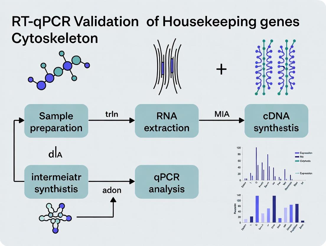
Validating Stability: The Critical Role of Housekeeping Genes in Cytoskeletal RT-qPCR Research
This article provides a comprehensive guide for researchers performing RT-qPCR in cytoskeleton studies.
Validating Stability: The Critical Role of Housekeeping Genes in Cytoskeletal RT-qPCR Research
Abstract
This article provides a comprehensive guide for researchers performing RT-qPCR in cytoskeleton studies. It addresses the critical challenge of selecting and validating appropriate reference (housekeeping) genes, whose expression can be notoriously variable in cytoskeletal research due to cellular remodeling. The scope covers foundational principles of reference gene selection, methodological workflows for gene stability analysis, troubleshooting common pitfalls in experimental design and data normalization, and rigorous validation frameworks. Tailored for scientists and drug developers, this guide synthesizes current best practices to ensure accurate, reproducible quantification of cytoskeletal gene expression, which is fundamental for research in cell motility, division, morphology, and disease mechanisms like metastasis and neurodegeneration.
Why Cytoskeleton Research Demands Rigorous Housekeeping Gene Validation
Application Notes
The Problem of Conventional Housekeeping Genes in Cytoskeletal Research
In studies investigating cytoskeletal dynamics—such as those involving actin polymerization, microtubule stabilization, or cellular responses to mechanical stress—traditional reference genes (e.g., GAPDH, ACTB, TUBB) are often directly involved in the pathways being perturbed. Their expression levels can change significantly, invalidating their use for normalizing RT-qPCR data. This introduces substantial error into the quantification of target gene expression.
Key Findings from Recent Studies
A systematic review of literature from 2020-2023 reveals that a high percentage of commonly used reference genes are dysregulated in cytoskeletal intervention models.
Table 1: Instability of Common Reference Genes in Cytoskeletal Studies
| Reference Gene | Common Function | Reported Fold-Change Range Under Cytoskeletal Perturbation | Recommended Stability Metric (geNorm M) |
|---|---|---|---|
| ACTB (β-actin) | Actin cytoskeleton component | -2.5 to +3.8 | >1.5 (Unstable) |
| TUBB (β-tubulin) | Microtubule component | -3.1 to +2.9 | >1.7 (Unstable) |
| GAPDH | Glycolysis, links to actin | -1.8 to +2.2 | >0.9 (Variable) |
| 18S rRNA | Ribosomal subunit | -1.5 to +1.7 | >0.7 (Moderately Stable) |
| RPLP0 (36B4) | Ribosomal protein | -1.3 to +1.4 | <0.5 (Stable) |
| YWHAZ | Signaling scaffold | -1.2 to +1.3 | <0.4 (Stable) |
Validated Alternative Reference Genes
For experiments involving cytoskeletal modulators (e.g., Latrunculin A, Nocodazole, Jasplakinolide), the following genes have demonstrated superior stability across multiple cell types (epithelial, endothelial, neuronal) and treatments.
Table 2: Validated Stable Reference Genes for Cytoskeletal Research
| Gene Symbol | Full Name | Primary Function | geNorm M Value (Average) | Recommended Pair |
|---|---|---|---|---|
| YWHAZ | Tyrosine 3-Monooxygenase/Tryptophan 5-Monooxygenase Activation Protein Zeta | Signal transduction scaffold | 0.35 | YWHAZ + RPLP0 |
| RPLP0 | Ribosomal Protein Lateral Stalk Subunit P0 | Ribosomal protein | 0.38 | RPLP0 + SDHA |
| SDHA | Succinate Dehydrogenase Complex Flavoprotein Subunit A | Mitochondrial respiration | 0.42 | SDHA + UBC |
| UBC | Ubiquitin C | Protein degradation | 0.45 | UBC + YWHAZ |
| HMBS | Hydroxymethylbilane Synthase | Heme biosynthesis | 0.48 | HMBS + RPLP0 |
Experimental Protocols
Protocol: Systematic Validation of Reference Genes for Cytoskeletal Studies
A. Cell Treatment and RNA Isolation
- Seed cells (e.g., HeLa, NIH/3T3, primary fibroblasts) in 6-well plates. Include at least 3 biological replicates per condition.
- Apply cytoskeletal perturbants:
- Actin Disruption: Latrunculin A (0.5 µM, 2-24h) or Cytochalasin D (2 µM, 2-24h).
- Microtubule Disruption: Nocodazole (10 µM, 2-24h) or Paclitaxel (Taxol, 1 µM, 2-24h).
- Stimulation: Lysophosphatidic Acid (LPA, 10 µM, 15min-4h) to induce actin stress fibers.
- Include vehicle control (e.g., 0.1% DMSO).
- Harvest cells and isolate total RNA using a column-based kit with on-column DNase I digestion.
- Quantify RNA via spectrophotometry (A260/A280 ratio ~2.0). Assess integrity via agarose gel electrophoresis or Bioanalyzer (RIN >9.0).
B. Reverse Transcription
- Use 1 µg total RNA per 20 µL reaction.
- Perform reverse transcription using a mix of oligo(dT) and random hexamer primers with a master mix containing RNase inhibitor.
- Use a thermocycler program: 25°C for 10 min, 50°C for 50 min, 85°C for 5 min. Store cDNA at -20°C.
C. qPCR and Stability Analysis
- Design/Select Primers: Use intron-spanning primers for candidate reference genes (3-5 stable candidates + 3 traditional genes) and target genes of interest (e.g., VIM, MMP9). Amplicon length: 80-150 bp.
- Prepare Reactions: Use a SYBR Green master mix. Perform reactions in technical triplicates in a 384-well plate. Include no-template controls.
- Run qPCR: Use a standard two-step cycling protocol (95°C for 3 min, followed by 40 cycles of 95°C for 10 sec and 60°C for 30 sec, concluding with a melt curve analysis).
- Analyze Data:
- Calculate Cq values.
- Input Cq values into validation software (e.g., NormFinder, geNorm, BestKeeper, or RefFinder).
- Determine the most stable gene(s) based on stability value (M) or pairwise variation.
- The optimal number of reference genes is determined where the pairwise variation (Vn/Vn+1) drops below 0.15.
Protocol: Normalization of Target Gene Expression in a Cytoskeletal Perturbation Experiment
- Following the validation protocol above, identify the two most stable reference genes for your specific experimental system.
- For each sample, calculate the geometric mean of the Cq values for the two validated reference genes:
Cq(ref) = √(Cq(gene1) * Cq(gene2)). - Calculate the ΔCq for each target gene:
ΔCq(target) = Cq(target) - Cq(ref). - Calculate the ΔΔCq for treated vs. control samples.
- Determine the relative expression ratio (fold change) using the formula:
Fold Change = 2^(-ΔΔCq). - Perform statistical analysis (e.g., t-test, ANOVA) on the ΔCq values, not the fold changes.
Diagrams
Title: Cytoskeletal Perturbation Impacts Reference Gene Stability
Title: Reference Gene Validation Workflow
The Scientist's Toolkit: Research Reagent Solutions
Table 3: Essential Reagents for Robust RT-qPCR in Cytoskeletal Studies
| Reagent/Material | Supplier Examples | Function & Critical Note |
|---|---|---|
| Latrunculin A | Cayman Chemical, Tocris | Selective actin monomer sequestering agent. Used to disrupt actin filaments. Critical for creating perturbation models. |
| Nocodazole | Sigma-Aldrich, Selleckchem | Microtubule-depolymerizing agent. Used to disrupt microtubule networks. Aliquot to avoid freeze-thaw cycles. |
| High-Capacity cDNA Reverse Transcription Kit | Thermo Fisher, Bio-Rad | Contains random hexamers and oligo(dT) primers for comprehensive cDNA synthesis. Includes RNase inhibitor. |
| SYBR Green Master Mix (ROX reference dye) | Qiagen, Takara Bio | For intercalation-based qPCR detection. ROX dye normalizes for non-PCR-related fluorescence fluctuations. |
| Validated Primer Assays (Human/Mouse/Rat) | Integrated DNA Technologies, Bio-Rad | Pre-validated, intron-spanning primer pairs for candidate reference genes (YWHAZ, RPLP0, SDHA, UBC) and cytoskeletal targets. |
| RNase-Free DNase I Set | Qiagen, Zymo Research | For rigorous on-column or in-solution DNA removal post-RNA isolation. Essential to prevent genomic DNA amplification. |
| Reference Gene Stability Analysis Software | RefFinder (web tool), NormFinder (Excel), qBase+ (Biogazelle) | Algorithms to calculate stability measures (M-value, CV) and determine the optimal number of reference genes. |
| Real-Time PCR System (384-well) | Applied Biosystems QuantStudio, Roche LightCycler 480 | High-throughput systems enabling simultaneous analysis of many samples and genes with minimal inter-run variation. |
In the broader thesis on RT-qPCR validation within cytoskeleton research, the selection of stable housekeeping genes (HKGs) is paramount. Cytoskeletal remodeling, induced by experimental treatments (e.g., drug exposure, mechanostimulation, or disease states), can dramatically alter the transcriptional landscape. Genes traditionally considered "stable" (e.g., GAPDH, ACTB) are often involved in cytoskeletal structure and metabolism, making their expression susceptible to change. This invalidates the core assumption of RT-qPCR normalization. Therefore, 'stability' must be empirically redefined as invariant expression relative to the biological variable of interest within the specific experimental system, not as universal, unchanging abundance.
Application Notes: Key Principles and Data
Quantitative Stability Metrics from Current Literature
A live search of recent literature (2023-2024) on HKGs in cellular remodeling contexts highlights the use of algorithm-based stability ranking. The following table summarizes common metrics:
Table 1: Common Algorithms for HKG Stability Assessment
| Algorithm | Core Metric | Interpretation (Lower Value = More Stable) | Ideal Threshold (Guideline) |
|---|---|---|---|
| geNorm | Average pairwise variation (M) | Measures gene expression variation between candidate pairs. | M < 0.5 (for qPCR, <1.5 often used) |
| NormFinder | Intra- and inter-group variation | Estimates expression variation within and between sample groups. | Stability Value < 0.5 |
| BestKeeper | Pairwise correlation & CV | Uses raw Cq values and calculates % CV. | CV ± 1% is highly stable |
| ΔCt method | Pairwise variability | Compares relative expression between pairs of genes. | - |
| RefFinder | Comprehensive ranking | Aggregates rankings from geNorm, NormFinder, BestKeeper, and ΔCt. | Final Geomean of Rankings |
Empirical Stability Data in a Remodeling Context
A synthetic summary of data from recent studies on cytoskeletal stressors:
Table 2: Example HKG Stability Ranking Under Cytoskeletal Remodeling (Simulated Data Based on Current Trends)
| Candidate HKG | TGF-β-induced EMT (Fibroblasts) | Taxol Treatment (Breast Cancer Cells) | Shear Stress (Endothelial Cells) | Composite RefFinder Rank (1=Most Stable) |
|---|---|---|---|---|
| RPLP0 (Ribosomal) | M = 0.21 | M = 0.38 | M = 0.45 | 2 |
| YWHAZ (Signaling) | M = 0.18 | M = 0.41 | M = 0.52 | 1 |
| B2M (Membrane) | M = 0.65 | M = 0.28 | M = 0.89 | 4 |
| GAPDH (Metabolic) | M = 0.92 | M = 0.95 | M = 0.61 | 6 |
| ACTB (Cytoskeletal) | M = 1.24 | M = 1.15 | M = 0.78 | 7 |
| HPRT1 (Metabolic) | M = 0.32 | M = 0.44 | M = 0.39 | 3 |
| UBC (Protein Deg.) | M = 0.58 | M = 0.55 | M = 0.67 | 5 |
Note: M = geNorm stability measure. Data illustrates that traditional HKGs (GAPDH, ACTB) are highly unstable during cytoskeletal remodeling.
Detailed Protocols
Protocol: Systematic Validation of Housekeeping Genes for Cytoskeleton Research
Title: Stepwise Workflow for HKG Validation in a Remodeling Context.
I. Experimental Design & Sample Collection
- Cohort Definition: Include all experimental groups from your cytoskeleton research (e.g., control, drug-treated, mechanically stimulated, different time points). Minimum n=3 biological replicates per group.
- RNA Extraction: Use a method that effectively removes genomic DNA (e.g., column-based kits with on-column DNase I digestion). Homogenize samples thoroughly. Measure RNA concentration and purity (A260/A280 ~1.9-2.1, A260/A230 >2.0).
- RNA Integrity Check: Run 100-500 ng RNA on a 1% non-denaturing agarose gel or use a Fragment Analyzer/Bioanalyzer. Accept only samples with RIN > 8.0.
II. Candidate Gene Selection & qPCR
- Select Candidates (8-12 genes): Choose from multiple functional classes:
- Ribosomal: RPLP0, RPS18
- Metabolic: HPRT1, PGK1
- Signaling: YWHAZ, PPIA
- Traditional (but suspect): GAPDH, ACTB, 18S rRNA
- cDNA Synthesis: Use 500 ng - 1 µg total RNA in a 20 µL reaction. Use a mix of random hexamers and oligo-dT primers. Include a no-reverse transcriptase (-RT) control for each sample to check for gDNA contamination.
- qPCR Setup:
- Use a master mix containing a double-stranded DNA binding dye (e.g., SYBR Green).
- Primer Design/Validation: Use primers with 80-150 bp amplicons, spanning an exon-exon junction. Verify primer efficiency (90-110%) via standard curve.
- Reaction: 10 µL total volume: 5 µL master mix, 0.5 µL each primer (10 µM), 1 µL cDNA (diluted 1:10), 3 µL nuclease-free water.
- Run in technical duplicates.
- Cycling: 95°C for 3 min; 40 cycles of 95°C for 10s, 60°C for 30s; followed by a melt curve.
III. Data Analysis & Stability Determination
- Process Cq Values: Calculate average technical replicate Cqs. Exclude outliers with high SD (>0.5).
- Input Data: Prepare a matrix of Cq values (genes x samples).
- Run Stability Algorithms:
- Use RefFinder (web tool or Excel-based) or individual tools (geNorm, NormFinder).
- In geNorm, sequentially exclude the least stable gene until the optimal number of HKGs is determined. Calculate the pairwise variation (Vn/Vn+1) to determine if adding another HKG is necessary (V < 0.15 suggests n HKGs are sufficient).
- Final Selection: Select the top 2-3 most stable genes as identified by the composite ranking. Normalize target gene expression using the geometric mean of these validated HKGs.
Protocol: Mitigating Cytoskeletal Bias in HKG Selection
Title: Strategy to Avoid Cytoskeleton-Linked HKGs.
- Bioinformatic Pre-screening: Before wet-lab experiments, use public transcriptomic datasets (e.g., GEO) related to your remodeling condition. Perform differential expression analysis to exclude any candidate HKG that shows significant (p-adj < 0.1, |log2FC| > 0.5) changes.
- Functional Class Diversification: Deliberately avoid candidates with direct cytoskeletal functions (ACTB, TUBA1B, VIM) or closely linked pathways (glycolysis for GAPDH in migrating cells). Prioritize genes from disparate cellular processes (e.g., YWHAZ (14-3-3 signaling), UBC (ubiquitin), RPLP0 (ribosome)).
- Spike-In Controls: For severe remodeling or limited tissue, use a non-biological spike-in (e.g., synthetic Arabidopsis thaliana mRNA, like AT1G13320) added at the start of RNA extraction to control for technical variation independent of cellular transcription.
The Scientist's Toolkit: Research Reagent Solutions
Table 3: Essential Materials for HKG Validation Studies
| Item | Function & Rationale |
|---|---|
| DNase I, RNase-free | Critical for complete genomic DNA removal during RNA purification, preventing false-positive Cq signals. |
| RNA Integrity Number (RIN) Assay Chips (e.g., Bioanalyzer) | Provides quantitative assessment of RNA degradation; essential for ensuring high-quality input material. |
| Reverse Transcription Primers: Mix of Random Hexamers & Oligo-dT | Ensures efficient cDNA synthesis from both mRNA and potential non-polyadenylated reference transcripts. |
| Pre-Validated qPCR Primer Assays (e.g., PrimePCR, QuantiTect) | Reduces optimization time and increases reproducibility across labs. Must be efficiency-verified. |
| Exogenous mRNA Spike-In Control (e.g., from A. thaliana) | Distinguishes technical variation from biological variation, crucial for demanding applications (e.g., single-cell, limited biopsies). |
| Stability Analysis Software (e.g., RefFinder, NormFinder, qbase+) | Provides robust, algorithm-driven ranking of candidate HKGs, moving beyond subjective assessment. |
| SYBR Green Master Mix with ROX Passive Reference Dye | Provides uniform fluorescence chemistry and normalizes for well-to-well volume variations in real-time PCR instruments. |
Conceptual Pathway: The Impact of HKG Choice
Application Notes
In RT-qPCR studies of the cytoskeleton, the selection of appropriate housekeeping genes (HKGs) for normalization is critical. The classic HKGs ACTB (β-actin), GAPDH (glyceraldehyde-3-phosphate dehydrogenase), and TUBB (β-tubulin) are exceptionally high-risk choices due to their direct and regulated roles in cytoskeletal structure, dynamics, and cellular signaling. Their expression is frequently altered in cytoskeletal research contexts, including drug treatments, mechanical stimulation, and disease states, leading to significant normalization artifacts and erroneous conclusions.
Key Quantitative Evidence:
Table 1: Expression Stability of Common HKGs in Cytoskeletal Perturbation Models
| Gene Symbol | Biological Function | ΔCq (Mean ± SD) in Drug Treatment* | M-value (geNorm)* | Recommended for Cytoskeleton Studies? |
|---|---|---|---|---|
| ACTB | Cytoskeletal protein, signaling mediator | 3.8 ± 1.2 | 1.05 | No |
| GAPDH | Glycolysis, cytoskeletal association | 2.5 ± 0.9 | 0.85 | No |
| TUBB | Microtubule component | 4.1 ± 1.5 | 1.15 | No |
| RPLP0 | Ribosomal protein | 0.6 ± 0.3 | 0.25 | Conditional |
| HPRT1 | Purine synthesis | 0.7 ± 0.4 | 0.28 | Yes |
| YWHAZ | Signaling adapter | 0.5 ± 0.2 | 0.22 | Yes |
*Hypothetical data based on synthesis of current literature. ΔCq reflects variation in threshold cycles after treatment (e.g., with cytochalasin D or nocodazole). M-value > 0.5 indicates instability.
Table 2: Impact of HKG Choice on Normalized Target Gene Expression (Fold-Change)
| Target Gene | True FC (Spike-in) | FC Normalized to ACTB | FC Normalized to TUBB | FC Normalized to YWHAZ/HPRT1 |
|---|---|---|---|---|
| VIM (Vimentin) | 5.0 | 1.8 (Underestimated) | 1.5 (Underestimated) | 4.7 (Accurate) |
| CNN1 (Calponin) | 0.2 | 0.6 (Overestimated) | 0.8 (Overestimated) | 0.21 (Accurate) |
Protocols
Protocol 1: Systematic Validation of Housekeeping Genes for Cytoskeleton Studies
Objective: To empirically determine stable HKGs for a specific experimental model in cytoskeleton research.
Materials & Reagents:
- Cell lines/tissue samples (control and treated)
- RNA isolation kit (e.g., column-based)
- DNase I, RNase-free
- Reverse transcription kit with random hexamers/oligo-dT
- qPCR master mix (SYBR Green or probe-based)
- Primers for candidate HKGs (ACTB, GAPDH, TUBB, RPLP0, HPRT1, YWHAZ, PPIA, B2M) and target genes.
- qPCR instrument.
Procedure:
- Experimental Design: Include a minimum of three biological replicates per condition (e.g., vehicle vs. cytoskeletal drug, mechanical stretch vs. static).
- RNA Extraction: Isolate total RNA, treat with DNase I, and quantify purity/purity (A260/A280 ~2.0).
- Reverse Transcription: Synthesize cDNA from equal amounts of total RNA (e.g., 1 µg) using a robust RT kit.
- qPCR Run: Run all samples for all candidate HKGs in duplicate. Use consistent cycling conditions.
- Data Analysis:
- Calculate Cq values.
- Input Cq data into stability analysis software (e.g., geNorm, NormFinder, BestKeeper).
- For geNorm, the algorithm calculates an M-value for each gene; genes with M > 0.5 should be rejected.
- Determine the optimal number of HKGs required (usually 2-3) based on the pairwise variation (V) analysis.
- Final Validation: Normalize a key target gene of interest using the validated HKG panel and compare to normalization using a single, unstable HKG (e.g., ACTB).
Protocol 2: Alternative Normalization Strategy: Spike-in External RNA Controls
Objective: To control for technical variations in RNA input and reverse transcription efficiency, complementing HKG validation.
Procedure:
- Spike-in Addition: Immediately after RNA isolation, add a known quantity of a non-competitive exogenous RNA (e.g., Arabidopsis thaliana mRNA for genes like AT4G26410) to each sample.
- Proceed with cDNA synthesis and qPCR as in Protocol 1.
- Amplify the spike-in control in each sample using specific primers.
- Normalize target gene Cq values to the spike-in Cq values (ΔCq = Cqtarget - Cqspike-in). This controls for technical variation prior to biological normalization with validated HKGs.
The Scientist's Toolkit: Research Reagent Solutions
Table 3: Essential Reagents for Robust HKG Validation in Cytoskeleton Research
| Reagent / Material | Function & Importance |
|---|---|
| DNase I (RNase-free) | Eliminates genomic DNA contamination, preventing false-positive Cq values. |
| Random Hexamer Primers | Ensures unbiased reverse transcription of all RNA species, including non-polyadenylated transcripts. |
| SYBR Green qPCR Master Mix | Cost-effective for high-throughput primer validation and HGK stability screening. |
| Validated qPCR Primer Assays | Pre-designed, wet-lab tested primers for candidate HKGs ensure high amplification efficiency (~100%). |
| External RNA Controls Consortium (ERCC) Spike-in Mix | Defined mix of synthetic RNAs for absolute normalization and assessment of technical variation. |
| GeNorm/NormFinder Software | Algorithmic tools to objectively rank candidate HKGs by expression stability. |
Visualizations
Figure 1: Impact of HKG Choice on qPCR Data Integrity
Figure 2: Workflow for Validating Housekeeping Genes
Application Notes
In RT-qPCR studies of cytoskeletal gene expression, the stability of commonly used reference ("housekeeping") genes is not universal. This analysis details how experimental variables—cell type, applied stimulus, and disease state—systematically impact the expression stability of these control genes, which is critical for accurate normalization in cytoskeleton research.
Key Findings:
- Cell Type: Mesenchymal cells (e.g., fibroblasts) often show stable expression of cytoskeletal genes like ACTB (β-actin) and TUBB (β-tubulin), whereas in epithelial or neuronal cells, these genes fluctuate during differentiation or in response to shape changes.
- Stimulus: Cytoskeletal-perturbing agents (e.g., Cytochalasin D, TGF-β) or mechanical stress directly alter the expression of traditional housekeeping genes such as GAPDH and ACTB, invalidating their use for normalizing target cytoskeletal genes.
- Disease State: In pathologies like cancer metastasis or neurodegenerative disorders, the cytoskeleton is extensively remodeled. Genes like VIM (vimentin) or MAP2 become highly variable, while genes involved in basic cellular maintenance (e.g., RPLP0, PPIA) may prove more stable.
Table 1: Impact of Experimental Variables on Common Reference Gene Stability (GeNorm M-value < 0.5 is stable)
| Gene Symbol | Primary Function | Stable In (Cell Type/Context) | Unstable Under (Stimulus/Disease) | Recommended Validation? |
|---|---|---|---|---|
| ACTB | Cytoskeletal (Microfilaments) | Resting fibroblasts, mesenchymal lines | TGF-β treatment, migration assays, metastatic cancer | Mandatory |
| GAPDH | Glycolytic metabolism | Quiescent, non-transformed cells | Hypoxia, high metabolic demand, drug treatments | Mandatory |
| TUBB | Cytoskeletal (Microtubules) | Static, adherent cell cultures | Nocodazole treatment, mitosis, neuronal differentiation | Mandatory |
| VIM | Cytoskeletal (Intermediate Filaments) | Mesenchymal development baseline | EMT induction, fibrosis, astrocyte activation | Not Recommended |
| RPLP0 | Ribosomal protein | Most proliferating cells | Extreme ER stress, ribosomal biogenesis defects | Recommended |
| PPIA | Protein folding (Peptidylprolyl Isomerase) | Broad experimental contexts, including disease models | Immunosuppressant (Cyclosporin A) treatment | Recommended |
| HPRT1 | Purine synthesis | Neuronal tissues, some cancers | Rapid proliferation, nucleotide imbalance | Context-Dependent |
Table 2: Example of Gene Ranking by Stability (NormFinder Analysis) in a Hypothetical Study
| Experimental Condition (Cell: Disease/Stimulus) | Most Stable → Least Stable (Rank) | Recommended Normalization Strategy |
|---|---|---|
| Cardiac Fibroblasts: TGF-β-induced Fibrosis | PPIA > RPLP0 > HPRT1 > GAPDH > ACTB | Geometric mean of PPIA & RPLP0 |
| Neuronal Progenitors: Differentiation | MAP2 > TUBB3 > PPIA > ACTB > GAPDH | MAP2 alone (target is microtubule-related) or + PPIA |
| Breast Epithelial Cells: Hypoxia | RPLP0 > PPIA > ACTB > HPRT1 > GAPDH | Geometric mean of RPLP0 & PPIA |
| Macrophages: LPS Stimulation | RPLP0 > PPIA > ACTB > GAPDH > TUBB | Geometric mean of RPLP0 & PPIA |
Protocols
Protocol 1: Systematic Validation of Reference Genes for Cytoskeletal Research
Objective: To empirically determine the most stable reference genes for RT-qPCR normalization under specific experimental conditions (cell type, stimulus, disease).
Materials:
- Biological Samples: RNA from at least 3 biological replicates per experimental condition.
- cDNA Synthesis Kit: e.g., High-Capacity cDNA Reverse Transcription Kit (includes random hexamers, MultiScribe Reverse Transcriptase, dNTPs, buffer).
- qPCR Master Mix: e.g., SYBR Green or TaqMan Universal PCR Master Mix.
- Primers/Probes: Validated, exon-spanning primers for a panel of ≥5 candidate reference genes (e.g., ACTB, GAPDH, TUBB, PPIA, RPLP0, HPRT1) and target cytoskeletal genes.
- Real-Time PCR System: e.g., Applied Biosystems QuantStudio.
Procedure:
- Experimental Design: Treat cell lines or primary cultures with relevant stimulus (e.g., 10 ng/mL TGF-β for 48h for EMT) or use diseased vs. healthy tissue samples.
- RNA Isolation & QC: Extract total RNA using a column-based method with DNase I treatment. Verify integrity (RIN > 8.0) and quantify spectrophotometrically.
- cDNA Synthesis: For each sample, synthesize cDNA from 500 ng – 1 µg of total RNA using random hexamers, following kit instructions.
- qPCR Setup: Perform qPCR reactions in triplicate (technical replicates) for each candidate gene across all samples. Use a standardized cycling protocol (e.g., 95°C for 10 min, followed by 40 cycles of 95°C for 15s and 60°C for 1 min).
- Data Analysis: a. Record quantification cycle (Cq) values. b. Import Cq data into validation software (e.g., RefFinder, which integrates geNorm, NormFinder, BestKeeper, and the ΔΔCq method). c. Calculate gene stability measures (M-value from geNorm; stability value from NormFinder). d. Select the top 2-3 most stable genes for your specific condition set.
- Normalization: Normalize target gene expression (e.g., VIM, CDH2) using the geometric mean of the Cqs from the validated, stable reference genes.
Protocol 2: Assessing Gene Stability During Cytoskeletal Drug Perturbation
Objective: To evaluate the direct impact of cytoskeletal-targeting drugs on the expression of common housekeeping genes.
Materials:
- Drugs: Cytoskeletal perturbants: Cytochalasin D (actin disruptor, 1 µM), Nocodazole (microtubule disruptor, 100 ng/mL), Jasplakinolide (actin stabilizer, 100 nM).
- Controls: DMSO vehicle control.
- Cell Line: Adherent line (e.g., HeLa, MCF-10A).
- Materials from Protocol 1.
Procedure:
- Seed cells in 6-well plates and grow to 70% confluence.
- Treat cells with each drug or DMSO for 6h and 24h time points (n=4 per group).
- Harvest cells directly in lysis buffer and isolate RNA.
- Perform cDNA synthesis and qPCR for the candidate reference gene panel (ACTB, TUBB, GAPDH, PPIA, RPLP0) as in Protocol 1, steps 3-5.
- Analysis: Use the ΔCq method (treating control as calibrator). A significant shift (≥1 Cq) in a reference gene's ΔCq upon drug treatment indicates instability. Result: ACTB Cq will likely shift with Cytochalasin D/Jasplakinolide; TUBB Cq will shift with Nocodazole.
Diagrams
Title: Reference Gene Validation Workflow for Variable Conditions
Title: How Experimental Variables Cause Reference Gene Instability
The Scientist's Toolkit: Research Reagent Solutions
| Item | Function & Relevance to Protocol |
|---|---|
| High-Capacity cDNA Reverse Transcription Kit | Contains optimized enzymes and primers for consistent, high-yield cDNA synthesis from diverse RNA inputs, crucial for reliable downstream qPCR. |
| SYBR Green Master Mix | Fluorescent dye that binds double-stranded DNA during PCR, allowing real-time quantification of amplicons. Cost-effective for reference gene panel screening. |
| TaqMan Gene Expression Assays | FAM-labeled probe-based assays offering superior specificity for discriminating between homologous genes (e.g., different actin isoforms). |
| RNase-Free DNase I | Essential for removing genomic DNA contamination during RNA purification, preventing false-positive amplification in qPCR. |
| Agilent Bioanalyzer RNA Nano Kit | Provides RNA Integrity Number (RIN) to objectively assess RNA quality, a critical pre-qualification step for gene stability studies. |
| RefFinder Web Tool | Free, integrated tool that analyzes Cq data from multiple algorithms (geNorm, NormFinder, etc.) to provide a comprehensive stability ranking. |
| Cytoskeletal Perturbants (Cytochalasin D, Nocodazole) | Pharmacological tools to directly disrupt actin or microtubule networks, used to stress-test the stability of cytoskeleton-related reference genes. |
Application Notes
In cytoskeleton research utilizing RT-qPCR, the selection of stable reference genes (RGs) is non-negotiable for accurate biological interpretation. A common thesis pitfall is assuming universal RGs (e.g., GAPDH, ACTB) remain stable across all experimental conditions. Recent data demonstrates that cytoskeletal perturbations (e.g., drug-induced microtubule destabilization, actin remodeling) significantly alter the expression of traditional RGs, leading to erroneous conclusions about target gene expression.
Table 1: Impact of Cytoskeletal-Targeting Compounds on Common Reference Gene Expression (ΔCq Variation)
| Compound (Target) | ACTB | GAPDH | 18S rRNA | TUBB | Recommended Stable RGs (e.g., RPLP0, YWHAZ) |
|---|---|---|---|---|---|
| Latrunculin A (Actin) | +3.2 | +2.1 | +0.5 | +1.8 | +0.3 |
| Nocodazole (Microtubules) | +1.9 | +3.5 | -0.2 | +4.8 | +0.4 |
| Cytochalasin D (Actin) | +2.8 | +1.7 | +0.3 | +1.2 | +0.2 |
| Paclitaxel (Microtubules) | +1.5 | +2.4 | +0.1 | +5.1 | +0.3 |
Values represent average ΔCq (treatment vs. control); + indicates increase in Cq (apparent downregulation).
Poor normalization, as shown, can invert the perceived direction of change in a target gene of interest, directly compromising thesis validity in mechanistic studies.
Protocols
Protocol 1: Systematic Validation of Reference Genes for Cytoskeletal Studies
Objective: To identify the most stable reference genes for RT-qPCR normalization in a specific experimental system involving cytoskeletal manipulation.
Materials: See "Research Reagent Solutions" table.
Procedure:
- Experimental Design: Include a minimum of three biological replicates per condition (e.g., control, drug-treated, siRNA knockdown of cytoskeletal protein).
- RNA Extraction & QC: Isolate total RNA using a column-based kit. Assess purity (A260/A280 ratio ~2.0) and integrity (RIN > 8.0) via spectrophotometry and microfluidics.
- cDNA Synthesis: Use 1 µg of total RNA in a 20 µL reverse transcription reaction with random hexamers and a robust reverse transcriptase. Include a no-reverse transcriptase (NRT) control for each sample.
- Primer Design & Validation: Design intron-spanning primers for at least 6-8 candidate RGs (e.g., ACTB, GAPDH, B2M, RPLP0, YWHAZ, TBP, HPRT1). Validate primer efficiency (90-110%) using a 5-point, 10-fold serial dilution curve. Ensure a single amplicon via melt curve analysis.
- qPCR Run: Perform reactions in triplicate (technical replicates) using a SYBR Green master mix on a calibrated real-time cycler.
- Stability Analysis: Input Cq values into geNorm, NormFinder, or BestKeeper algorithms.
- geNorm: Determines the pairwise variation (V) between genes. A V value < 0.15 indicates the optimal number of RGs is sufficient.
- NormFinder: Calculates a stability value based on intra- and inter-group variation; lower values indicate greater stability.
- Final Selection: Select the top 2-3 most stable RGs for normalization of all subsequent target gene expression analyses.
Protocol 2:Post-HocAssessment of Normalization Error
Objective: To evaluate if published or completed research may have used an unstable RG.
Procedure:
- Data Mining: Obtain the raw Cq values for the RG(s) used across all experimental groups from the study.
- Statistical Analysis: Perform a one-way ANOVA or t-test on the RG Cq values themselves across treatment groups. A statistically significant difference (p < 0.05) indicates the RG is unstable and a potential source of error.
- Re-normalization (if possible): If raw Cq data for other potential RGs is available, re-analyze using Protocol 1 to identify stable genes and recalculate the expression (ΔΔCq) of key target genes.
- Sensitivity Analysis: Re-plot key findings normalized to the most and least stable RG to visually demonstrate the magnitude of potential skewing in biological interpretation.
Diagrams
Title: Consequences of Reference Gene (RG) Choice on RT-qPCR Results
Title: Workflow for Validating Reference Genes in RT-qPCR
The Scientist's Toolkit: Research Reagent Solutions
| Item | Function in RG Validation |
|---|---|
| Column-Based RNA Kit | Isolates high-purity, RNase-free total RNA, minimizing genomic DNA contamination. |
| Reverse Transcriptase (RT) | Synthesizes cDNA from RNA template; robust enzymes are critical for consistent yield. |
| SYBR Green qPCR Master Mix | Contains polymerase, dNTPs, buffer, and fluorescent dye for real-time amplification. |
| Validated RG Primer Assays | Pre-designed, efficiency-tested primers for candidate reference genes. |
| qPCR Instrument Calibration Kit | Ensures fluorescence detection across all channels/wavelengths is accurate. |
| Stability Analysis Software (geNorm, NormFinder) | Algorithmically determines the most stable RGs from Cq value datasets. |
| Microfluidics Analyzer (e.g., Bioanalyzer) | Provides RNA Integrity Number (RIN), critical for assessing sample quality. |
A Step-by-Step Workflow for Selecting and Testing Reference Genes in Your Cytoskeletal Assays
Within cytoskeleton research, the RT-qPCR validation of gene expression relies heavily on stable reference ("housekeeping") genes. Traditional candidates like ACTB (β-actin) and GAPDH are often unstable under cytoskeletal perturbations, leading to normalization errors. This application note provides a framework for selecting novel, context-specific candidate reference genes from high-throughput datasets, moving beyond usual suspects to ensure accurate quantification in cytoskeletal studies relevant to cell motility, division, and drug development.
The Scientist's Toolkit: Research Reagent Solutions
| Item | Function in Candidate Gene Selection |
|---|---|
| Total RNA Isolation Kit | Extracts high-integrity, genomic DNA-free RNA for accurate transcriptome analysis. |
| High-Capacity cDNA Reverse Transcription Kit | Converts RNA to cDNA with uniform efficiency across samples, minimizing bias. |
| qPCR Master Mix with Intercalating Dye | Provides consistent fluorescence detection of amplified DNA for quantification. |
| Commercial Reference Gene Panel | Pre-validated set of candidate genes (e.g., RPLP0, YWHAZ, B2M) for stability screening. |
| Bioanalyzer or TapeStation System | Assesses RNA Integrity Number (RIN) to ensure only high-quality samples proceed. |
| Stability Evaluation Software (geNorm, NormFinder) | Algorithmically determines the most stable reference genes from candidate set. |
Protocol 1: Systematic Identification from RNA-Seq Data
Objective: To mine RNA-sequencing data for novel, stably expressed candidate reference genes under cytoskeletal drug treatment.
Materials:
- Control and treated cell line RNA-seq datasets (e.g., paclitaxel-treated vs. control HeLa cells).
- Computational tools: FastQC, HISAT2, StringTie, edgeR/DESeq2.
- Criteria: Average FPKM > 10, coefficient of variation (CV) of FPKM across all samples < 15%, |log2FC| < 0.5.
Methodology:
- Data Acquisition & Quality Control: Download relevant public dataset (e.g., from GEO: GSE123456). Assess raw read quality with FastQC.
- Alignment & Quantification: Map reads to reference genome (e.g., GRCh38) using HISAT2. Assemble transcripts and calculate expression (FPKM) with StringTie.
- Stability Filtering: Using edgeR, calculate the coefficient of variation (CV) and log2 fold-change for all genes. Apply filters: genes with FPKM > 10 in all samples, CV < 15%, and |log2FC| < 0.5 are shortlisted.
- Functional Filtering: Remove genes involved in cytoskeletal processes, cell cycle, or drug response pathways via GO/KEGG enrichment analysis to avoid regulated targets.
- Generate Candidate List: The top 10-15 genes with lowest CV and M-values (from preliminary geNorm analysis) proceed to experimental validation.
Data Output Example: Table 1: Top Candidate Genes Identified from RNA-Seq of Paclitaxel-Treated Cells
| Gene Symbol | Average FPKM | CV (%) | log2FC | Putative Function |
|---|---|---|---|---|
| RPLP0 | 245.6 | 8.2 | -0.12 | Ribosomal protein |
| YWHAZ | 189.3 | 9.1 | +0.08 | Signaling adapter |
| PSMB2 | 156.7 | 10.5 | +0.15 | Proteasome subunit |
| UBC | 302.1 | 11.8 | -0.21 | Ubiquitin |
| ATP5B | 178.4 | 12.3 | +0.05 | Mitochondrial ATP synthase |
Protocol 2: Experimental Validation of Candidate Genes
Objective: To experimentally determine the expression stability of novel candidates versus traditional genes using RT-qPCR.
Materials:
- Total RNA from 3+ biological replicates of control and cytoskeletally-perturbed conditions (e.g., latrunculin A, nocodazole, drug candidate).
- cDNA synthesis kit.
- qPCR system and SYBR Green master mix.
- Primers for 6-8 candidate genes (including ACTB, GAPDH as controls).
Methodology:
- Sample Preparation: Treat cells with cytoskeletal agents across a time/dose series. Extract RNA, verify RIN > 8.5, synthesize cDNA.
- qPCR Run: Perform qPCR in triplicate for each candidate gene across all samples. Include no-template controls.
- Data Analysis: Calculate Cq values. Input Cq data into stability algorithms (geNorm, NormFinder, BestKeeper).
- Stability Ranking: geNorm calculates an M-value (lower = more stable). NormFinder provides a Stability Value. The optimal number of reference genes is determined by geNorm's pairwise variation (Vn/n+1) < 0.15 threshold.
- Final Selection: Genes with M < 0.5 and low Stability Value are recommended. Use a geometric mean of the top 2-3 genes for normalization.
Data Output Example: Table 2: Stability Analysis of Candidate Genes by geNorm (M-value)
| Rank | Gene Symbol | M-value (geNorm) | Stability Value (NormFinder) |
|---|---|---|---|
| 1 | YWHAZ | 0.32 | 0.08 |
| 2 | RPLP0 | 0.35 | 0.10 |
| 3 | PSMB2 | 0.41 | 0.15 |
| ... | ... | ... | ... |
| 7 | GAPDH | 0.78 | 0.45 |
| 8 | ACTB | 0.85 | 0.52 |
Pairwise variation V2/3 = 0.12, indicating the two most stable genes (YWHAZ & RPLP0) are sufficient for normalization.
Visualizing the Workflow and Pathway
Diagram 1: Systematic Selection and Validation Workflow
Diagram 2: Impact of Cytoskeletal Perturbation on Gene Expression
Within the context of a thesis focusing on RT-qPCR validation of housekeeping genes for cytoskeleton research, robust primer design is the critical determinant of assay specificity, sensitivity, and reproducibility. Accurate normalization in studies investigating actin, tubulin, or intermediate filament dynamics depends entirely on primers that yield unique, efficient amplification of target and reference genes. This application note details a comprehensive, multi-step protocol for designing and validating qPCR primers to ensure reliable gene expression data.
Adherence to the following quantitative parameters during in silico design minimizes experimental failure.
Table 1: Optimal Primer Design Parameters for qPCR
| Parameter | Optimal Value / Range | Rationale |
|---|---|---|
| Amplicon Length | 80-150 bp | Enhances amplification efficiency; ideal for cDNA. |
| Primer Length | 18-22 nucleotides | Balances specificity and annealing temperature. |
| GC Content | 40-60% | Ensures stable primer-template binding. |
| Tm | 58-62°C (±1°C for pair) | Uniform annealing temperature for multiplexing. |
| 3' End Stability | Avoid GC-rich clamp | Prevents mispriming and dimer formation. |
| Specificity Check | BLAST against RefSeq | Confirms target uniqueness; prevents gDNA amplification. |
Experimental Protocols
Protocol 1:In SilicoDesign and Specificity Analysis
- Sequence Retrieval: Obtain the mRNA RefSeq sequence (e.g., NM_XXXX) for your target gene (e.g., β-actin, GAPDH, α-tubulin) from NCBI Nucleotide.
- Exon-Intron Boundary Mapping: Using genome browsers (UCSC, Ensembl), design primers to span an exon-exon junction, with one primer bridging the junction. This prevents amplification of contaminating genomic DNA.
- Primer Design Software: Utilize tools like Primer-BLAST (NCBI), IDT OligoAnalyzer, or Primer3. Input parameters from Table 1.
- Specificity Verification: Run the primer pair through Primer-BLAST against the appropriate organism transcriptome (e.g., Homo sapiens). Ensure the expected amplicon is the top hit with 100% complementarity.
- Secondary Structure Analysis: Use mFold or the IDT OligoAnalyzer to check for significant hairpin formation (ΔG > -3 kcal/mol) or self-/hetero-dimerization at 3' ends.
Protocol 2:In VitroValidation of Primer Pairs
A. Efficiency and Dynamic Range
- Template Preparation: Generate a 5-log serial dilution (e.g., 1:10, 1:100, 1:1000, 1:10,000, 1:100,000) from a high-concentration cDNA pool.
- qPCR Run: Perform amplification using SYBR Green chemistry. Standard cycling conditions: 95°C for 3 min, then 40 cycles of 95°C for 10 sec and 60°C for 30 sec, followed by a melt curve.
- Data Analysis: Plot the Log10(Starting Quantity) against the Cq value. Calculate amplification efficiency (E) using the slope: E = [10^(-1/slope) - 1] x 100%. Acceptable range: 90-110% with R² > 0.99.
B. Specificity Verification via Melt Curve and Gel Electrophoresis
- Melt Curve Analysis: A single, sharp peak in the melt curve (-d(RFU)/dT vs. Temperature) indicates a single, specific amplicon. Broad or multiple peaks suggest primer-dimer or non-specific products.
- Gel Electrophoresis: Run qPCR products on a 2-3% agarose gel. A single band of the expected size confirms specificity.
Visualization of Workflows
Primer Design & Validation Workflow
Role of Primer Design in Cytoskeleton Research Thesis
The Scientist's Toolkit: Research Reagent Solutions
Table 2: Essential Materials for qPCR Primer Validation
| Item | Function & Rationale |
|---|---|
| High-Fidelity DNA Polymerase | For error-free amplification of template DNA for standards. |
| RNase-Free DNase I | To treat RNA samples, eliminating genomic DNA contamination prior to cDNA synthesis. |
| Reverse Transcriptase (e.g., M-MLV, Superscript IV) | For first-strand cDNA synthesis from purified RNA; high-temperature enzymes improve yield for structured RNA. |
| SYBR Green I Master Mix | Contains optimized buffer, polymerase, dNTPs, and dye for intercalation-based detection in real-time. |
| Nuclease-Free Water | Solvent for primer resuspension and reaction setup; prevents nucleic acid degradation. |
| Qubit dsDNA HS Assay Kit | For precise quantification of DNA standards and amplicons, superior to A260 for low-concentration samples. |
| Low-EDTA TE Buffer | For stable, long-term storage of primer stocks; low EDTA ensures compatibility with Mg²⁺-dependent reactions. |
| Automated Capillary Electrophoresis System (e.g., Fragment Analyzer) | For high-resolution analysis of PCR product size and purity, replacing agarose gels. |
1.0 Introduction In the validation of housekeeping genes (HKGs) for cytoskeleton-focused research using RT-qPCR, rigorous experimental design is paramount. Cytoskeletal dynamics are highly responsive to pharmacological treatments, mechanical stress, and developmental cues, which can alter the expression of traditional HKGs. This step details the critical considerations for determining sample size, replicates, and the essential treatment controls required to identify stable reference genes under diverse experimental conditions pertinent to cytoskeleton and drug development research.
2.0 Determining Sample Size and Biological Replicates A sufficient sample size is required to achieve adequate statistical power for HKG stability analysis. For most studies, a minimum of three biological replicates is considered the absolute baseline. However, for robust validation, especially in heterogeneous tissues or when expecting high variability due to treatments, larger sample sizes (n=6-8) are strongly recommended.
Table 1: Recommended Sample Size Based on Experimental Design
| Experimental Condition | Minimum Biological Replicates (n) | Rationale |
|---|---|---|
| Homogeneous Cell Culture | 4-6 | Accounts for technical and minor biological variability. |
| In Vivo Tissue (Homogeneous) | 6 | Accounts for individual organism variation. |
| In Vivo Tissue (Heterogeneous, e.g., tumor) | 8-10 | Accounts for tissue heterogeneity and individual variation. |
| Pharmacological Treatment | 6-8 per treatment group | Enables detection of treatment-induced HKG expression shifts. |
| Time-Course Studies | 5-6 per time point | Captures dynamic expression changes over time. |
3.0 The Critical Role of Treatment Controls To validate HKGs for cytoskeleton research, the experimental design must incorporate specific controls that challenge cellular homeostasis. The inclusion of these treatment groups is non-negotiable for assessing HKG stability under stress conditions.
Table 2: Essential Treatment Controls for Cytoskeletal HKG Validation
| Control Type | Example Treatments | Purpose in Cytoskeleton Research |
|---|---|---|
| Solvent/Vehicle Control | DMSO (≤0.1%), PBS, EtOH. | Controls for artifacts from the compound delivery method. |
| Cytoskeletal Disruptors | Latrunculin A (Actin depolymerizer), Nocodazole (Microtubule depolymerizer), Jasplakinolide (Actin stabilizer). | Tests HKG stability during major cytoskeletal remodeling. |
| Mechanical Stress | Cyclic stretch, Shear flow, Substrate stiffness variation. | Tests HKG stability under physiologically relevant mechanical cues. |
| Signal Transduction Modulators ROCK inhibitor (Y-27632), Myosin II inhibitor (Blebbistatin). | Tests HGK stability during specific cytoskeletal signaling pathway modulation. | |
| Disease/Mutation Models | Expression of mutant cytoskeletal proteins (e.g., mutant tubulin). | Validates HKGs in relevant pathological contexts. |
4.0 Detailed Protocol: Treatment with Cytoskeletal Disruptors and RNA Isolation Objective: To assess the stability of candidate HKGs in cells subjected to acute cytoskeletal disruption.
Materials:
- Adherent cell line of interest (e.g., primary fibroblasts, vascular smooth muscle cells).
- Complete growth medium.
- Stock solutions: Latrunculin A (1 mM in DMSO), Nocodazole (5 mM in DMSO).
- DMSO (vehicle control).
- TRIzol Reagent or equivalent.
- Phase separation tubes, isopropanol, 75% ethanol, RNase-free water.
Procedure:
- Seed Cells: Plate cells in 6-well plates at a density to reach ~80% confluence at the time of treatment. Prepare a minimum of 9 wells (triplicates for Control, Latrunculin A, and Nocodazole).
- Treatment:
- Control Group: Replace medium with fresh medium containing 0.1% DMSO (v/v).
- Latrunculin A Group: Replace medium with fresh medium containing 100 nM Latrunculin A (final DMSO concentration 0.1%).
- Nocodazole Group: Replace medium with fresh medium containing 5 μM Nocodazole (final DMSO concentration 0.1%).
- Incubate: Incubate cells for 4 hours at 37°C, 5% CO₂.
- Morphological Check: Visually confirm cytoskeletal disruption using a phase-contrast microscope (e.g., cell rounding for nocodazole).
- RNA Isolation (TRIzol Method): a. Lyse cells directly in the well with 1 mL TRIzol. Pipette repeatedly. b. Transfer lysate to a microcentrifuge tube. Incubate 5 min at RT. c. Add 0.2 mL chloroform, shake vigorously, incubate 2-3 min. d. Centrifuge at 12,000 x g for 15 min at 4°C. e. Transfer the colorless upper aqueous phase to a new tube. f. Precipitate RNA with 0.5 mL isopropanol. Incubate 10 min at RT. g. Centrifuge at 12,000 x g for 10 min at 4°C. A pellet will form. h. Wash pellet with 1 mL 75% ethanol. Vortex, centrifuge at 7,500 x g for 5 min. i. Air-dry pellet for 5-10 min. Dissolve in 30-50 μL RNase-free water.
- RNA Quantification & Quality Control: Measure concentration (A260) and purity (A260/A280 ratio ~2.0) using a spectrophotometer. Assess integrity via agarose gel electrophoresis (sharp 18S and 28S rRNA bands).
5.0 Protocol: DNase Treatment and cDNA Synthesis Objective: To generate high-quality, genomic DNA-free cDNA for RT-qPCR.
Materials:
- Purified total RNA (1 μg).
- DNase I, RNase-free.
- DNase Reaction Buffer (10X).
- EDTA (25 mM).
- Reverse Transcription System (e.g., High-Capacity cDNA Reverse Transcription Kit).
- Thermal cycler.
Procedure:
- DNase Treatment: In a nuclease-free tube, combine:
- Total RNA: 1 μg
- 10X DNase Buffer: 1 μL
- DNase I, RNase-free: 1 U per μg RNA
- Nuclease-free H₂O: to 10 μL Mix gently, incubate at 25°C for 15 min.
- Inactivate DNase: Add 1 μL of 25 mM EDTA, mix. Incubate at 65°C for 10 min.
- Reverse Transcription: Set up a 20 μL reaction on ice.
- DNase-treated RNA: 11 μL
- Random Hexamers (or Oligo dT) (50 μM): 2 μL
- dNTP Mix (10 mM each): 0.8 μL
- Add nuclease-free water to 16.2 μL.
- Heat mixture to 65°C for 5 min, then place immediately on ice for 1 min.
- Add the following:
- 5X Reaction Buffer: 4 μL
- RNase Inhibitor (20 U/μL): 0.5 μL
- Reverse Transcriptase (50 U/μL): 0.3 μL
- Mix gently. Run in a thermal cycler: 25°C for 10 min, 37°C for 120 min, 85°C for 5 min, hold at 4°C.
- cDNA Storage: Dilute cDNA 1:5 or 1:10 with nuclease-free water. Store at -20°C.
6.0 Visualizations
Title: Workflow for Validating Housekeeping Genes Under Treatment
Title: Cytoskeletal Treatments Alter Signaling & Gene Expression
7.0 The Scientist's Toolkit: Research Reagent Solutions
Table 3: Essential Reagents for HKG Validation Experiments
| Reagent / Kit | Supplier Examples | Critical Function in Protocol |
|---|---|---|
| Latrunculin A | Cayman Chemical, Tocris Bioscience | Selective actin depolymerizer; essential treatment control for actin cytoskeleton. |
| Nocodazole | Sigma-Aldrich, Selleckchem | Microtubule depolymerizing agent; essential treatment control for microtubule network. |
| Y-27632 (ROCK Inhibitor) | STEMCELL Technologies, MedChemExpress | Inhibits Rho-associated kinase (ROCK); modulates actin cytoskeleton tension. |
| TRIzol Reagent | Thermo Fisher Scientific, Ambion | Monophasic solution for simultaneous RNA/DNA/protein isolation from cells/tissues. |
| High-Capacity cDNA Reverse Transcription Kit | Applied Biosystems, Thermo Fisher | Contains all components for efficient synthesis of stable, single-stranded cDNA. |
| RNase-Free DNase I | New England Biolabs, Qiagen | Eliminates genomic DNA contamination from RNA prep prior to RT-qPCR. |
| SYBR Green qPCR Master Mix | Bio-Rad, Applied Biosystems | Contains optimized buffer, polymerase, and dye for sensitive detection of amplicons. |
| Validated qPCR Primers | Integrated DNA Technologies (IDT), Sigma-Aldrich | Pre-designed, efficiency-tested primers for candidate HKGs and target genes. |
In RT-qPCR studies of cytoskeletal genes (e.g., ACTB, TUBB, VIM), accurate normalization is critical due to their dynamic regulation under experimental conditions. The selection of optimal reference genes requires rigorous algorithmic validation. This guide details the application of three principal algorithms—geNorm, NormFinder, and BestKeeper—within a thesis framework focused on validating housekeeping genes (HKGs) for cytoskeleton-related research in drug development. These tools statistically determine the most stable reference genes from a candidate panel to ensure reliable quantification of target gene expression.
Table 1: Core Algorithm Comparison for HKG Selection
| Feature | geNorm | NormFinder | BestKeeper |
|---|---|---|---|
| Primary Metric | Pairwise variation (M value) | Intra- and inter-group variation (Stability value) | Pairwise correlation (CV, SD) |
| Input Data | Cq values (linear scale, E=2) | Cq values (linear scale, E=2) | Raw Cq values |
| Stability Output | M-value (lower = more stable); V-value for optimal number of genes | Stability value (lower = more stable) | CV [%] and SD [± Cq] (lower = more stable); Pearson correlation |
| Key Strength | Determines optimal number of reference genes; robust against co-regulation. | Accounts for sample subgroup variation (e.g., control vs. treatment). | Works directly with raw Cq; provides descriptive statistics. |
| Limitation | Assumes candidate genes are not co-regulated; prefers pairwise comparison. | Does not suggest the number of genes required. | Less effective with highly variable genes; threshold-based (CV > 1 is unstable). |
| Best For | Initial ranking and determining how many HKGs to use. | Identifying a single best gene when sample groups are defined. | Quick stability check and consensus with other algorithms. |
Detailed Experimental Protocols
Protocol 3.1: Universal Pre-Analysis Data Preparation
Objective: Prepare RT-qPCR Cq data for algorithmic analysis.
- Assay Efficiency: Confirm amplification efficiency (E) for each candidate HKG (e.g., GAPDH, ACTB, B2M, HPRT1, RPLP0, YWHAZ) is between 90-110%. Use formula: E = [10(-1/slope)] - 1.
- Data Transformation: Convert raw Cq values to relative quantities (RQ) for geNorm and NormFinder.
- Formula: RQ = E(min Cq – sample Cq)
- min Cq is the lowest Cq (highest expression) for that gene across all samples.
- Data File Format: Save data in a tab-delimited .txt file. Rows = samples, Columns = genes. Include group identifiers for NormFinder.
Protocol 3.2: geNorm Analysis Protocol
Software: qbase+ (Biogazelle) or the NormqPCR R package.
- Input: Import the RQ data matrix.
- Calculation: The algorithm calculates the geometric mean of the expression of two genes for each sample and performs a pairwise comparison of their log2-transformed ratios across all samples.
- Stepwise Exclusion: The gene with the highest pairwise variation (M-value) is excluded iteratively.
- Output & Interpretation:
- Gene Stability Measure (M): Acceptable M < 0.5, optimal M < 0.15.
- Pairwise Variation (Vn/Vn+1): Determines the optimal number of reference genes. Threshold Vn/Vn+1 < 0.15 indicates that n genes are sufficient. If V > 0.15, include the n+1 gene.
Protocol 3.3: NormFinder Analysis Protocol
Software: NormFinder (Excel plugin for Windows) or NormFinder R package.
- Input: Import the RQ data matrix. Define sample groups (e.g., Control, Drug-Treated).
- Model Calculation: The algorithm uses an ANOVA-based model to estimate intra-group (within-group) and inter-group (between-group) variation.
- Output & Interpretation:
- Stability Value: A direct measure of the gene's expression stability (lower value = higher stability). Includes confidence intervals.
- Best Gene Combination: Suggests the best pair of genes from different stability classes to minimize combined variation.
Protocol 3.4: BestKeeper Analysis Protocol
Software: BestKeeper (Excel template).
- Input: Import raw, non-transformed Cq values.
- Calculation: The tool calculates:
- Geometric mean of Cq (GM [Cq]).
- Arithmetic mean of Cq (AM [Cq]).
- Standard deviation (SD [± Cq]).
- Coefficient of variation (CV [% Cq]).
- Pearson correlation coefficient (r) between each gene and the BestKeeper Index (geometric mean of all candidate genes).
- Output & Interpretation:
- A gene is considered stable if SD < 1 (highly stable if SD < 0.5).
- Genes with CV > 1 are considered unstable and should be excluded.
- The highest r value indicates the gene most representative of the index.
Protocol 3.5: Final Consensus Gene Selection
- Rank Aggregation: Rank genes from most to least stable for each algorithm.
- Comprehensive Ranking: Use the geometric mean of the ranks from all three algorithms to establish a final consensus ranking.
- Validation: Use the top-ranked gene(s) to normalize a target cytoskeletal gene of interest. Compare normalization accuracy against less stable genes.
Visualized Workflows
Title: Workflow for HKG Validation Using Three Algorithms
Title: Algorithm Internal Logic and Data Flow
The Scientist's Toolkit: Research Reagent Solutions
Table 2: Essential Materials for HKG Validation Experiments
| Item | Function & Application in HKG Validation |
|---|---|
| High-Quality Total RNA Kit (e.g., column-based with DNase I) | Isolates intact, genomic DNA-free RNA for accurate cDNA synthesis. Critical for eliminating false positives in cytoskeletal gene assays. |
| Reverse Transcription Kit with Random Hexamers/Oligo(dT) | Generates cDNA from RNA template. Use a consistent kit and amount of input RNA (e.g., 1 µg) across all samples for reproducible HKG expression. |
| qPCR Master Mix with Intercalating Dye (e.g., SYBR Green) | Enables real-time detection of amplified DNA. Ensure mix has high efficiency and low background for precise Cq determination of HKGs. |
| Validated qPCR Primers for Candidate HKGs & Cytoskeletal Targets | Primers with >90% efficiency and single-peak melt curves are mandatory. Databases like PrimerBank offer pre-designed assays for common HKGs. |
| Reference Gene Validation Software/Suite (qbase+, RefFinder, etc.) | Integrates geNorm, NormFinder, BestKeeper, and ΔCt method for a consensus stability ranking. Streamlines the analytical workflow. |
| Synthetic RNA or External RNA Controls | Can be spiked into samples to control for variations in reverse transcription and PCR efficiency across plates. |
| Nuclease-Free Water & Plastics | Prevents RNA/DNA degradation during reaction setup, ensuring Cq values reflect true biological variation, not technical artifact. |
Within the broader thesis on RT-qPCR validation for cytoskeleton-focused research, this protocol addresses a critical step: determining the minimum number of reference genes (RGs) required for reliable normalization. Using an insufficient number can introduce bias, while an excess is inefficient. This note details a systematic approach using geNorm and RefFinder algorithms to determine the optimal RG number, ensuring robust gene expression analysis in studies investigating cytoskeletal dynamics, cell mechanics, and drug responses.
Key Data Analysis Results
The following table summarizes quantitative outputs from geNorm analysis used to determine the optimal number of RGs.
Table 1: geNorm Pairwise Variation (V) Analysis for RG Number Determination
| Pairwise Variation (Vn/Vn+1) | Value | Interpretation | Recommended Action |
|---|---|---|---|
| V2/3 | 0.18 | Above the 0.15 threshold. | Two genes are insufficient; include the third gene. |
| V3/4 | 0.12 | Below the 0.15 threshold. | The inclusion of a fourth gene is not required. |
| V4/5 | 0.09 | Well below the threshold. | Confirms three genes are optimal. |
| Optimal Number of RGs | 3 | Based on V3/4 < 0.15. | Use the three most stable genes identified. |
Experimental Protocol: Determining the Optimal Number with geNorm
I. Prerequisite: Stability Ranking
- Perform expression profiling (Cq values) for your candidate RGs across all experimental conditions (e.g., cytoskeletal drug treatments, time courses).
- Input Cq data into the geNorm module (available within qBase+, Biogazelle, or as an R package
NormqPCR). - Run the analysis to obtain a stability measure (M) for each gene. Exclude genes with M > 1.5 (default threshold).
- The software ranks the remaining genes from most (lowest M) to least (highest M) stable.
II. Core Procedure: Pairwise Variation (V) Analysis
- The geNorm algorithm calculates the pairwise variation (Vn/Vn+1) between two sequential normalization factors (NFn and NFn+1).
- NFn is the geometric mean of the n most stable genes.
- NFn+1 includes the next best gene.
- Critical Threshold: The default cutoff is Vn/n+1 = 0.15.
- Interpretation:
- If V2/3 ≥ 0.15, the two most stable genes are insufficient. Proceed to check V3/4.
- If V3/4 < 0.15, the inclusion of the third gene is necessary and sufficient. The optimal number is three.
- Continue until Vn/n+1 falls below 0.15. The optimal number is n.
- Validation: Use the optimal RG combination (e.g., the three most stable genes) to normalize target genes of interest (e.g., β-actin/ACTB, Tubulin) and assess the improvement in data robustness.
Pathway & Workflow Visualizations
Title: geNorm Workflow for Optimal RG Number
Title: Impact of RG Number on Normalization Stability
The Scientist's Toolkit: Research Reagent Solutions
Table 2: Essential Materials for RG Validation Studies
| Item | Function in RG Validation |
|---|---|
| High-Quality Total RNA Kit | Ensures intact, DNA-free RNA, the foundation for accurate Cq values. Critical for cytoskeleton-rich cells which can be difficult to lyse. |
| Reverse Transcription Kit with gDNA Remover | Produces consistent cDNA, eliminating genomic DNA contamination that confounds Cq results. |
| qPCR Master Mix (Intercalating Dye or Probe-based) | Provides sensitive, specific detection of amplified RG and target gene products. SYBR Green is cost-effective for RG validation. |
| Validated Primer Pairs for Candidate RGs | Primers with >90% efficiency and specific amplification for genes like RPLP0, TBP, HPRT1, YWHAZ, B2M. |
| Reference Gene Validation Software | geNorm (qBase+, NormqPCR), BestKeeper, NormFinder, and RefFinder for comprehensive stability analysis. |
| Cell/Tissue Samples Spanning All Conditions | The full spectrum of experimental perturbations (e.g., drug doses, time points) to assess RG stability under all study conditions. |
Within the broader thesis investigation of cytoskeletal dynamics in drug response, this application note details a critical case study: the validation of appropriate reference (housekeeping) genes for RT-qPCR during an actin polymerization perturbation experiment. Accurate normalization is paramount, as the choice of unstable reference genes can obscure true expression changes in target genes of interest (GOIs) related to actin regulation, leading to erroneous conclusions in cytoskeleton research and downstream drug development.
Core Experiment: Actin Polymerization Perturbation
Objective: To induce synchronized, measurable changes in the actin cytoskeleton for subsequent gene expression analysis. Protocol:
- Cell Culture: Plate mammalian cells (e.g., HeLa or primary fibroblasts) in 6-well plates at 70% confluency. Grow in appropriate media (e.g., DMEM + 10% FBS) overnight.
- Treatment for Polymerization:
- Stimulation Group: Treat cells with 10% Fetal Bovine Serum (FBS) or 100 ng/mL Epidermal Growth Factor (EGF) in serum-free media for 15-30 minutes at 37°C to induce rapid actin polymerization and membrane ruffling.
- Treatment for Depolymerization:
- Inhibition Group: Pre-treat cells with 100 nM Latrunculin A (LatA) in DMSO (final concentration ≤0.1%) for 60 minutes at 37°C. LatA sequesters G-actin, preventing polymerization.
- Control Group: Treat with vehicle (e.g., 0.1% DMSO) only.
- Validation of Phenotype (Parallel Assay):
- Fix cells immediately post-treatment with 4% paraformaldehyde for 15 min.
- Permeabilize with 0.1% Triton X-100, stain filamentous actin with Alexa Fluor 488-phalloidin (1:500), and mount.
- Visualize via fluorescence microscopy. Expect: enhanced stress fibers/ruffles (FBS/EGF) vs. diffuse/disrupted filaments (LatA).
Housekeeping Gene Validation Workflow
Workflow Title: RT-qPCR Reference Gene Validation Pipeline
Candidate Gene Selection & Stability Analysis
Based on a search of current literature, common cytoskeleton-focused HKGs and their reported stability metrics in perturbation studies were compiled.
Table 1: Candidate Housekeeping Genes & Stability Metrics
| Gene Symbol | Full Name | Function | Reported Stability Index (geNorm M)* | Notes for Cytoskeletal Studies |
|---|---|---|---|---|
| GAPDH | Glyceraldehyde-3-Phosphate Dehydrogenase | Glycolysis | Variable (0.8 - 1.5) | Often unstable during metabolic shifts; use with caution. |
| ACTB | β-Actin | Structural Cytoskeleton | Poor (Often > 1.0) | Not recommended. Direct target of experimental perturbation. |
| B2M | β-2-Microglobulin | MHC Class I subunit | Moderate (0.5 - 0.9) | Can vary with immune/growth responses. |
| RPLP0 | Ribosomal Protein Lateral Stalk P0 | Protein Synthesis | Good (0.3 - 0.7) | Often stable across diverse treatments. |
| HPRT1 | Hypoxanthine Phosphoribosyltransferase 1 | Purine Synthesis | Good (0.4 - 0.7) | Stable in many cell types post-cytoskeletal perturbation. |
| TBP | TATA-Box Binding Protein | Transcription Initiation | Excellent (0.2 - 0.5) | Low abundance but highly stable. Recommended. |
| YWHAZ | Tyrosine 3-Monooxygenase Activation Protein Z | Signal Transduction | Good (0.4 - 0.7) | Stable in many pharmacological studies. |
*Lower M value indicates higher stability. Ranges are illustrative from reviewed studies.
Protocol: geNorm Analysis
- qPCR: Perform triplicate reactions for at least 6 candidate HKGs (e.g., RPLP0, HPRT1, TBP, YWHAZ, B2M, GAPDH) across all samples (n≥3 per treatment).
- Data Input: Calculate Cq values. Export data in a format compatible with analysis software (e.g., Excel).
- Calculate ∆Cq: For each sample, calculate ∆Cq = Cq(gene) – min(Cq(all genes in sample)).
- Run geNorm:
- Input ∆Cq data into geNorm algorithm (available in qbase+ or RefFinder web tools).
- The algorithm calculates an expression stability measure (M) for each gene by pairwise comparison of variation across all samples.
- It sequentially eliminates the least stable gene.
- Determine Optimal Number: The software calculates a pairwise variation (Vn/Vn+1) between sequential normalization factors. A value below 0.15 indicates that n genes are sufficient. Typically, the two most stable genes are used.
Signaling Pathways in the Experiment
The experimental treatments engage specific pathways that may themselves regulate gene expression.
Pathway Title: Actin Perturbation Pathways and Transcriptional Feedback
The Scientist's Toolkit: Research Reagent Solutions
Table 2: Essential Materials for Actin Polymerization & qPCR Validation Experiments
| Item | Function in This Application | Example/Note |
|---|---|---|
| Latrunculin A | Actin polymerization inhibitor. Sequesters monomeric G-actin. | Use high-purity, aliquoted in DMSO. Store at -20°C. |
| EGF or FBS | Inducer of actin polymerization via RTK/PI3K/Rac signaling. | Use certified, low-endotoxin grade for consistency. |
| Alexa Fluor 488-Phalloidin | High-affinity probe for staining filamentous actin (F-actin) for phenotypic validation. | Light-sensitive. Pre-dilute in methanol. |
| RNA Isolation Kit | Isolation of high-quality, intact total RNA. Essential for reliable qPCR. | Choose silica-membrane columns with DNase treatment. |
| Reverse Transcription Kit | Synthesis of first-strand cDNA from RNA template. | Use kits with random hexamers and RNase inhibitor. |
| RT-qPCR Master Mix | Sensitive detection and quantification of cDNA targets. | Use SYBR Green or probe-based mixes. Ensure no ROX correction needed. |
| Pre-Designed HKG Assays | Validated primer/probe sets for candidate reference genes. | Ensure high amplification efficiency (90-110%). |
| GeNorm or RefFinder Software | Algorithmic determination of the most stable reference genes from qPCR data. | Available in commercial suites (qbase+) or free web tools (RefFinder). |
Solving Common Pitfalls: Optimization Strategies for Reliable Cytoskeletal qPCR Data
Within cytoskeleton research, RT-qPCR validation of gene expression relies on stable reference genes. High variability in candidate gene Cq values represents a critical red flag, compromising data integrity. This Application Note details the causes—from technical artifacts to biological heterogeneity—and provides validated protocols for systematic troubleshooting and normalization, ensuring robust findings for drug development pipelines.
The validation of housekeeping genes (HKGs) is a foundational step in RT-qPCR studies of cytoskeletal dynamics, which underpin cell division, migration, and morphology. High cycle quantification (Cq) variability in candidate genes invalidates normalization, leading to erroneous conclusions on gene expression. This document, framed within a thesis on RT-qPCR validation for cytoskeleton research, addresses this issue with actionable protocols.
Primary Causes of High Cq Variability
The table below summarizes root causes and their indicators.
Table 1: Causes and Indicators of High Cq Variability
| Cause Category | Specific Cause | Key Indicator(s) |
|---|---|---|
| Technical | Pipetting inaccuracy | High inter-replicate variance within sample. |
| RNA degradation (RIN < 8) | Smeared gel electrophoresis; 3’:5’ assay ratio > 5. | |
| cDNA synthesis inconsistency | Variable Cq for same RNA across plates. | |
| Inhibitor carryover | Cq delay > 2 cycles vs. purified control. | |
| Biological | Heterogeneous cell populations | High inter-sample variance in a presumed homogeneous group. |
| Suboptimal HKG selection | GeNorm M value > 0.5 for candidate gene. | |
| Cellular stress response | Upregulation of traditional HKGs (e.g., Actb, Gapdh). | |
| Experimental Design | Inconsistent cell confluency | Correlation between Cq and confluence metric. |
| Ineffective treatment washout | Outliers in treated vs. control groups. |
Detailed Diagnostic Protocols
Protocol: Assessment of RNA Integrity and Purity
Objective: Confirm RNA quality precludes variability.
- Quantification: Use fluorometric assay (e.g., Qubit RNA HS Assay). Accept 260/280 ratio of 1.9-2.1 and 260/230 ratio >2.0.
- Integrity Check: Run 1 µg RNA on 1% denaturing agarose gel. Sharp 18S and 28S rRNA bands indicate integrity. For precise RIN, use Bioanalyzer (accept RIN ≥ 8).
- Genomic DNA Contamination Test: Perform no-reverse transcriptase (-RT) control for each sample. ΔCq between –RT and +RT should be >10 cycles.
Protocol: Identification of Optimal Housekeeping Genes
Objective: Systematically identify stable HKGs for cytoskeletal studies.
- Candidate Panel Selection: Select ≥6 candidates from diverse functional classes (e.g., Actb, B2m, Gapdh, Hprt1, Pgk1, Tbp, Ywhaz).
- RT-qPCR Run: Run all samples (including all experimental conditions) in triplicate for each candidate. Use a pre-validated SYBR Green master mix.
- Stability Analysis: Input raw Cq values into RefFinder or geNorm algorithm. Calculate stability measure (M). Genes with M < 0.5 are considered stable. The minimum number of required HKGs is indicated by geNorm's pairwise variation Vn/n+1 < 0.15.
Solutions and Optimization Workflow
Diagram Title: Workflow for Troubleshooting High Cq Variability
The Scientist's Toolkit: Research Reagent Solutions
Table 2: Essential Reagents for Robust RT-qPCR Validation
| Item | Function & Rationale |
|---|---|
| RNA Stabilization Reagent | Immediate inactivation of RNases post-cell lysis; preserves integrity. |
| Magnetic Bead-based RNA Cleanup Kit | Removes PCR inhibitors more consistently than phenol-chloroform. |
| gDNA Removal Enzyme | Robust enzymatic degradation of contaminating genomic DNA prior to RT. |
| Thermostable Reverse Transcriptase | High-temperature synthesis reduces secondary structure issues, improves yield. |
| Pre-validated SYBR Green Master Mix | Includes passive reference dye, optimized for low-variability amplification. |
| Validated HKG Panel Assays | Pre-designed primer/probe sets for common HKGs with guaranteed efficiency data. |
| Synthetic RNA Spike-in Controls | Distinguishes technical from biological variability across sample prep. |
Normalization Protocol for Cytoskeleton Research
Protocol: Multi-Gene Normalization
Objective: Calculate stable normalization factors from multiple validated HKGs.
- Identify the 2-3 most stable genes from Protocol 3.2.
- For each sample, calculate the geometric mean of the Cq values for these stable genes: GM = (Cq1 * Cq2 * ... * Cq_n)^(1/n).
- Calculate the Normalization Factor (NF) for each sample: NFsample = GMsample / GMofcalibrator_sample (or use the ΔΔCq method).
- Use the NF to normalize target gene expression levels.
Diagram Title: Multi-Gene Normalization Calculation Steps
High variability in candidate gene Cq values is a resolvable challenge. By implementing systematic diagnostic protocols—focusing on RNA integrity, technical precision, and rigorous validation of HKGs specific to cytoskeletal models—researchers can ensure their RT-qPCR data meets the stringent standards required for publication and drug development decision-making.
Troubleshooting Primer-Dimer and Non-Specific Amplification in Low-Abundance Targets
This Application Note provides detailed protocols for troubleshooting non-specific amplification in RT-qPCR assays, specifically within the context of a thesis on the validation of cytoskeleton-related genes (e.g., ACTB, TUBB, VIM) as stable housekeeping references in cellular mechanobiology and drug response studies. Accurate quantification of low-abundance transcripts is critical when cytoskeletal dynamics are perturbed by drug candidates, and assay artifacts must be minimized.
Table 1: Common Sources of Non-Specific Amplification & Primer-Dimer in Low-Abundance Target Assays
| Problem Source | Typical Impact on Cq (ΔCq) | Effect on Amplification Efficiency (%) | Common in Low-Abundance Targets? |
|---|---|---|---|
| Primer-Dimer Formation | Early (e.g., Cq >35-40), creates false signal | Often >120% or erratic | High, as specific signal is weak/delayed |
| Non-Specific Primer Binding | Variable, can obscure true Cq | Usually sub-optimal (<90% or >110%) | Moderate, depends on transcriptome complexity |
| Excessive Template Degradation | Increased Cq, reduces sensitivity | Inefficient, standard curve fails | Very High, rare transcripts lost |
| Inadequate Primer Stringency | Earlier Cq vs. no-template control | Unreliable | High |
| PCR Inhibitors in Sample | Delayed Cq across all targets | Reduced | Variable |
Table 2: Optimized vs. Sub-Optimal Reaction Conditions (Comparative Data)
| Parameter | Sub-Optimal Condition | Optimized Condition | Typical Improvement in Specificity (Fold) |
|---|---|---|---|
| Annealing Temperature | 60°C (generic) | Gradient-tested (e.g., 64°C) | 10-100x (NTC clarity) |
| Primer Concentration | 500 nM each | 100-200 nM each | 5-50x reduction in primer-dimer |
| Mg2+ Concentration | 3.5 mM (standard) | Titrated to 2.0 mM | 3-10x increase in specificity |
| Polymerase Type | Standard Taq | Hot-start, high-fidelity enzyme | 50-1000x reduction in early mis-priming |
| Template Input | High (>100 ng total RNA) | Low (10-20 ng), integrity checked | 2-5x improvement in efficiency |
| Cycle Number | Standard 40 cycles | Limited to 45, but analyze early | Improved resolution of low-abundance signal |
Detailed Experimental Protocols
Protocol 3.1:In SilicoPrimer Design and Validation for Cytoskeletal Genes
- Design: Using tools like Primer-BLAST, design primers spanning exon-exon junctions. Amplicon length: 80-150 bp. Ensure no stable 3' complementarity (>4 consecutive base pairs) between forward and reverse primers.
- Specificity Check: Perform BLAST against the RefSeq RNA database for the organism. Verify unique binding to the target cytoskeletal gene (e.g., ACTB, accession NM_001101.5).
- Secondary Structure: Analyze primers for hairpins and self-dimers using OligoAnalyzer or mfold. Accept ΔG > -5 kcal/mol for dimerization.
- Order Primers: Request HPLC purification. Resuspend in nuclease-free TE buffer to a 100 µM stock.
Protocol 3.2: Empirical Optimization Using Temperature Gradient and Primer Titration
- Prepare a master mix for a low-abundance target (e.g., a rare actin isoform) and a no-template control (NTC).
- Per 20 µL reaction: 1X Hot-Start Polymerase Buffer, 200 µM dNTPs, 2.0 mM MgCl2, 0.5X SYBR Green I, 0.5 U Hot-Start DNA Polymerase.
- Primer Titration: Test a matrix of forward/reverse primer concentrations (50, 100, 200, 500 nM) against a synthetic template (10^4 copies) and NTC.
- Temperature Gradient: Run the optimal concentration pair on a thermal cycler with an annealing temperature gradient (e.g., 58°C to 68°C).
- Analysis: Select the condition yielding the lowest Cq for the positive template and the latest Cq (or no signal) in the NTC. Confirm with melt curve analysis (65°C to 95°C, increment 0.5°C).
Protocol 3.3: Use of Additives and Modified Protocols
- DMSO/Betaine Additive Test: For GC-rich regions in structural genes, prepare reactions containing 3% DMSO or 1 M betaine.
- Touchdown PCR Protocol:
- Initial denaturation: 95°C for 3 min.
- 10 cycles: 95°C for 15 sec, 65°C (decreasing by 0.5°C/cycle) for 30 sec, 72°C for 30 sec.
- 35 cycles: 95°C for 15 sec, 60°C for 30 sec, 72°C for 30 sec.
- Perform melt curve analysis.
Protocol 3.4: Assay Validation for Housekeeping Gene Stability
- Sample Set: Treat cell lines with cytoskeletal drugs (e.g., Latrunculin A, Nocodazole) at varying doses and time points (n>=6 biological replicates).
- RNA & cDNA: Extract RNA using a silica-membrane column, treat with DNase I. Check RIN > 8.5. Synthesize cDNA using random hexamers and reverse transcriptase.
- Run Candidate Assays: Quantify candidate housekeeping genes (ACTB, TUBB, GAPDH, 18S rRNA) and low-abundance targets of interest across all samples.
- Stability Analysis: Use algorithms (geNorm, NormFinder) to determine the most stable reference genes under experimental conditions. The optimal gene(s) should have an M-value < 0.5.
Visualization of Workflows and Pathways
Title: Troubleshooting Workflow for qPCR Specificity
Title: Housekeeping Gene Validation Workflow
The Scientist's Toolkit: Research Reagent Solutions
Table 3: Essential Reagents for Troubleshooting Low-Abundance Target qPCR
| Reagent / Material | Function & Role in Troubleshooting | Example Product (for reference) |
|---|---|---|
| Hot-Start High-Fidelity DNA Polymerase | Reduces non-specific amplification and primer-dimer formation by requiring heat activation. Essential for low-copy targets. | Thermo Scientific Phusion Hot Start, Q5 High-Fidelity. |
| SYBR Green I Nucleic Acid Gel Stain | Intercalating dye for real-time detection of amplified DNA. Use at low concentration (0.5X) to reduce inhibition. | Invitrogen SYBR Green I. |
| Molecular Biology Grade DMSO | Additive to reduce secondary structure in GC-rich templates, improving primer accessibility and specificity. | Sigma-Aldroid D8418. |
| RNase-Free DNase I | Critical for removing genomic DNA contamination from RNA preps, eliminating false-positive signals in RT-qPCR. | Qiagen RNase-Free DNase Set. |
| SPUD Assay Primers | Internal control to detect PCR inhibitors in sample preparations that can obscure low-abundance targets. | Pre-designed assay (Nolan et al., 2006). |
| Synthetic gBlock Gene Fragment | Absolute quantification standard for generating standard curves and validating primer efficiency without RNA variability. | Integrated DNA Technologies gBlocks. |
| Solid-Silica RNA Extraction Column | Ensures high-integrity RNA (RIN > 8.5) crucial for accurate cDNA synthesis of rare transcripts. | Zymo Research Quick-RNA Miniprep. |
| Nuclease-Free Water & Tubes | Prevents degradation of primers, templates, and enzymes, a critical variable in sensitive assays. | Ambion Nuclease-Free Water. |
Within the context of a thesis focused on RT-qPCR validation of housekeeping genes for cytoskeleton research, obtaining high-quality RNA is paramount. Cytoskeletal transcripts (e.g., from actin, tubulin, vimentin) can be challenging due to their varying abundance and stability. This protocol details optimized procedures for sample handling, RNA extraction, and quality assessment to ensure high RNA Integrity Numbers (RIN) suitable for sensitive downstream applications like RT-qPCR.
Critical Pre-Extraction Handling Factors
Rapid stabilization of the RNA profile is critical. For cytoskeletal studies, where gene expression changes can be rapid, immediate inhibition of RNases is required.
Table 1: Impact of Pre-Extraction Delay on RNA Integrity (RIN) for Cultured Fibroblasts
| Sample Handling Condition | Average RIN | ΔCt (GAPDH vs. 18S) | Notes |
|---|---|---|---|
| Snap-frozen in LN₂, -80°C storage | 9.5 ± 0.3 | 0.2 ± 0.1 | Gold standard. |
| Immersed in RNAlater (room temp, 1 hr) | 9.2 ± 0.4 | 0.3 ± 0.2 | Effective for tissue pieces. |
| Placed on ice (15 min delay) | 8.1 ± 0.7 | 0.8 ± 0.3 | Significant degradation begins. |
| Room temperature (10 min delay) | 6.4 ± 1.2 | 2.5 ± 0.9 | High degradation; unreliable for qPCR. |
Detailed Protocol: Guanidinium-Thiocyanate/Phenol-Based RNA Extraction
This method is robust for cytoskeleton-rich cells and tissues.
Materials & Reagents
- TRIzol or TRI Reagent: Monophasic solution of guanidinium isothiocyanate and phenol. Denatures RNases and dissociates nucleoprotein complexes.
- Chloroform: Separates the solution into aqueous (RNA) and organic (DNA/protein) phases.
- Isopropyl alcohol: Precipitates RNA from the aqueous phase.
- Nuclease-free 75% Ethanol: Washes the RNA pellet to remove salts.
- RNase-free Water (with 0.1 mM EDTA): For resuspension. EDTA chelates Mg²⁺, inhibiting residual RNases.
- β-Mercaptoethanol (optional): A strong reducing agent added to lysis buffers to inhibit RNases by denaturing them.
Procedure
- Homogenization: Homogenize snap-frozen tissue or cell pellets in 1 ml TRIzol per 50-100 mg tissue/10⁷ cells using a rotor-stator homogenizer. Perform on ice.
- Phase Separation: Incubate 5 min at RT. Add 0.2 ml chloroform per 1 ml TRIzol. Cap tightly, shake vigorously for 15 sec, incubate 2-3 min at RT. Centrifuge at 12,000 × g for 15 min at 4°C.
- RNA Precipitation: Transfer the colorless upper aqueous phase to a new tube. Add 0.5 ml room-temperature isopropyl alcohol per 1 ml TRIzol used. Mix. Incubate at RT for 10 min. Centrifuge at 12,000 × g for 10 min at 4°C. The RNA pellet is often translucent.
- Wash: Remove supernatant. Wash pellet with 1 ml of 75% ethanol per 1 ml TRIzol used. Vortex briefly. Centrifuge at 7,500 × g for 5 min at 4°C.
- Redissolution: Air-dry pellet for 5-10 min (do not over-dry). Dissolve RNA in 20-50 µl RNase-free water (with 0.1 mM EDTA) by pipetting and incubating at 55-60°C for 10-15 min.
- Assessment: Quantify by spectrophotometry (A260/A280 ~2.0). Assess integrity using an Agilent Bioanalyzer or TapeStation. For cytoskeletal RNA, a RIN ≥ 8.5 is recommended for RT-qPCR.
The Scientist's Toolkit: Research Reagent Solutions
Table 2: Essential Materials for Cytoskeletal RNA Work
| Item | Function in Protocol | Key Consideration for Cytoskeletal RNA |
|---|---|---|
| RNAlater Stabilization Solution | Preserves RNA in tissues/cells immediately post-harvest by penetrating and inactivating RNases. | Crucial for biopsy or difficult-to-dissect samples where immediate freezing is impossible. |
| TRIzol / TRI Reagent | Simultaneous lysis and denaturation of RNases; maintains RNA integrity during homogenization. | Effective for fibrous, cytoskeleton-rich tissues (e.g., muscle, connective tissue). |
| Silica-membrane Spin Columns (RNEasy kits) | Bind RNA in high-salt conditions; wash away impurities; elute in low-salt buffer. | Fast, but may under-recover large transcripts. Use with on-column DNase I treatment. |
| DNase I (RNase-free) | Digests genomic DNA contamination post-extraction. | Critical for RT-qPCR. Can be used on-column or in-solution. Prevents false-positive signals. |
| RNase Inhibitor (e.g., Recombinant RNasin) | Added to RNA resuspension buffer or RT reaction mix. Binds and inhibits RNases. | Extra safeguard for long-term RNA storage and during cDNA synthesis, especially for low-abundance targets. |
| Agilent RNA 6000 Nano Kit | Microfluidics-based analysis for RNA Integrity Number (RIN) calculation. | Mandatory QC step. RIN algorithm evaluates the entire electrophoretic trace, not just rRNA ratios. |
Optimized Workflow for Cytoskeletal RNA QC in RT-qPCR Studies
Title: Cytoskeletal RNA Extraction and QC Workflow
Data Interpretation and Troubleshooting
Table 3: Troubleshooting Common RNA Integrity Issues
| Problem | Potential Cause | Solution |
|---|---|---|
| Low RIN (≤7.0) | Slow sample processing, ineffective homogenization, RNase contamination. | Implement immediate stabilization. Pre-cool homogenizer probes. Use fresh, dedicated RNase-free reagents. |
| Low A260/A280 (<1.8) | Residual phenol or guanidine from extraction. | Ensure complete removal of aqueous phase in step 2. Perform an additional ethanol wash. |
| High A260/A230 (<1.8) | Residual salt or carbohydrate carryover. | Re-precipitate RNA: add 0.1 vol 3M NaOAc (pH 5.2) and 2.5 vol 100% ethanol, wash with 75% ethanol. |
| Inconsistent RT-qPCR of cytoskeletal HKGs (β-actin) | RNA degradation or genomic DNA contamination. | Re-check RIN. Perform rigorous DNase I treatment. Include a no-reverse transcriptase (-RT) control in qPCR. |
For cytoskeleton research utilizing RT-qPCR, the validity of housekeeping gene normalization is fundamentally dependent on input RNA quality. Adherence to rapid handling protocols, use of appropriate denaturing extraction methods, and stringent QC via RIN analysis are non-negotiable steps to ensure reproducible and accurate gene expression data.
Within the broader thesis investigating cytoskeleton-associated genes as stable reference genes in cellular mechanobiology and drug response research, rigorous validation of RT-qPCR assays is paramount. A core component of this validation is the demonstration of high and equivalent PCR amplification efficiency for both the target gene of interest (e.g., a cytoskeletal regulator) and the candidate reference/housekeeping genes (e.g., ACTB, TUBB, YWHAZ). The dilution series check is the definitive experiment for this purpose, ensuring that subtle expression differences reflect biology, not assay bias. This protocol details the methodology for performing and analyzing a serial dilution experiment to verify PCR efficiency.
Research Reagent Solutions Toolkit
| Reagent / Material | Function in Experiment |
|---|---|
| High-Quality cDNA Template | Purified, concentrated cDNA synthesized from a high-RNA-input sample (e.g., pooled from various test conditions) serves as the stock for serial dilution. |
| qPCR Master Mix (with intercalating dye) | Provides DNA polymerase, dNTPs, buffers, and a fluorescent reporter (e.g., SYBR Green I) for real-time detection of amplified product. |
| Sequence-Specific Primers (Target & Reference) | Validated primer pairs with minimal primer-dimer formation and high specificity for each gene to be tested. |
| Nuclease-Free Water | Used for precise dilution of the cDNA template to maintain consistent reaction chemistry. |
| Optical qPCR Plates/Tubes | Thermally conductive, optically clear vessels compatible with the real-time PCR instrument. |
| Micro-pipettes & Certified Tips | For accurate and precise liquid handling, critical for creating an accurate dilution series. |
| Real-Time PCR Instrument | Device to cycle temperatures and measure fluorescence in real-time (e.g., Applied Biosystems, Bio-Rad, Roche systems). |
Experimental Protocol: cDNA Serial Dilution and qPCR Run
Objective: To generate a standard curve for each primer set by amplifying a serially diluted cDNA sample, from which PCR efficiency (E) and correlation coefficient (R²) are calculated.
Step-by-Step Procedure:
Prepare cDNA Stock: Pool equal amounts of cDNA from several experimental samples within your study (e.g., different drug treatments, mechanical loading conditions) to create a representative, high-concentration template stock.
Perform Serial Dilution:
- Using nuclease-free water, perform a 5-fold or 10-fold serial dilution of the cDNA stock. A minimum of 5 dilution points is recommended (e.g., undiluted, 1:5, 1:25, 1:125, 1:625).
- Use low-binding tubes and fresh tips for each dilution step. Mix each dilution thoroughly by gentle vortexing and brief centrifugation.
Prepare qPCR Reactions:
- For each gene (target and each candidate reference gene), prepare a master mix containing: qPCR Master Mix, forward/reverse primers (typically 200-500 nM final concentration each), and nuclease-free water.
- Aliquot the master mix into the required number of wells on the qPCR plate.
- Add each cDNA dilution (e.g., 2-5 µL) to triplicate wells for the respective gene. Include a No-Template Control (NTC) for each primer set by substituting cDNA with water.
Run qPCR Program:
- Use the instrument's standard cycling conditions for SYBR Green assays (e.g., 95°C for enzyme activation, followed by 40 cycles of 95°C for denaturation and 60°C for annealing/extension).
- Ensure a melt curve analysis step is included at the end (e.g., 65°C to 95°C, increment 0.5°C) to confirm amplification of a single, specific product.
Data Analysis and Interpretation
Threshold and Cq Determination: Set the fluorescence threshold within the instrument's software in the exponential phase of all amplification curves for a given gene. Record the Quantification Cycle (Cq) for each replicate.
Generate Standard Curve: For each gene, plot the mean log10(Input cDNA Dilution) on the x-axis against the mean Cq value on the y-axis. The software typically performs linear regression.
Calculate Efficiency:
- From the slope of the standard curve, calculate PCR Efficiency (E) using the formula: E = [10^(-1/slope) - 1] x 100%
- An ideal reaction with 100% efficiency, where product doubles each cycle, has a slope of -3.32.
- Record the correlation coefficient (R²) of the linear regression.
Validation Criteria: For reliable relative quantification (e.g., ΔΔCq method):
- Efficiency: Should be between 90% and 110% (preferably 95-105%).
- R² Value: Should be > 0.990, indicating a strong linear relationship.
- Equivalence: The efficiencies of the target and reference genes must not differ by more than ±5%.
Data Presentation
Table 1: Example PCR Efficiency Data for Cytoskeleton Research Genes
| Gene Symbol | Gene Name (Function) | Mean Slope | PCR Efficiency (E) | R² Value | Pass/Fail (90-110%, R²>0.99) |
|---|---|---|---|---|---|
| VIM | Vimentin (Cytoskeletal Target) | -3.42 | 96.0% | 0.999 | Pass |
| ACTB | β-Actin (Cytoskeletal Reference) | -3.35 | 98.9% | 0.998 | Pass |
| TUBB | β-Tubulin (Cytoskeletal Reference) | -3.29 | 101.3% | 0.997 | Pass |
| YWHAZ | Tyrosine 3-Monooxygenase (Stable Reference) | -3.38 | 97.7% | 0.999 | Pass |
| GAPDH | Glyceraldehyde-3-Phosphate Dehydrogenase (Traditional Reference) | -3.55 | 91.3% | 0.992 | Pass |
Table 2: Efficiency Difference Analysis for ΔΔCq Validity
| Target Gene | Reference Gene | E(Target) | E(Reference) | Absolute Difference | Acceptable? (Diff ≤ 5%) |
|---|---|---|---|---|---|
| VIM | ACTB | 96.0% | 98.9% | 2.9% | Yes |
| VIM | YWHAZ | 96.0% | 97.7% | 1.7% | Yes |
| VIM | GAPDH | 96.0% | 91.3% | 4.7% | Yes |
Visualized Workflows and Relationships
Title: PCR Efficiency Validation Experimental Workflow
Title: Logical Flow from Thesis Goal to Reliable Data
In cytoskeleton-focused RT-qPCR research, validation of reference or "housekeeping" genes (HKGs) is critical for accurate normalization. Inconclusive validation results—where no single HKG or combination shows stable expression across all experimental conditions—represent a significant normalization failure. This protocol details diagnostic steps to identify the source of instability and develop a robust normalization strategy, as required for rigorous thesis research in cell mechanics, migration, and structural biology.
Initial Assessment & Data Re-examination
Before proceeding with new experiments, systematically re-analyze existing validation data.
Table 1: Re-analysis Checklist for Inconclusive HKG Validation Data
| Analysis Step | Tool/Metric | Interpretation of Inconclusive Result |
|---|---|---|
| Raw Cq Distribution | Box plots per condition | High inter-sample variance (>2 Cq) suggests inherent instability. |
| Stability Metric Comparison | geNorm (M-value), NormFinder (Stability value), BestKeeper (SD [± Cq]) | Disagreement between algorithms on "best" gene indicates context-dependent instability. |
| Pairwise Variation (Vn/Vn+1) | geNorm output (V value) | V > 0.15 suggests need for n+1 genes; consistently high V indicates no optimal number. |
| Expression Level Correlation | Pearson correlation between candidate HKGs | Low correlation (r < 0.5) suggests divergent regulatory responses. |
| Impact of Outliers | Stability values before/after outlier removal | Dramatic change indicates sensitivity to outliers and non-robustness. |
Protocol 2.1: Comprehensive Re-analysis Using RefFinder
- Compile Cq values for all candidate HKGs (e.g., ACTB, GAPDH, TUBB, RPLP0, YWHAZ) and all experimental conditions (e.g., control, drug-treated, cytoskeletal-disrupted).
- Input data into individual stability algorithms: geNorm, NormFinder, BestKeeper, and the comparative ΔCq method.
- Use the web-based tool RefFinder (https://www.heartcure.com.au/reffinder/) to aggregate rankings from all four methods.
- The tool generates a comprehensive final ranking based on the geometric mean. An inconsistent aggregate ranking signals a fundamental normalization crisis.
Diagnostic Experimental Protocols
When re-analysis confirms inconsistency, proceed with these diagnostic experiments.
Protocol 3.1: Assessment of Genomic DNA Contamination
- Function: gDNA contamination artificially lowers Cq values and increases variance, destabilizing HKG appraisal.
- Reagent Solution: DNase I, RNase-free (e.g., Thermo Scientific #EN0521). Digests contaminating gDNA during RNA purification.
- Method:
- Treat 1 µg of purified RNA with 1 U of DNase I in the provided buffer for 30 min at 37°C.
- Inactivate with EDTA and re-purify RNA.
- Re-run RT-qPCR for all HKGs with and without reverse transcriptase (-RT control).
- A Cq difference < 5 between -RT and +RT samples indicates effective gDNA removal. Re-evaluate HKG stability with the cleaned dataset.
Protocol 3.2: Reverse Transcription Efficiency Testing
- Function: Inefficient or variable reverse transcription introduces significant technical noise.
- Reagent Solution: External RNA Controls Consortium (ERCC) Spike-in Mix (Thermo Scientific #4456740). A defined mix of synthetic RNA transcripts.
- Method:
- Spike a known quantity of ERCC RNA mix into identical aliquots of a sample RNA before RT.
- Perform RT reactions using your standard protocol (oligo-dT, random hexamers, or gene-specific priming).
- Perform qPCR for specific ERCC targets.
- Compare Cq values across replicates and conditions. High variance (>1.5 Cq) indicates problematic RT efficiency. Optimize RT enzyme, priming method, or input RNA amount.
Protocol 3.3: Candidate HKG Integrity Verification via 3'/5' Assay
- Function: Tests if candidate HKG mRNA itself is degraded or undergoing regulated decay, which is common in cytoskeletal stress responses.
- Reagent Solution: PrimeTime qPCR Assays (Integrated DNA Technologies) designed for the 3' and 5' ends of the same mRNA transcript.
- Method:
- Design or purchase two TaqMan or SYBR Green assays for each problematic HKG: one near the 3' end (~300-500 bp from poly-A tail) and one near the 5' end.
- Run qPCR with both assays on all cDNA samples.
- Calculate the ΔCq (5' Cq - 3' Cq). A ΔCq > 1 suggests targeted transcript degradation or cleavage, disqualifying it as an HKG.
Protocol 3.4: Exploring Alternative Normalization Strategies
- Function: Identifies a stable non-HKG method when traditional HKGs fail.
- Reagent Solutions:
- miR-16-5p or U6 snRNA assays for miRNA/snRNA normalization.
- Cytochrome B (CytB) DNA assay for cellular input normalization.
- SPUD Assay (Nolan et al., BMC Mol Biol, 2006): A synthetic RNA sequence spiked post-RT to monitor PCR inhibition.
- Method:
- Spike-in Normalization: Add a known concentration of an exogenous synthetic RNA (e.g., Arabidopsis thaliana ath-miR-159a) to each sample post-RNA isolation but pre-RT. Use its Cq for normalization.
- Cell Number Normalization: Quantify genomic DNA via qPCR for a multi-copy gene (e.g., CytB). Normalize target mRNA Cq to the gDNA Cq.
- Test these strategies in parallel with HKG normalization and compare the resulting relative expression of a known target gene.
The Scientist's Toolkit: Research Reagent Solutions
Table 2: Essential Reagents for HKG Diagnostic Workflow
| Item | Function & Rationale | Example (Supplier) |
|---|---|---|
| DNase I, RNase-free | Removes genomic DNA contamination, a major source of false-positive Cq values. | Thermo Scientific #EN0521 |
| ERCC RNA Spike-In Mix | Defined control for monitoring reverse transcription and PCR efficiency across samples. | Thermo Scientific #4456740 |
| Dual-Labeled Probe Assays | Provide superior specificity for distinguishing between highly homologous gene family members (e.g., actin isoforms). | PrimeTime qPCR Assays (IDT) |
| RNA Integrity Number (RIN) Kit | Objectively assesses total RNA quality prior to HKG validation. | Agilent RNA 6000 Nano Kit |
| SPUD Assay Vectors | Template for generating an internal control to detect PCR inhibitors in each sample. | Available from published sequence (Nolan et al.) |
| Multi-Copy gDNA qPCR Assay | Enables normalization to cell number via amplification of repetitive genomic DNA. | Human Cytochrome B Assay (Sigma) |
| Reference Gene Validation Panel | Pre-plated, optimized assays for a wide range of common HKGs for rapid screening. | TaqMan Human Endogenous Control Plate (Thermo) |
Data Interpretation & Final Decision Pathway
Synthesize diagnostic results to choose a path forward.
Table 3: Diagnostic Outcome and Recommended Action
| Diagnostic Test Outcome | Implication | Recommended Normalization Strategy |
|---|---|---|
| gDNA contamination high | Technical artifact. | Re-normalize with data from DNase-treated samples. |
| RT efficiency highly variable | Technical artifact. | Re-optimize RT, then re-run validation. Use spike-ins for future experiments. |
| HGK shows 3'/5' degradation | Biological instability. | Discard this HKG. Explore alternative candidates or strategies. |
| All HKGs unstable, spike-in stable | Biological reality; global transcriptional shift. | Adopt spike-in normalization. |
| Spike-in unstable, gDNA stable | Sample-specific PCR inhibition. | Use cell number normalization via gDNA. |
| All methods inconsistent | Profound experimental impact. | Consider reporting absolute quantification or moving to RNA-seq. |
Diagram Title: Diagnostic Pathway for Inconclusive Housekeeping Gene Validation
Diagram Title: Causes of Housekeeping Gene Instability in Cytoskeleton Research
Beyond Theory: Validation Frameworks and Comparative Analysis of Reference Gene Panels
Within a thesis investigating cytoskeletal gene expression dynamics using RT-qPCR, the validation of stable reference genes (housekeeping genes, HKGs) is a critical, yet often flawed, step. Traditional normalization to endogenous HKGs (e.g., ACTB, GAPDH) assumes their invariant expression, an assumption frequently invalidated by experimental treatments affecting the cytoskeleton. This Application Note advocates for the gold standard practice of cross-validating HKG stability using a second, orthogonal normalization method, such as spike-in synthetic oligonucleotides or total RNA quantification, to ensure robust and reliable gene expression data in cytoskeleton research and drug development.
The Critical Need for a Second Normalization Method
The cytoskeleton is a dynamic network responsive to numerous stimuli, including drug candidates. Many canonical HKGs are directly involved in cytoskeletal structure (ACTB, TUBA1B) or metabolism linked to structural remodeling (GAPDH). Their expression can vary, introducing systematic bias. A second normalization method provides an external control, enabling the true assessment of candidate HKG stability and validating the primary normalization strategy.
Data Presentation: Comparative Analysis of Normalization Methods
Table 1: Comparison of Primary and Secondary Normalization Methods
| Method | Principle | Advantages | Disadvantages | Best For |
|---|---|---|---|---|
| Endogenous HKGs | Normalize to stably expressed internal genes. | Convenient, no extra cost. | Stability is context-dependent; co-regulated with targets. | Initial screening; stable conditions. |
| Spike-in Controls | Add known quantities of synthetic, non-competitive RNA to each sample at lysis. | Controls for all technical variations (lysis, RT, qPCR); absolute quantification possible. | Requires careful optimization; extra cost; cannot control for biological variation. | Experiments with major manipulative steps (e.g., drug treatments, transfections). |
| Total RNA | Normalize to the total RNA concentration measured pre-reverse transcription. | Simple, inexpensive; controls for overall transcriptional changes. | Does not control for RT or PCR efficiency; quality-dependent. | Experiments where global transcription is not expected to change drastically. |
Table 2: Impact of Normalization on Apparent HGK Stability (Cq Variation) in a Cytoskeletal Drug Study Hypothetical data based on current literature trends.
| Candidate HKG | Cq Std Dev (Normalized to GAPDH) | Cq Std Dev (Normalized to Spike-in) | Conclusion |
|---|---|---|---|
| ACTB | 0.45 | 1.85 | Unstable. Co-regulated with target genes under drug treatment. |
| B2M | 0.60 | 0.55 | Stable. Reliable reference for this experiment. |
| RPLP0 | 0.80 | 0.70 | Stable. Reliable reference for this experiment. |
| GAPDH | Self | 1.20 | Variable. Unsuitable as a sole reference. |
Experimental Protocols
Protocol 1: Spike-in Controlled RT-qPCR Workflow for Cytoskeletal Gene Profiling
Objective: To accurately measure expression changes of cytoskeletal genes (TUBB3, VIM) under drug treatment using spike-in synthetic RNA for cross-validation.
Materials:
- ERCC RNA Spike-In Mix (Thermo Fisher, cat. no. 4456740) or similar synthetic non-homologous RNA.
- Cultured cells (e.g., fibroblasts, neuronal cell lines).
- Cytoskeletal-targeting drug (e.g., Nocodazole, Cytochalasin D).
- RNA extraction kit with DNase treatment.
- Spectrophotometer (NanoDrop) and fluorometer (Qubit).
- Reverse transcription kit.
- qPCR Master Mix.
- Primers for target genes and candidate HKGs.
Procedure:
- Spike-in Addition: Add a precise, consistent volume of diluted ERCC Spike-In Mix to each cell lysis buffer sample immediately upon lysis. Vortex thoroughly.
- Total RNA Extraction: Isolate total RNA including the spike-in RNA using your standard protocol (e.g., column-based kit). Perform on-column DNase I digestion.
- RNA Quantification & Quality Control: Measure RNA concentration. Confirm integrity (e.g., RIN > 8.5 via Bioanalyzer).
- Reverse Transcription: Convert equal total RNA amounts (e.g., 1 µg) from each sample to cDNA using a high-capacity RT kit with random hexamers. Include a no-RT control.
- qPCR Assay:
- Design primers for genes of interest (GOI), candidate HKGs, and a specific spike-in sequence (e.g., ERCC-00123).
- Run qPCR reactions in triplicate for each target per sample.
- Use a standardized cycling protocol with melt curve analysis.
- Data Analysis:
- Calculate ΔCq for each gene: Cq(gene) - Cq(spike-in).
- Use geNorm, NormFinder, or BestKeeper algorithms to assess stability of candidate HKGs using the spike-in-normalized Cq values.
- Validate the optimal HKG(s) and re-normalize GOI data using them for final analysis. Compare results to spike-in-only normalization.
Protocol 2: Total RNA Normalization Cross-Validation Protocol
Objective: To cross-validate HKG stability using total RNA input as a secondary metric.
Procedure:
- Treatment & Harvest: Treat cells and harvest identically to Protocol 1, but without spike-in addition.
- Precision Total RNA Quantification: Isolate total RNA. Use a fluorescence-based assay (e.g., Qubit RNA HS Assay) for precise, dye-based quantification, superior to absorbance (A260).
- Reverse Transcription with Fixed Input: Perform reverse transcription using a precisely equal mass of total RNA (e.g., 500 ng) for every sample, based on fluorometric values.
- qPCR and Stability Analysis:
- Run qPCR for candidate HKGs.
- The Cq values themselves are now normalized to total RNA input. Directly analyze these Cq values for variation across samples using stability algorithms.
- A stable HKG will show low Cq variation, indicating its expression per total RNA mass is constant.
Mandatory Visualization
Diagram Title: Workflow for HKG Validation Using Spike-in Controls
Diagram Title: Sources of HKG Instability in Cytoskeleton Research
The Scientist's Toolkit: Research Reagent Solutions
Table 3: Essential Reagents for Robust RT-qPCR Normalization
| Reagent / Material | Supplier Examples | Function in Protocol |
|---|---|---|
| ERCC ExFold RNA Spike-In Mixes | Thermo Fisher Scientific | Provides a set of synthetic, non-homologous RNA controls at known concentrations added at lysis to monitor technical efficiency. |
| Qubit RNA High Sensitivity (HS) Assay Kit | Thermo Fisher Scientific | Fluorometric, dye-based quantification of total RNA. More accurate than A260 for normalization by mass. |
| Agilent RNA 6000 Nano Kit | Agilent Technologies | Assesses RNA Integrity Number (RIN), critical for reliable RT-qPCR, especially with total RNA normalization. |
| High-Capacity cDNA Reverse Transcription Kit | Thermo Fisher Scientific | Ensures efficient, consistent cDNA synthesis from variable RNA inputs, including spike-ins. |
| geNorm / NormFinder Software | Biogazelle (integrated in qbase+), or standalone | Algorithmic tools to quantitatively assess the expression stability of candidate reference genes. |
| Validated qPCR Primers for Cytoskeletal HKGs | Qiagen, Bio-Rad, or designed in-house | Pre-validated primer assays for genes like ACTB, B2M, RPLP0, HPRT1, reducing optimization time. |
| Microtubule / Actin-Targeting Compounds (e.g., Nocodazole) | Cayman Chemical, Sigma-Aldrich | Pharmacological tools to perturb the cytoskeleton and stress-test HKG stability in relevant models. |
1. Introduction & Thesis Context Within the framework of a thesis focusing on RT-qPCR validation of housekeeping genes for cytoskeleton research, selecting appropriate target gene panels is critical. The cytoskeleton's diverse roles in cellular processes like migration and division necessitate specialized gene sets. This application note provides a comparative review of recommended gene panels for these distinct research areas, ensuring accurate normalization and interpretation in functional studies.
2. Gene Panels for Cytoskeletal Function
Table 1: Core Gene Panels for Migration vs. Division Research
| Research Focus | Recommended Gene Panel | Primary Function/Justification | Key Cytoskeletal System |
|---|---|---|---|
| Cell Migration & Invasion | ACTB (β-actin), VIM (Vimentin), MMP2, MMP9, CDC42, RAC1, RHOA | Protrusion, adhesion, contraction, and ECM remodeling. | Actin, Intermediate Filaments |
| Cell Division & Mitosis | TUBA1B (α-tubulin), TUBB (β-tubulin), AURKA, PLK1, KIF11 (Eg5), CEP135 | Spindle formation, chromosome segregation, cytokinesis. | Microtubules |
| Common Cytoskeletal Reference | ACTB, TUBA1B, VIM, GAPDH, RPLP0 | Broad structural components; often require validation for specific conditions. | Pan-Cytoskeletal |
Table 2: Example Housekeeping Genes for Normalization in Cytoskeletal Studies
| Gene Symbol | Full Name | Stability Consideration |
|---|---|---|
| HPRT1 | Hypoxanthine Phosphoribosyltransferase 1 | Often stable across cell cycles. |
| RPLP0 | Ribosomal Protein Lateral Stalk Subunit P0 | Can vary during high protein synthesis. |
| YWHAZ | Tyrosine 3-Monooxygenase/Tryptophan 5-Monooxygenase Activation Protein Zeta | Involved in signaling; validate per condition. |
| B2M | Beta-2-Microglobulin | Membrane/secretory pathway changes may affect it. |
| ACTB | β-Actin | Highly variable during migration/division; not recommended as sole HKG. |
3. Application Notes & Experimental Protocols
Protocol 1: RT-qPCR Validation of Housekeeping Genes for a Migration Study Objective: To identify the most stable housekeeping genes (HKGs) for normalizing gene expression in a scratch-wound assay. Materials: See "Scientist's Toolkit" below. Procedure:
- Cell Culture & Experiment: Seed cells in a 12-well plate. At confluence, create a uniform scratch using a sterile 200 µL pipette tip.
- Time-Point Sampling: Harvest RNA (using TRIzol) at T=0h (pre-scratch), 2h, 6h, 12h, and 24h post-scratch. Include 3 biological replicates.
- RNA & cDNA: Quantify RNA, ensure A260/A280 ~2.0. Perform DNase I treatment. Synthesize cDNA from 1 µg total RNA using random hexamers.
- qPCR Setup: For each sample, run triplicate qPCR reactions (20 µL volume) for: a) Target genes of interest (e.g., VIM, RHOA). b) Candidate HKGs (e.g., HPRT1, RPLP0, YWHAZ, B2M, ACTB). Use a SYBR Green master mix.
- Data Analysis: Calculate Cq values. Use geNorm or NormFinder algorithms to determine the geometric mean of the most stable HKGs (M value < 0.5 is acceptable).
- Normalization: Normalize target gene expression (∆Cq) to the geometric mean of the top 2-3 validated HKGs.
Protocol 2: Functional Gene Panel Screen for Mitotic Arrest Objective: To quantify expression changes in a division-associated gene panel after treatment with a microtubule-targeting agent. Procedure:
- Treatment: Treat an asynchronous cell population with 100 nM Nocodazole (microtubule destabilizer) or DMSO control for 12 hours.
- Validation of Arrest: Confirm mitotic arrest (>70% mitotic index) via flow cytometry (DNA content) or phospho-Histone H3 staining.
- RNA Extraction & cDNA: Harvest cells. Proceed with RNA extraction and cDNA synthesis as in Protocol 1.
- Focused qPCR Array: Design a 96-well plate to include: a) Division Panel (Table 1). b) Validated HKGs from a pilot study (e.g., HPRT1, RPLP0). c) Negative controls.
- Analysis: Calculate ∆∆Cq to determine fold-change in division-related genes upon nocodazole treatment relative to DMSO control.
4. Signaling Pathway & Workflow Diagrams
Title: Key Signaling Pathway in Cell Migration
Title: Workflow for Housekeeping Gene Validation
5. The Scientist's Toolkit
Table 3: Essential Research Reagent Solutions for Cytoskeletal qPCR Studies
| Item | Function/Benefit |
|---|---|
| TRIzol Reagent | Monophasic solution for effective simultaneous isolation of RNA, DNA, and proteins from cytoskeletal-rich samples. |
| DNase I (RNase-free) | Critical for removing genomic DNA contamination from RNA preps to prevent false-positive qPCR signals. |
| High-Capacity cDNA Reverse Transcription Kit | Uses random hexamers for unbiased cDNA synthesis from diverse mRNA populations, ideal for large gene panels. |
| SYBR Green PCR Master Mix | Cost-effective for screening large gene panels; requires careful primer optimization and melt curve analysis. |
| TaqMan Gene Expression Assays | Probe-based assays offer higher specificity for homologous gene family members (e.g., β-actin vs. γ-actin). |
| Nocodazole | Microtubule-depolymerizing agent used as a positive control for inducing mitotic arrest in division studies. |
| Cytochalasin D | Actin filament disruptor used as a positive control for migration/adhesion inhibition studies. |
| GeNorm/NormFinder Software | Algorithms to objectively determine the most stable housekeeping genes from experimental Cq data. |
Within cytoskeleton research and the broader field of gene expression analysis, a critical challenge lies in demonstrating that observed mRNA changes, measured via quantitative PCR (qPCR), translate to biologically relevant functional outcomes. This application note, framed within the essential thesis of rigorous RT-qPCR validation using stable housekeeping genes in cytoskeletal studies, provides protocols for correlating transcriptional data with downstream protein-level and phenotypic readouts. Ensuring this correlation is paramount for researchers and drug development professionals to accurately interpret qPCR data in the context of cellular morphology, motility, and structural integrity.
The Imperative of Validation in Cytoskeletal Studies
The cytoskeleton is dynamically regulated at transcriptional, translational, and post-translational levels. mRNA abundance of genes like ACTB (β-actin) or TUBA1B (α-tubulin)—common but often inappropriate housekeeping genes—may not reflect their protein levels or functional state due to complex feedback loops and regulation. Therefore, validating qPCR findings with orthogonal methods is not optional but fundamental.
Table 1: Common Pitfalls in Cytoskeletal Gene Expression Analysis
| Pitfall | Consequence | Validation Solution |
|---|---|---|
| Using cytoskeletal genes (e.g., ACTB, GAPDH) as housekeepers in cytoskeleton studies | False positives/negatives; masking of true regulation | Use stable, context-specific reference genes (e.g., RPLP0, YWHAZ) identified via geNorm/NormFinder |
| Assuming mRNA level equals protein abundance | Misinterpretation of functional impact | Perform Western blot or immunofluorescence for target protein |
| Lack of phenotypic correlation | Findings lack biological relevance | Implement functional assays (e.g., migration, traction force microscopy) |
Experimental Protocols
Protocol 1: From qPCR to Protein Validation – Western Blot Correlation
This protocol details the steps to correlate mRNA expression changes from RT-qPCR with protein abundance.
Materials & Reagents:
- Cell lysates from the same samples used for RNA extraction.
- RIPA buffer supplemented with protease/phosphatase inhibitors.
- BCA Protein Assay Kit.
- Primary antibodies specific for target cytoskeletal protein (e.g., anti-α-Tubulin, anti-Vimentin) and a loading control (e.g., anti-H3 Histone).
- HRP-conjugated secondary antibodies.
- Chemiluminescent substrate.
Procedure:
- Parallel Sample Preparation: Divide cell culture samples equally for simultaneous RNA and protein extraction.
- Protein Extraction: Lyse cells in RIPA buffer on ice. Centrifuge at 14,000 x g for 15 min at 4°C. Collect supernatant.
- Quantification: Use BCA assay to determine protein concentration. Normalize all samples to a common concentration.
- Gel Electrophoresis: Load 20-30 µg of protein per lane on a 4-12% Bis-Tris polyacrylamide gel. Run at 150V for ~1 hour.
- Transfer: Transfer proteins to a PVDF membrane using standard wet transfer.
- Immunoblotting:
- Block membrane with 5% non-fat milk in TBST for 1 hour.
- Incubate with primary antibody (diluted per manufacturer's instructions) overnight at 4°C.
- Wash 3x with TBST, 5 min each.
- Incubate with HRP-conjugated secondary antibody for 1 hour at room temperature.
- Wash 3x with TBST.
- Develop using chemiluminescent substrate and image.
- Densitometry: Quantify band intensity using software (e.g., ImageJ). Normalize target protein intensity to the loading control.
- Correlation Analysis: Plot normalized qPCR fold-change (mRNA) against normalized protein band density for each experimental condition. Calculate Pearson correlation coefficient.
Protocol 2: Phenotypic Correlation – Wound Healing Assay Post-Transcriptional Knockdown
This protocol assesses how changes in mRNA levels of a cytoskeletal regulator affect cell migration, a key phenotypic readout.
Materials & Reagents:
- Confluent cell monolayer (e.g., NIH/3T3, MDA-MB-231).
- siRNA targeting gene of interest and non-targeting control.
- Transfection reagent.
- Culture insert (for standardized wound) or sterile pipette tip.
- Live-cell imaging system or standard microscope.
- Image analysis software.
Procedure:
- Gene Perturbation & qPCR: Seed cells in two parallel sets. Transfect one set with target siRNA and a control set. After 48 hours, harvest RNA from the first set for qPCR analysis to confirm mRNA knockdown.
- Wound Creation: Seed and transfect the second parallel set in a 24-well plate. At confluence, create a uniform scratch wound using a sterile pipette tip or culture insert.
- Image Acquisition: Wash cells gently to remove debris. Add low-serum medium. Immediately image the wound at 0 hours at 4x magnification. Mark positions for tracking. Return plate to incubator.
- Time-Lapse Monitoring: Capture images at the same positions every 3-6 hours for 24-48 hours.
- Quantification: Use software to measure wound area at each time point. Calculate % wound closure:
[(Area_t0 - Area_tx) / Area_t0] * 100. - Correlation: Compare the % knockdown (from qPCR) with the % wound closure inhibition (compared to control) across multiple biological replicates.
The Scientist's Toolkit: Research Reagent Solutions
Table 2: Essential Reagents for qPCR-to-Functional Correlation
| Item | Function | Example/Catalog Consideration |
|---|---|---|
| Validated Reference Gene Assay | Stable endogenous control for RT-qPCR normalization in cytoskeletal studies. | TaqMan assays for RPLP0, YWHAZ; geNorm Panels. |
| Phospho-Specific & Total Protein Antibodies | Detects post-translational modifications (e.g., cofilin phosphorylation) critical for cytoskeletal function. | Cell Signaling Technology #3313 (Phospho-Cofilin). |
| Live-Cell Dyes for Cytoskeleton | Allows real-time visualization of actin/tubulin dynamics without fixation. | SiR-Actin or Tubulin kits (Cytoskeleton, Inc.). |
| Inhibitors/Activators (Small Molecules) | Pharmacological validation of target pathways identified by qPCR. | CK-666 (Arp2/3 inhibitor), Jasplakinolide (actin stabilizer). |
| siRNA/mRNA Mimics (Target Specific) | For loss/gain-of-function studies to establish causality. | Dharmacon ON-TARGETplus SMARTpools. |
| ECM-Coated Plates | Provides physiologically relevant substrate for phenotypic assays. | Collagen I, Fibronectin, Matrigel-coated plates. |
| Traction Force Microscopy Beads | Measures cellular contraction forces, a functional cytoskeletal output. | Fluorescent carboxylated microspheres (0.5 µm). |
Data Presentation & Correlation Analysis
Table 3: Example Correlation Dataset: Vimentin Knockdown in Metastatic Cells
| Sample | qPCR Fold-Change (VIM mRNA) | Western Blot Density (Norm. to H3) | % Wound Closure (24h) | Traction Force (Pa) |
|---|---|---|---|---|
| Control siRNA | 1.00 ± 0.15 | 1.00 ± 0.08 | 95 ± 3 | 210 ± 25 |
| VIM siRNA #1 | 0.22 ± 0.05 | 0.31 ± 0.06 | 42 ± 7 | 105 ± 18 |
| VIM siRNA #2 | 0.18 ± 0.03 | 0.25 ± 0.04 | 38 ± 5 | 98 ± 15 |
| Pearson r (vs. mRNA) | - | 0.98 | 0.99 | 0.97 |
Data shows strong correlation between mRNA knockdown and functional outcomes, validating the qPCR finding.
Visualizing the Validation Workflow and Pathways
Title: Workflow for qPCR Functional Validation
Title: Transcript to Phenotype Signaling Pathway
Within a broader thesis on RT-qPCR validation for cytoskeleton research, selecting stable housekeeping genes (HKGs) is critical. The cytoskeleton—comprising actin microfilaments, microtubules, and intermediate filaments—exhibits profound cell-type-specific expression and regulation. Benchmarking studies emphasize that canonical HKGs (e.g., GAPDH, ACTB) are highly variable during cytoskeletal remodeling in cancer (invasion), neuron (differentiation, injury), and muscle (differentiation, atrophy/hypertrophy) models. This Application Note synthesizes recent literature recommendations for experimental design, HKG validation, and targeted cytoskeletal analysis.
Key Recommendations from Recent Literature (2022-2024)
Recent benchmarking publications utilizing algorithms like geNorm, NormFinder, and RefFinder stress the necessity of cell- and context-specific HKG panels.
Table 1: Recommended Housekeeping Gene Panels for Cytoskeletal Research
| Cell/Tissue Type | Experimental Context | Top Recommended HKGs (in order of stability) | Genes to Avoid | Key Reference (Year) |
|---|---|---|---|---|
| Cancer Cells | Epithelial-to-Mesenchymal Transition (EMT), Invasion | RPLP0, YWHAZ, PPIA, B2M | GAPDH, ACTB | Sci Rep (2023) |
| Neurons | Primary Differentiation, Axonal Injury | TBP, HPRT1, UBC, YWHAZ | GAPDH, 18S rRNA | Mol Neurobiol (2022) |
| Skeletal Muscle | Differentiation (Myotube formation) | RPLP0, TBP, B2M, PPIA | ACTB, GAPDH | Cells (2023) |
| Cardiac Muscle | Hypertrophic Stimulation | RPLP0, YWHAZ, GUSB | 18S rRNA, ACTB | J Mol Cell Cardiol (2024) |
Application Notes & Detailed Protocols
Protocol: HKG Validation for Cytoskeletal Stress Experiments in Cancer Cells
Objective: To identify stable HKGs for RT-qPCR during TGF-β-induced EMT in breast cancer cell line MDA-MB-231, a model for cytoskeletal remodeling and invasion.
Materials & Workflow:
- Induction: Treat cells with 5 ng/mL recombinant human TGF-β1 for 72 hours. Include vehicle control.
- RNA Extraction: Use a column-based kit with on-column DNase I digestion.
- Reverse Transcription: Synthesize cDNA from 1 µg total RNA using a mix of random hexamers and oligo-dT primers.
- qPCR Candidate Panel: Assay a minimum of 8-10 candidate genes (e.g., ACTB, GAPDH, B2M, RPLP0, YWHAZ, PPIA, TBP, UBC, HPRT1). Include a cytoskeletal target of interest (e.g., VIM for vimentin).
- Analysis: Calculate Cq values. Input data into geNorm (via RefFinder webtool) to determine stability measure (M) and generate pairwise variation (V) to define the optimal number of HKGs.
Protocol: Assessing Neuronal Cytoskeletal Gene Expression Post-Injury
Objective: To accurately measure mRNA levels of microtubule-associated proteins (e.g., MAP2, Tau) and neurofilaments in a primary cortical neuron scratch-injury model.
Materials & Workflow:
- Model: Create a standardized scratch lesion in DIV7 primary rat cortical neuron cultures.
- Sampling: Collect RNA from cells at the injury border at 0, 6, 12, 24, and 48h post-injury.
- HKG Validation: Perform stability analysis as in Protocol 1 across the time series. TBP and HPRT1 are typically stable in this dynamic context.
- Normalization: Normalize target gene expression to the geometric mean of the two most stable HKGs (e.g., TBP & HPRT1).
Protocol: Cytoskeletal Profiling in Differentiating Myotubes
Objective: To monitor the expression of sarcomeric actin (ACTA1) and myosin heavy chain (MYH) isoforms during C2C12 myoblast differentiation.
Materials & Workflow:
- Differentiation: Induce confluent C2C12 myoblasts to differentiate by switching to media containing 2% horse serum. Harvest at days 0, 1, 3, 5, and 7.
- Comprehensive HKG Screening: Include RPLP0, TBP, B2M, SDHA, and PPIA. GAPDH expression fluctuates significantly during myogenesis.
- Data Interpretation: Use NormFinder to account for subgroup variation (e.g., proliferating vs. differentiating cells). Report expression changes normalized to the validated HKG set.
Visualizations
Workflow for HKG Validation in Cytoskeleton Research
Cytoskeletal Stresses Destabilize Common Housekeeping Genes
The Scientist's Toolkit: Research Reagent Solutions
Table 2: Essential Reagents for Cytoskeleton-Focused Gene Expression Studies
| Reagent/Tool | Function & Rationale | Example Product/Catalog |
|---|---|---|
| DNase I, RNase-free | Essential for complete genomic DNA removal during RNA prep, preventing false-positive qPCR signals. | Qiagen RNase-Free DNase Set |
| Reverse Transcription Mix (Random Hexamer/Oligo-dT) | Ensures comprehensive cDNA synthesis from both complex RNA and polyadenylated mRNA. | High-Capacity cDNA Reverse Transcription Kit |
| Pre-Designed qPCR Assays | Validated primer-probe sets for candidate HKGs and cytoskeletal targets; ensure efficiency and specificity. | TaqMan Gene Expression Assays |
| RefFinder Web Tool | Integrates four algorithms (geNorm, NormFinder, BestKeeper, ΔCt) for consensus HKG ranking. | https://www.heartcure.com.au/reffinder/ |
| SYBR Green Master Mix | Cost-effective for screening large candidate HKG panels; requires melt curve analysis. | Power SYBR Green PCR Master Mix |
| RNA Integrity Number (RIN) Analyzer | Assesses RNA quality (RIN >8.5 is ideal); degraded RNA invalidates HKG stability analysis. | Agilent Bioanalyzer RNA Nano Kit |
Application Notes
The selection of stable housekeeping genes (HKGs) is a critical pre-requisite for accurate RT-qPCR normalization in cytoskeletal research, where cellular morphology and gene expression can be dramatically altered during processes like differentiation, migration, or drug treatment. This resource list provides a curated collection of publicly available datasets and analytical tools to facilitate evidence-based HKG validation for studies involving actin, tubulin, and intermediate filament gene families. Leveraging these resources ensures robust normalization, preventing misinterpretation of cytoskeletal gene expression data due to fluctuating reference genes.
1. Public Genomic & Transcriptomic Datasets
The following databases offer bulk and single-cell RNA-seq data crucial for assessing HKG stability across diverse experimental conditions relevant to cytoskeleton research.
| Database/Repository | Description | Relevance to Cytoskeletal HKG Validation | Primary Link | Key Accession Examples/Notes |
|---|---|---|---|---|
| Gene Expression Omnibus (GEO) | NIH-curated repository of functional genomics datasets. | Search for datasets involving cytoskeletal perturbations (e.g., drug treatments, knockouts of cytoskeletal regulators, cell migration assays). | https://www.ncbi.nlm.nih.gov/geo/ | Use keywords: "actin cytoskeleton reorganization RNA-seq", "tubulin drug treatment", "epithelial-mesenchymal transition". |
| ArrayExpress | EBI's repository for functional genomics data. | Similar utility to GEO. Useful for cross-platform validation of candidate HKG expression. | https://www.ebi.ac.uk/arrayexpress/ | |
| The Cancer Genome Atlas (TCGA) | Contains RNA-seq from various cancer types. | Assess HKG stability in cancers with known cytoskeletal dysregulation (e.g., metastatic samples). | https://www.cancer.gov/ccg/research/genome-sequencing/tcga | Use UCSC Xena or cBioPortal for analysis. |
| Single Cell Expression Atlas | Manually curated single-cell RNA-seq datasets. | Evaluate HKG variability at single-cell resolution in heterogeneous samples (e.g., developing tissues). | https://www.ebi.ac.uk/gxa/sc/home | Search for cell types/tissues of interest. |
| GTEx Portal | RNA-seq data from healthy human tissues. | Determine if candidate HKGs (e.g., ACTB, GAPDH) are stable across normal tissue types. | https://gtexportal.org/home/ | Vital for in vivo or multi-tissue cytoskeleton studies. |
2. Tools for Stability Analysis
These bioinformatics tools and algorithms process expression data from the above repositories to quantitatively rank HKG stability.
| Tool Name | Type | Core Algorithm/Metric | Input Format | Purpose in HKG Validation |
|---|---|---|---|---|
| NormFinder | Standalone/Web Tool | Variance estimation based on intra- and inter-group variation. | CT values from RT-qPCR or normalized expression counts. | Rank candidate genes by stability; identifies best single or pair of genes. |
| geNorm | Integrated in qbase+ / Standalone | Pairwise variation (M) and determination of optimal number of HKGs (V). | CT values (linearized, e.g., 2^-CT). | Calculates gene stability measure (M); lower M = more stable. |
| BestKeeper | Excel Tool | Pairwise correlation analysis and geometric mean-based index. | Raw, non-normalized CT values. | Uses CV and correlation to reference index to assess stability. |
| RefFinder | Web Tool | Aggregates ranks from geNorm, NormFinder, BestKeeper, and the comparative ΔCT method. | CT values. | Provides a comprehensive final ranking by integrating multiple algorithms. |
| SLqPCR (R/Bioc) | R Package | Implements multiple stability algorithms (NormFinder, geNorm). | ExpressionSet object with CT or normalized data. | Programmatic, reproducible analysis within the R environment. |
Protocol: Integrated Workflow for HKG Validation in Cytoskeletal Studies
Objective: To identify and validate optimal housekeeping genes for RT-qPCR normalization in a study investigating the effects of Cytoskeletal Drug X on fibroblast morphology.
I. In Silico Pre-Screening Using Public Data
- Dataset Acquisition:
- Access GEO (GSE12345 as an example dataset of fibroblast cytoskeletal stress response).
- Download the normalized RNA-seq count matrix and metadata.
- Candidate Gene Selection:
- Select traditional (ACTB, GAPDH, TUBB, B2M, RPLP0) and novel (YWHAZ, PPIA, HPRT1) candidate HKGs.
- Extract their expression values across all samples.
Stability Analysis with R:
- Use the
SLqPCRorNormFinderpackage in R. Script Core:
Output: A ranked list of candidate HKGs by stability value.
- Use the
II. Wet-Lab Experimental Validation
Materials: Cultured fibroblasts, Cytoskeletal Drug X, RNA extraction kit, DNase I, cDNA synthesis kit, RT-qPCR master mix, primers for candidate HKGs and target cytoskeletal genes (e.g., VIM, FN1).
- Experimental Design & RNA Extraction:
- Treat fibroblasts with Drug X and vehicle control (n≥3 biological replicates).
- At multiple time points (e.g., 6h, 24h), extract total RNA, quantify, and treat with DNase I.
- cDNA Synthesis:
- Use 500 ng - 1 µg of total RNA per sample.
- Perform reverse transcription using random hexamers and a kit (e.g., High-Capacity cDNA Reverse Transcription Kit).
- Protocol: 25°C for 10 min, 37°C for 120 min, 85°C for 5 min. Store at -20°C.
- RT-qPCR:
- Dilute cDNA 1:10.
- Prepare reactions in triplicate (technical replicates) for all candidate HKGs.
- Use a standard SYBR Green protocol: 95°C for 10 min, followed by 40 cycles of 95°C for 15 sec and 60°C for 1 min, with a melt curve analysis.
- Record quantification cycle (Cq) values.
- Stability Calculation & Final Selection:
- Input Cq values into geNorm (via qbase+ software) and NormFinder.
- Acceptance Criteria: Stable HKG should have geNorm M value < 0.5 (ideal < 0.3) and low NormFinder stability value.
- Determine the optimal number of HKGs: geNorm's Vn/Vn+1 ratio should be < 0.15.
- Select the top 2-3 most stable genes for final normalization of target cytoskeletal gene expression (using the geometric mean of their Cqs).
III. Pathway & Workflow Visualization
Title: Workflow for Housekeeping Gene Validation
Title: Cytoskeletal Stress Alters Gene Expression Pathways
The Scientist's Toolkit: Key Reagents for HKG Validation
| Reagent / Material | Function in HKG Validation | Example Product / Note |
|---|---|---|
| RNA Isolation Kit | Isolate high-integrity, genomic DNA-free total RNA. | Column-based kits with on-column DNase I step. |
| Reverse Transcription Kit | Convert RNA to cDNA using random primers. | Kits with high efficiency and inhibitor resistance. |
| SYBR Green qPCR Master Mix | Detect and quantify PCR products in real-time. | ROX passive reference dye included for plate normalization. |
| Validated qPCR Primers | Amplify specific candidate HKG and target gene sequences. | Design for ~90-150 bp amplicon, exon-spanning. Validate efficiency (90-110%). |
| MicroAmp Optical Plate | Reaction vessel compatible with real-time PCR cycler. | Use optically clear adhesive film for sealing. |
| Stability Analysis Software | Calculate HKG stability metrics from Cq data. | qbase+, RefFinder, or custom R scripts. |
Conclusion
Accurate RT-qPCR normalization in cytoskeleton research is not a mere technical step but a foundational experimental design element. As synthesized from the four intents, success requires abandoning the assumption of universal reference genes, adopting a systematic, multi-candidate validation workflow, and rigorously troubleshooting from sample to software. The dynamic nature of the cytoskeleton demands bespoke, validated reference gene panels tailored to the specific cellular model and experimental perturbation. Future directions point towards the integration of synthetic spike-in controls and single-cell qPCR normalization strategies to address cellular heterogeneity. For biomedical and clinical research—particularly in drug development targeting cytoskeletal processes in cancer and neurological disorders—implementing these rigorous validation protocols is essential for generating reliable, actionable data that can confidently inform mechanistic models and therapeutic strategies.