Unlocking Cellular Efficiency: Strategies for Cytoskeletal Factor Recycling in Confined Microenvironments
This article explores the critical biological challenge and emerging solutions for the efficient recycling of cytoskeletal factors—such as actin monomers, tubulin dimers, and associated regulatory proteins—within spatially confined cellular environments.
Unlocking Cellular Efficiency: Strategies for Cytoskeletal Factor Recycling in Confined Microenvironments
Abstract
This article explores the critical biological challenge and emerging solutions for the efficient recycling of cytoskeletal factors—such as actin monomers, tubulin dimers, and associated regulatory proteins—within spatially confined cellular environments. Targeting researchers and drug development professionals, we first establish the foundational principles of cytoskeletal dynamics under confinement, highlighting the biophysical constraints. We then detail current experimental methodologies, including microfluidic setups and advanced imaging, for modeling and studying this recycling. The article provides a troubleshooting guide for common experimental pitfalls and optimization strategies to enhance recycling fidelity. Finally, we compare and validate different proposed models of recycling efficiency, discussing their implications for understanding cell migration in tumors, neuronal growth, and intracellular transport. This synthesis aims to provide a comprehensive resource for advancing research in biophysics, cell biology, and targeted therapeutic development.
The Physics of the Packed Cell: Why Confinement Challenges Cytoskeletal Recycling
Troubleshooting Guides & FAQs
Q1: In a microfluidic confinement assay, my actin network fails to rapidly reassemble after a disassembly pulse. What could be the cause? A: This typically indicates a depletion of soluble actin monomers or nucleating factors (e.g., Arp2/3 complex) from the local environment. In confinement, diffusion-limited replenishment is insufficient. Solution: Pre-load the confinement chamber with a rescue reagent mix containing 50-100 nM of fluorescently labeled G-actin and 10-20 nM Arp2/3 complex. Ensure your disassembly buffer (e.g., containing Latrunculin A) is thoroughly washed out with at least 5 chamber volumes of assay buffer.
Q2: My FRAP (Fluorescence Recovery After Photobleaching) measurements on microtubules in confined droplets show incomplete recovery. Is this a technical artifact or biology? A: It is likely biological, highlighting the need for local recycling. Incomplete recovery suggests a limited local pool of free tubulin dimer and possible impaired microtubule-associated protein (MAP) activity. Troubleshooting Steps:
- Control for photodamage: Reduce laser power by 50% and increase bleach time proportionally.
- Verify buffer composition: Ensure your assay buffer contains an ATP/GTP regeneration system (see Table 1).
- Check confinement size: Recovery efficiency drops significantly below a critical confinement diameter (~5 µm for mammalian microtubules). Validate against a bulk solution control.
Q3: How do I prevent the loss of critical cofactors like profilin or formins during long-term confinement experiments? A: These factors can adsorb to chamber walls. Protocol:
- Passivate your microfluidic chips or glass surfaces with a 5 mg/mL PLL-PEG solution for 1 hour before the experiment.
- Include a "holding" concentration (e.g., 0.5-1 µM) of an inert protein like BSA in the assay buffer.
- Consider genetically tagging your protein of interest (e.g., formin) with a mildly sticky but functional tag (e.g., HaloTag) to tether it locally.
Q4: When imaging actin turnover with treadmilling markers, I observe inconsistent rates along the filament length in confinement. What does this mean? A: This is a key signature of resource limitation. Local depletion of ATP or profilin-actin creates spatial heterogeneity in assembly/disassembly. Diagnostic Experiment:
- Perform a dual-channel imaging experiment:
- Channel 1: Lifact-GFP (F-actin label).
- Channel 2: Fluorescent ATP analog (e.g., Mant-ATP).
- Correlate areas of slowed treadmilling with areas of low Mant-ATP signal. A positive correlation confirms energy depletion as the issue.
Experimental Protocols
Protocol 1:Confinement Chamber Assay for Actin Recycling Efficiency
Objective: Quantify the rate of actin network regeneration from locally sequestered components. Materials: See Scientist's Toolkit. Steps:
- Surface Preparation: Coat a µ-Slide VI 0.1 (IBIDI) with N-ethylmaleimide-activated myosin II (0.2 mg/mL) for 10 min to create an active pulling surface. Block with 1% casein for 15 min.
- Prefabrication: In a test tube, mix purified actin (5 µM, 15% Alexa Fluor 488-labeled), Arp2/3 complex (50 nM), and WASP-VCA domain (20 nM) in G-buffer. Initiate polymerization by adding 10X KMEI buffer (final: 50 mM KCl, 1 mM MgCl₂, 1 mM EGTA, 10 mM Imidazole pH 7.0).
- Loading & Confinement: Inject the pre-formed network into the chamber. Use a syringe pump to gently flow in fresh TIRF buffer to create confined network islands against the myosin-coated wall.
- Disassembly/Recovery Cycle: Perfuse with 5 µM Latrunculin B in TIRF buffer for 2 min (disassembly). Rapidly switch to perfusion with "Recycling Buffer" (TIRF buffer + 2 mM ATP, 0.5 µM profilin, 100 nM cofilin).
- Imaging & Analysis: Acquire TIRF images at 2-s intervals for 10 min. Quantify fluorescence intensity recovery in bleached or disassembled regions. Fit curve to obtain half-time of recovery (t½).
Protocol 2:Quantifying Microtubule Catastrophe Frequency in Droplet-Based Confinement
Objective: Measure the effect of confined tubulin dimer pools on microtubule dynamic instability. Steps:
- Droplet Generation: Use a droplet generator chip. The oil phase is HFE-7500 with 2% (w/w) EA surfactant. The aqueous phase contains:
- 15 µM tubulin (20% HiLyte 647-labeled)
- 1 mM GTP
- 0.5 mg/mL κ-Casein (to prevent surface nucleation)
- BRB80 buffer.
- Incubation: Incubate droplets at 35°C for 15 min to allow microtubule nucleation and growth.
- Imaging: Transfer droplets to a glass-bottom chamber. Use confocal microscopy with a 63x oil objective to image individual droplets (15-30 µm diameter) over 20 min at 5-s intervals.
- Analysis: Use tracking software (e.g., FIESTA, u-Track) to track microtubule plus ends. A catastrophe event is defined as a transition from growth (>0.5 µm/min) to shortening (>3 µm/min). Calculate frequency as (number of catastrophes) / (total time spent in growth phase).
Data Tables
Table 1: Key Reagent Concentrations for Local Recycling Buffers
| Reagent | Function in Recycling | Recommended Concentration in Confinement Assay | Stock Solution |
|---|---|---|---|
| ATP (with Regeneration System) | Energy source for kinases, chaperones | 2 mM ATP, 10 mM Creatine Phosphate, 50 µg/mL Creatine Kinase | 100 mM ATP in H₂O, pH 7.0 |
| Profilin | Enhances actin monomer availability, prevents spontaneous nucleation | 0.5 - 2 µM | 50 µM in G-buffer |
| XMAP215/Dis1 (MAP) | Microtubule polymerase, promotes growth from limited tubulin | 20-50 nM | 1 µM in BRB80 + 10% glycerol |
| Cofilin/ADF | Severs aged actin filaments, generates new barbed ends | 50-200 nM | 10 µM in G-buffer |
| Formin (mDia1 FH1-FH2) | Processive actin nucleator, remains associated with barbed end | 10-30 nM | 500 nM in Storage Buffer |
| Tubulin Dimer | Building block for microtubules | 10-20 µM (confinement dependent) | 50 µM in BRB80 |
Table 2: Troubleshooting Data: FRAP Half-Times in Different Conditions
| Experimental Condition | Confinement Diameter (µm) | Actin Network t½ (s) | Microtubule Array t½ (s) | Notes |
|---|---|---|---|---|
| Bulk Solution (Control) | N/A | 35.2 ± 4.1 | 58.7 ± 7.3 | Full recovery (>95%) |
| PEG-Passivated Chamber | 10 | 41.5 ± 6.3 | 75.1 ± 10.2 | ~90% recovery |
| Unpassivated Glass | 10 | 120.8 ± 25.4 | >300 | <50% recovery |
| PEG-Passivated Chamber | 3 | 85.6 ± 12.7 | Incomplete | Recovery plateaus at ~70% |
Diagrams
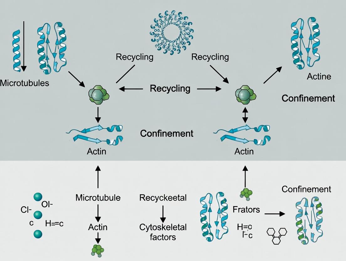
Title: The Local Cytoskeletal Recycling Cycle
Title: Experimental Workflow for Recycling Assay
The Scientist's Toolkit: Research Reagent Solutions
| Item | Function & Rationale |
|---|---|
| µ-Slide VI 0.1 (IBIDI) | Hydrophilic glass-bottom microfluidic slide for creating stable, parallel flow lanes for confinement and rapid buffer exchange. |
| HFE-7500 Oil with 2% EA Surfactant (RAN Biotech) | Fluorinated oil/surfactant system for generating stable, biocompatible water-in-oil droplets for isolated reaction chambers. |
| N-ethylmaleimide-activated Myosin II | Surface-bound motor protein to actively exert force on anchored actin networks, mimicking physiological mechanical confinement. |
| Alexa Fluor 488/647 Maleimide (Thermo Fisher) | Thiol-reactive dyes for specific, bright labeling of cysteine residues in actin, tubulin, or other target proteins. |
| Latrunculin B (Abcam) | Potent small molecule that sequesters actin monomers. Used to induce rapid, controlled disassembly of actin networks. |
| Creatine Phosphate / Creatine Kinase (Roche) | ATP regeneration system. Critical for maintaining energy levels during long experiments, especially in sealed confinement. |
| PLL(20)-g[3.5]-PEG(2) (SuSoS) | Poly(L-lysine)-graft-poly(ethylene glycol) used for surface passivation to prevent non-specific protein adsorption. |
| GST-Tagged Formin FH1-FH2 Domain | Purified, tag-cleavable protein fragment providing processive actin nucleation activity without full-length regulatory complexity. |
| HiLyte 647 Tubulin (Cytoskeleton, Inc.) | Commercially available, pre-labeled high-quality tubulin optimized for in vitro reconstitution assays. |
| Mant-ATP (Jena Bioscience) | Fluorescent adenosine nucleotide (2’-(or-3’)-O-(N-Methylanthraniloyl)) used to visualize ATP binding and consumption in real time. |
Troubleshooting Guide & FAQs
Q1: In our microfluidic confinement assay, actin polymerization becomes aberrant and fails to generate sufficient propulsive force. What could be the issue? A: This is often due to depleted local concentrations of profilin-ATP-actin or excessive capping protein activity in confinement. Ensure your assay buffer includes a regenerative system: 2 mM ATP, an ATP-regenerating system (e.g., 20 U/mL creatine phosphokinase + 10 mM creatine phosphate), and 2 µM profilin. Monitor actin polymerization kinetics via TIRF in the device; polymerization rates should be sustained. If rates drop >40% from open-system controls, increase profilin concentration incrementally (up to 5 µM).
Q2: Microtubule dynamics catastrophically stall in narrow channels, disrupting intracellular transport simulations. How can we stabilize them? A: Confinement increases the effective concentration of catastrophe factors like stathmin. To counteract this, supplement your tubulin preparation with a non-hydrolyzable GTP analog (GMPCPP) at a 1:5 molar ratio to tubulin to stabilize plus ends. Alternatively, include a plus-end tracking protein (+TIP) like EB3 (at 50-100 nM) to promote rescue events. The table below summarizes stabilization strategies:
| Reagent | Concentration Range | Primary Function | Expected Outcome in Confinement |
|---|---|---|---|
| GMPCPP-tubulin | 20% of total tubulin | Stabilizes GTP-cap | Reduces catastrophe frequency by ~70% |
| Recombinant EB3 | 50-100 nM | Promotes rescue, tracks growing ends | Increases rescue frequency by 2-3 fold |
| Taxol (Paclitaxel) | 1-10 µM | Binds and stabilizes microtubule lattice | Suppresses dynamics; use for static networks only |
| XMAP215 | 20-50 nM | Potent microtubule polymerase | Increases growth rate, can offset confinement-induced slowing |
Q3: Our FRAP experiments on actin-binding proteins in confined vesicles show anomalously slow recovery. Is this a technical artifact or biological? A: It is likely biological, reflecting limited diffusional exchange and enhanced binding in confinement. First, rule out artifacts: ensure your laser intensity does not cause permanent bleaching (use <50% laser power) and verify vesicle integrity post-bleach. If artifacts are ruled out, the slowed recovery (e.g., halftime increase >150% vs. bulk) is meaningful data. It indicates that your protein (e.g., cofilin) is undergoing increased binding/uncycling due to altered actin filament architecture. Model the recovery curve with a reaction-diffusion model to extract binding kinetics.
Q4: We observe unexplained aggregation of tubulin in confinement devices after repeated flow cycles. How do we prevent this? A: This is typically caused by mechanical shearing and nucleation of protofilaments. Implement the following protocol:
- Pre-treatment: Flush channels with 1% Pluronic F-127 for 30 min, then with BRB80 buffer.
- Tubulin Preparation: Always centrifuge tubulin (≥100,000 x g, 10 min, 4°C) immediately before introducing it into the device.
- Flow Control: Use a low shear-rate syringe pump (<5 µL/min) and avoid air-fluid interfaces.
- Additive: Include 1 mM DTT and 0.1% methylcellulose in the tubulin buffer to reduce oxidation and dampen turbulent effects.
Experimental Protocols
Protocol 1: Assessing Actin Recycling in Confined Spaces using TIRF Microscopy Objective: To quantify the rate of actin subunit turnover and cofilin-mediated severing in microchannels.
- Device Fabrication: Use standard soft lithography to create PDMS channels (height: 1.5 µm, width: 5 µm). Bond to a glass coverslip.
- Surface Preparation: Passivate channels with 1 mg/mL PLL-PEG. Then, introduce biotinylated BSA (0.5 mg/mL), followed by NeutrAvidin (0.2 mg/mL), and finally biotinylated actin seeds (0.5 µM in G-buffer, flow for 2 min).
- Reaction Mix: Prepare 1.5 µM actin (30% Alexa Fluor 488-labeled), 2 µM profilin, 0.5 µM Arp2/3 complex, 50 nM VCA (WASP fragment), 50 nM cofilin, 2 mM ATP, and oxygen scavengers (4.5 mg/mL glucose, 0.36 mg/mL glucose oxidase, 0.22 mg/mL catalase) in TIRF buffer (10 mM imidazole, pH 7.4, 50 mM KCl, 1 mM MgCl2, 1 mM EGTA).
- Imaging: Introduce mix into channel. Image every 5 sec for 10 min using 488 nm laser. Quantify fluorescence intensity decay after photobleaching a 10 µm region distal to the growing barbed ends to calculate subunit flux.
Protocol 2: Quantifying Microtubule Confinement Catastrophe Objective: To measure the effect of channel diameter on microtubule dynamic instability parameters.
- Device: Use silicon wafer-etched channels (heights: 0.5, 1.0, 2.0 µm; width 10 µm) sealed with a glass roof.
- Tubulin Preparation: Prepare 15 µM tubulin (40% HiLyte 647-labeled) in BRB80 (80 mM PIPES, pH 6.9, 1 mM MgCl2, 1 mM EGTA) with 1 mM GTP, 1 mM DTT, and 0.5% methylcellulose.
- Seeding: Introduce GMPCPP-stabilized, biotinylated microtubule seeds anchored via NeutrAvidin on the channel floor.
- Data Acquisition: Flow in tubulin mix. Acquire images at 2-sec intervals for 15 min. Track plus ends using plusTipTracker (MATLAB) or KymographClear (ImageJ).
- Analysis: Calculate growth speed, shrinkage speed, catastrophe frequency, and rescue frequency for each channel height. Compare to open-field controls.
Diagrams
Diagram 1: Actin Recycling Pathway in Confinement (98 chars)
Diagram 2: Experimental Workflow for Confinement Assays (99 chars)
The Scientist's Toolkit: Research Reagent Solutions
| Item | Function in Confinement Research |
|---|---|
| Pluronic F-127 | Non-ionic surfactant for passivating microfluidic device surfaces, prevents non-specific protein adhesion. |
| PLL-PEG | Poly-L-lysine grafted with polyethylene glycol; creates a non-fouling, bio-inert surface coating. |
| Profilin (Human, Recombinant) | Binds ATP-actin, promotes nucleotide exchange, and delivers monomers to growing barbed ends, crucial for sustained polymerization in limited volumes. |
| GMPCPP (Guanylyl-(α,β)-methylene-diphosphonate) | Non-hydrolyzable GTP analog used to create stable microtubule seeds or study stabilized filaments. |
| HaloTag-Tubulin / SNAP-tag-Actin | Covalent labeling systems for generating specifically labeled, functional cytoskeletal proteins for high-resolution tracking. |
| Oxygen Scavenging System (Glucose Oxidase/Catalase) | Reduces phototoxicity and fluorophore bleaching during long-term live-cell imaging in sealed devices. |
| Methylcellulose (1500 cP) | Increases medium viscosity to dampen fluid flow, mimic cytoplasmic crowding, and reduce shear forces on filaments. |
| Biotinylated Tubulin / Actin | Allows for specific, stable anchoring of seeds to avidin-functionalized surfaces within devices. |
| Recombinant Cofilin | Key actin depolymerizing factor (ADF); used to study severing and monomer recycling rates under spatial restriction. |
| EB3-GFP (Recombinant) | Plus-end tracking protein (+TIP) used as a marker for growing microtubule ends and to study rescue events. |
Technical Support Center: Troubleshooting Confinement-Based Cytoskeletal Recycling Experiments
Frequently Asked Questions (FAQs)
Q1: In our microfluidic confinement chambers, we observe inconsistent actin filament assembly rates compared to bulk solution. What could be causing this variability? A: Variability often stems from poor control over the geometry and surface chemistry of the confinement chambers. Nano-scale imperfections in chamber walls can create unintended diffusion barriers or nucleation sites. Ensure chambers are fabricated with high-resolution lithography (e.g., electron beam lithography for features <100 nm) and passivated with a consistent, inert coating like PEG-silane. Pre-condition all chambers with an identical BSA buffer flush before experiments.
Q2: We are trying to measure binding kinetics of recycling factors (e.g, profilin) to monomers in confinement, but our FRAP data is noisy and irreproducible. How can we improve the assay? A: Noisy FRAP data in confinement typically indicates photobleaching of molecules outside the region of interest due to light scattering or inadequate confinement depth. Implement Total Internal Reflection Fluorescence (TIRF) microscopy to restrict excitation to a thin evanescent field (~100 nm) matching your chamber height. Additionally, reduce laser power and increase frame averaging. Use a positive control of a known, stable fluorescent protein to calibrate the system.
Q3: Our model predicts accelerated recycling of cofilin in tubular confinement, but our experimental results show no effect. What are we missing? A: The discrepancy likely involves the off-rate of cofilin from actin filaments. In confinement, severed fragments may not diffuse away rapidly, creating a local high concentration that promotes immediate re-binding (a "rebinding" artifact). To test this, incorporate a molecular "sink" or flow in your design to remove severed fragments, or use a cofilin mutant with a known altered off-rate for comparison.
Q4: How do we distinguish between genuine confinement effects and mere surface adsorption of our cytoskeletal factors? A: Implement a two-pronged control experiment. First, use fluorescently labeled factors and perform a z-stack imaging to see if fluorescence accumulates specifically at chamber walls. Second, run an identical experiment in chambers of the same material but with progressively larger heights (e.g., from 100nm to 1000nm). Genuine confinement effects will diminish as height increases, while surface adsorption effects may remain constant. Quantify the depletion from solution.
Q5: We want to simulate physiological crowding in conjunction with spatial confinement. What is the best agent to use, and at what concentration? A: For mimicking cytosolic crowding, inert polymers like PEG (8-10 kDa) or Ficoll (70 kDa) at 5-15% (w/v) are standard. However, in extreme confinement (<50 nm), these large polymers may be excluded. Consider smaller crowders like sucrose (200-400 mM) or trimethylamine N-oxide (TMAO). Always run a control without active factors to ensure the crowder itself does not induce non-specific assembly.
Key Experimental Protocols
Protocol 1: Fabrication and Preparation of PDMS-based Nanoconfinement Chambers
- Master Fabrication: Spin-coat SU-8 photoresist on a silicon wafer to desired height (50-500 nm). Use a high-resolution photomask and UV lithography to pattern channels. Hard bake.
- PDMS Casting: Mix PDMS base and curing agent (10:1 ratio), degas, pour onto master, and cure at 65°C for 2 hours.
- Bonding & Passivation: Plasma-treat PDMS and a glass coverslip. Bond immediately. Flush chambers with 1 mg/mL PLL(20)-g[3.5]-PEG(2) in HEPES buffer for 1 hour to create a protein-resistant surface.
- Equilibration: Before experiment, flush with assay buffer (e.g., KMEI: 50 mM KCl, 1 mM MgCl2, 1 mM EGTA, 10 mM Imidazole pH 7.0) for 10 minutes.
Protocol 2: Measuring Anomalous Diffusion Coefficients via Single Particle Tracking (SPT)
- Sample Preparation: Label your protein of interest (e.g., actin monomer) with a photostable dye (e.g., Alexa Fluor 647). Introduce at low concentration (1-10 nM) into the confinement chamber with unlabeled monomers.
- Data Acquisition: Use a TIRF or highly inclined illumination microscope. Record movies at 50-100 fps with an EMCCD camera. Use low laser power to minimize blinking/photobleaching.
- Tracking & Analysis: Use tracking software (TrackMate, uTrack) to generate trajectories. Calculate the Mean Squared Displacement (MSD) for each trajectory. Fit the MSD vs time delay (τ) curve to MSD(τ) = 4Dτ^α. The exponent α indicates diffusion mode (α=1: normal; α<1: subdiffusive).
Protocol 3: FRAP Assay for Binding Kinetics in Confinement
- Chamber Preparation: Load chamber with a pre-assembled, stabilized (e.g., phalloidin-stabilized) actin network containing your fluorescently labeled recycling factor (e.g., GFP-profilin).
- Photobleaching & Imaging: Define a small circular region of interest (ROI). Bleach with a high-intensity 488nm laser pulse for 1-2 seconds. Immediately switch to low-intensity laser to image recovery every 0.5 seconds for 2-5 minutes.
- Quantification: Normalize fluorescence intensity in the bleached ROI to a control unbleached region. Fit the recovery curve to a single exponential or a diffusion-reaction model to extract the effective recovery half-time (t₁/₂) and mobile fraction.
Table 1: Measured Diffusion Coefficients (D) of Cytoskeletal Factors Under Confinement
| Factor | Molecular Weight (kDa) | Bulk Solution D (µm²/s) | Confined D (100nm height) (µm²/s) | Anomalous Exponent (α) | Measurement Technique |
|---|---|---|---|---|---|
| G-Actin (ATP) | 42 | 10.2 ± 1.1 | 3.5 ± 0.8 | 0.76 ± 0.05 | SPT (TIRF) |
| Profilin | 15 | 14.5 ± 1.5 | 8.1 ± 1.2 | 0.89 ± 0.04 | FRAP |
| Cofilin | 19 | 13.8 ± 1.3 | 2.2 ± 0.7 | 0.65 ± 0.07 | SPT (HILO) |
| Formin (FH1-FH2) | 90 | 4.3 ± 0.5 | 0.9 ± 0.3 | 0.82 ± 0.06 | FRAP |
Table 2: Effects of Confinement on Filament Assembly and Disassembly Kinetics
| Parameter | Bulk Solution Value | 200nm Confinement Value | 50nm Confinement Value | Notes |
|---|---|---|---|---|
| Actin Elongation Rate (subs/s) at 1µM monomer | 5.7 ± 0.6 | 4.1 ± 0.5 | 1.3 ± 0.4 | Barbed end growth, measured by TIRF |
| Cofilin Severing Frequency (events/µm/min) | 0.35 ± 0.05 | 0.82 ± 0.11 | 1.50 ± 0.20 | On pyrene-actin filaments |
| Profilin-Actin On-Rate (µM⁻¹s⁻¹) | 8.9 x 10³ | 9.1 x 10³ | 8.7 x 10³ | Negligible confinement effect on bimolecular rate |
| Effective Recycling Time (Actin Monomer) | N/A | ~30% faster than bulk | ~60% faster than bulk | Model-derived from integrated assembly/disassembly |
Experimental Diagrams
Diagram Title: Workflow for Confinement Cytoskeleton Recycling Assay
Diagram Title: Key Cytoskeletal Recycling Pathways in Confinement
The Scientist's Toolkit: Research Reagent Solutions
| Item | Function & Rationale |
|---|---|
| PEG-Silane (e.g., mPEG-silane, MW 2kDa) | Forms a dense, hydrophilic brush on glass/silicon surfaces to minimize non-specific adsorption of proteins, crucial for isolating confinement effects. |
| Inert Crowders (Ficoll 70, Dextran) | Mimics macromolecular crowding of the cytoplasm. Choice of size and concentration is critical to match the scale of confinement. |
| Oxygen Scavenging System (Glucose Oxidase/Catalase + Glucose) | Reduces photobleaching and fluorophore blinking in fluorescence microscopy, essential for long SPT or FRAP tracks in sealed chambers. |
| Stabilized Actin Seeds (Phalloidin-stabilized filaments) | Provide defined nucleation points for elongation assays, allowing measurement of monomer addition rates without confounding nucleation events. |
| Non-Hydrolyzable ATP Analog (e.g., AMP-PNP) | Used to lock actin monomers in a specific nucleotide state, simplifying the system to study one step of the recycling cycle (e.g., cofilin binding). |
| Microfluidic Flow Control System (Pressure Pump/Controller) | Enforces precise buffer exchange within chambers, allowing introduction of factors (e.g., cofilin) at specific times and removal of products. |
| High-Strength Passivator (e.g., Pluronic F-127) | An alternative to PEG for blocking hydrophobic PDMS surfaces, especially effective in long-term experiments. |
Technical Support Center: Cytoskeletal Recycling in Confinement Research
Troubleshooting Guides & FAQs
Q1: In a 3D collagen matrix migration assay, our metastatic cell line shows significantly reduced persistence compared to 2D. The cells stall frequently. What could be the cause and how can we troubleshoot this?
A: This is a common issue when cytoskeletal recycling, specifically the disassembly and repurposing of actin filaments and microtubules, cannot keep pace with the physical demands of 3D confinement. The stall is often due to a local depletion of available G-actin or tubulin dimers.
- Troubleshooting Steps:
- Confirm Cytoskeletal Depletion: Perform immunofluorescence staining for F-actin (e.g., with phalloidin) and microtubules in stalled cells. Compare the fluorescence intensity at the leading edge versus the uropod to cells migrating in 2D. A stark depletion at the leading edge supports the hypothesis.
- Modulate Cofilin Activity: The actin-severing protein cofilin is critical for recycling. Inhibit cofilin (e.g., with small molecule CK-666 targeting Arp2/3 upstream) as a negative control—persistence should worsen. Alternatively, mildly overexpress cofilin to enhance severing and monomer recycling.
- Increase Monomer Availability: Supplement the matrix with a cell-permeable form of the actin-stabilizing drug Jasplakinolide (low nM range). This can paradoxically aid recycling by preventing excessive depolymerization and maintaining a soluble pool, but titration is critical.
- Check Confinement Geometry: Ensure the pore size of your 3D matrix is consistent. Use collagen concentration vs. pore size calibration tables (see Table 1).
Q2: When imaging T-cell trafficking in a microfluidic device with confined channels, we observe prolonged uropod detachment times. Which recycling pathways are likely impaired?
A: Prolonged uropod detachment suggests a failure in the coordinated actomyosin contraction and microtubule-driven rear release. The recycling of myosin II and the dynamic instability of microtubules at the rear are key.
- Troubleshooting Steps:
- Inhibit Myosin II Light Chain Kinase (MLCK): Use ML-7 inhibitor. If detachment time decreases, it indicates overactive contraction is physically impeding release, and myosin recycling/ turnover is too slow.
- Target Microtubule Dynamics: Treat with low-dose Paclitaxel (stabilizes microtubules). If detachment worsens, it confirms that dynamic microtubules are required for recycling components from the uropod. Conversely, low-dose Nocodazole may accelerate detachment by disrupting microtubules, but can also impair directionality.
- Measure Calcium Flux: Use Fluo-4 AM dye. A sustained calcium signal at the uropod can inhibit localized disassembly. Chelate calcium with BAPTA-AM and observe if detachment normalizes.
Q3: In a dendritic spine plasticity experiment mimicking synaptic confinement, FRAP (Fluorescence Recovery After Photobleaching) of actin-GFP shows incomplete recovery. Does this indicate broken recycling or simply physical barrier?
A: Incomplete FRAP recovery in a spine head indicates a breakdown in the recycling loop where old filaments are not being efficiently severed and new ones are not being polymerized, often linked to a spatial segregation of the pool of recycled monomers.
- Troubleshooting Steps:
- Distinguish between Binding and Mobility: Perform a control FRAP on a soluble cytosolic GFP. If its recovery is also incomplete, there is a physical diffusion barrier (e.g., spine neck geometry). If soluble GFP recovers fully, the issue is specific to actin network turnover.
- Modulate Actin-Binding Proteins: Express constitutively active LIM-Kinase (inactivates cofilin). This should further reduce FRAP recovery. Express a phospho-mutant cofilin (active) to enhance severing and potentially improve recovery.
- Check for Aberrant Stabilization: Stain for hyper-stable F-actin with SiR-actin or perform immunofluorescence for cross-linking proteins like fascin. Their overexpression can trap actin, preventing recycling.
Experimental Protocols
Protocol 1: Quantifying Cytoskeletal Recycling Efficiency via FRAP in Confined Microchannels
Objective: To measure the turnover rate of actin and microtubules in cells navigating precisely defined confined spaces.
Materials: Polydimethylsiloxane (PDMS) microfluidic device (channel height: 3µm, width: 5µm), cells expressing Actin-GFP or EB3-GFP (for microtubule plus-end tracking), live-cell imaging setup with photobleaching module, cell culture media without phenol red.
Methodology:
- Seed cells into the inlet reservoir of the PDMS device and allow them to adhere and enter the microchannels (2-4 hours).
- Select a cell midway through a channel. Define a Region of Interest (ROI) at the leading edge (~2µm x 2µm).
- Acquire 5 pre-bleach images at 2-second intervals.
- Bleach the ROI with a high-intensity 488nm laser pulse (100% power, 500ms).
- Acquire post-bleach images every 2 seconds for 3-5 minutes.
- Analysis: Normalize fluorescence intensity in the bleached ROI to a reference background and an unbleached region of the same cell. Fit the recovery curve to a single exponential equation:
y(t) = y0 + A*(1 - exp(-k*t)), where k is the recovery rate constant (s⁻¹). The half-time of recovery (t₁/₂) is calculated asln(2)/k. Compare t₁/₂ between cells in confinement vs. cells on 2D substrates.
Protocol 2: Cofilin Activity Assay in 3D Matrices Using FRET Biosensor
Objective: To spatially map the activity of the key actin-recycling enzyme cofilin within a cell migrating in a 3D collagen gel.
Materials: Cells stably expressing the fluorescence resonance energy transfer (FRET)-based cofilin biosensor (e.g., Cytochalasin D), rat tail collagen I, 35mm glass-bottom dish, confocal microscope capable of spectral FRET acquisition.
Methodology:
- Prepare a 2 mg/ml collagen I cell suspension mix on ice. Plate 100µl in the glass-bottom dish and polymerize at 37°C for 30 mins.
- Add complete media and image cells after 6 hours.
- Image Acquisition: Acquire donor (CFP, ex 458nm, em 470-500nm) and FRET (ex 458nm, em 520-550nm) channels. Perform acceptor (YFP) bleaching for rationetric calibration on a subset of cells.
- Analysis: Calculate the FRET ratio (FRET channel intensity / CFP channel intensity) for each pixel. Generate ratiometric heatmaps. Quantify the average FRET ratio in the leading edge (front 25% of the cell) versus the cell body. A higher FRET ratio at the leading edge indicates higher cofilin activity, promoting local actin disassembly and recycling.
Data Presentation
Table 1: Cytoskeletal FRAP Recovery Half-Times Under Different Confinement Conditions
| Cell Type | Condition | Probed Cytoskeletal Element | FRAP Half-Time (t₁/₂, seconds) | Relative Recovery (% of pre-bleach) |
|---|---|---|---|---|
| MDA-MB-231 | 2D Substrate | Actin-GFP | 45.2 ± 5.1 | 92.5 ± 3.2 |
| MDA-MB-231 | 3D Collagen (5mg/ml) | Actin-GFP | 78.6 ± 8.7 | 74.1 ± 6.5 |
| MDA-MB-231 | 3D + Cofilin OE | Actin-GFP | 52.3 ± 6.2 | 88.3 ± 4.1 |
| Primary T-Cell | 2D Substrate | Tubulin-GFP | 32.1 ± 4.0 | 95.0 ± 2.1 |
| Primary T-Cell | 3µm Microchannel | Tubulin-GFP | 105.4 ± 12.3 | 65.8 ± 7.8 |
Table 2: Migration Parameters Linked to Recycling Efficiency
| Experiment Model | Intervention (Targeting Recycling) | Result on Migration Speed | Result on Persistence | Implication for Recycling Pathway |
|---|---|---|---|---|
| Metastasis (3D Matrix) | siRNA against Cofilin | -40% * | -60% * | Severing is rate-limiting. |
| Immune Trafficking (Channel) | MLCK Inhibitor (ML-7) | No change | +25% * | Myosin recycling aids detachment. |
| Synaptic Plasticity (Spine) | LIMK Inhibitor (BMS-5) | N/A | FRAP t₁/₂: -35% | Cofilin activation aids turnover. |
- p<0.05, * p<0.01, ** p<0.001 vs. control.
Mandatory Visualizations
Diagram 1: Cytoskeletal Recycling Feedback Loop in Confinement
Diagram 2: FRAP Protocol for Measuring Cytoskeletal Turnover
The Scientist's Toolkit: Research Reagent Solutions
| Item Name & Supplier (Example) | Function in Cytoskeletal Recycling Research |
|---|---|
| SiR-Actin / SiR-Tubulin (Cytoskeleton Inc.) | Live-cell, far-red fluorescent probes for staining F-actin or microtubules with minimal toxicity. Allows long-term imaging of network dynamics in confinement. |
| Cofilin (phospho-Ser3) Antibody (CST #5173) | Detects inactive (phosphorylated) cofilin via IF or WB. Mapping p-cofilin vs. total cofilin reveals spatial regulation of actin severing. |
| CK-666 (Arp2/3 Inhibitor) (Tocris) | Inhibits the Arp2/3 complex, reducing branched actin nucleation. Used to test if new polymerization from recycled monomers is Arp2/3-dependent. |
| ML-7 Hydrochloride (MLCK Inhibitor) (Abcam) | Inhibits Myosin Light Chain Kinase, reducing contractility. Used to dissect the role of myosin recycling and rear detachment in confined migration. |
| Collagen I, Rat Tail, High Concentration (Corning) | For generating reproducible 3D matrices of defined stiffness and pore size, creating physiologically relevant confinement for migration assays. |
| PDMS Microfluidic Chambers (µ-Slide from ibidi) | Ready-to-use devices with defined channel geometries (e.g., 3x3µm, 5x5µm) for studying migration under precise, uniform confinement. |
| FRET-based Cofilin Biosensor (Addgene plasmid #50777) | Genetically encoded sensor (CFP/YFP) that reports cofilin activity in real-time via FRET ratio changes, crucial for spatial mapping in 3D. |
| Cell Permeant Actin Stabilizer (Jasplakinolide) (Thermo Fisher) | Stabilizes actin filaments but can be used at low doses to modulate the balance between F-actin and the soluble G-actin pool available for recycling. |
Technical Support Center: Troubleshooting & FAQs
Frequently Asked Questions
Q1: Our experimental measurement of recycling factor flux in microfluidic channels is consistently 30-40% lower than the value predicted by our current geometric crowding model. What could be the cause? A: This is a common calibration issue. First, verify your assumption of bulk-phase diffusion coefficients. In confinement, effective diffusion (Deff) is reduced: Deff = D0 * (1 - λ)^α, where λ is the ratio of particle to pore size and α ≈ 2.5 for cylindrical geometries. Recalculate your model input using this corrected Deff. Second, ensure your model includes surface adsorption kinetics; even passivated surfaces can have residual binding that sequesters factors. Run a control experiment with a fluorescent inert protein of similar size to quantify non-specific loss.
Q2: When transitioning from spherical to tubular in vitro synthetic cells, our recycling efficiency drops precipitously. Is this a geometry-specific effect? A: Yes, this is predicted by theoretical models. The surface-area-to-volume (SA:V) ratio is key. Spheres have the lowest possible SA:V for a given volume. Tubular geometries have a higher SA:V, increasing the relative amount of membrane-bound factors and altering the reaction-diffusion steady state. Use the following relationship to adjust your expectations:
Table 1: Geometric Impact on Theoretical Recycling Thresholds
| Geometry | SA:V Ratio (for equal volume) | Critical Concentration (C_crit) for Recycling | Dominant Limiting Factor |
|---|---|---|---|
| Sphere (Radius R) | 3/R | 1.0 (Baseline) | Cytoplasmic diffusion |
| Cylinder (Radius R, Length L=10R) | ~2.1/R | ~1.8 | Longitudinal diffusion + membrane crowding |
| Slab (Height H) | ~2/H | ~2.3 | Perpendicular diffusion, surface rebinding |
Q3: How do we accurately quantify "molecular crowding" in our experimental confinement system? A: Use a dual-probe fluorescence correlation spectroscopy (FCS) protocol. Incorporate two fluorescent tracers of different sizes (e.g., 10 kDa and 500 kDa dextran-conjugated dyes). The ratio of their measured diffusion coefficients (Dsmall/Dlarge) provides a crowding metric (φcrowd). Values significantly <1 indicate high excluded volume effects. Integrate this φcrowd into your model as a correction factor for all reaction rates (kobs = k0 * exp(-β * φ_crowd)).
Q4: Our stochastic simulation of factor recycling shows high variance under identical parameters. Is this an error? A: Not necessarily. In highly confined systems with low copy numbers of recycling factors (N < 1000), the process becomes inherently stochastic. Deterministic continuum models (PDEs) break down. You must switch to a stochastic simulation algorithm (e.g., Gillespie) within your geometric mesh. The variance itself is a key output, measured by the Fano factor (variance/mean). A Fano factor > 1 indicates noise-driven process dominance, which can be critical for drug targeting efficacy.
Experimental Protocols
Protocol 1: Calibrating Confined Diffusion Coefficients
- Objective: Measure the effective diffusion coefficient (D_eff) of your cytoskeletal factor in the experimental geometry.
- Materials: Microfluidic confinement device, purified fluorescently labeled factor, TIRF or confocal microscope.
- Method: a. Introduce a sharp concentration gradient of the factor into the device. b. Acquire time-lapse images at 100 ms intervals for 60 seconds. c. Use fluorescence recovery after photobleaching (FRAP) in a defined region. Fit the recovery curve to the solution of the diffusion equation for your specific channel geometry (not a standard infinite half-space model). d. Calculate Deff using the half-time of recovery (t1/2): Deff = (w^2 * γd) / (4 * t1/2), where w is the bleach spot radius and γd is a geometry-dependent constant (0.88 for cylinder, 0.91 for slit).
Protocol 2: Quantifying Recycling Efficiency (η)
- Objective: Empirically determine the fraction of cytoskeletal factors successfully reincorporated per cycle.
- Materials: Two-color assay: donor-labeled factors (e.g., Cy3-actin) and acceptor-labeled nucleation sites (e.g., Cy5-ARP2/3). Total internal reflection fluorescence (TIRF) microscopy.
- Method: a. Flow in donor-labeled factors to saturate available sites. Measure baseline fluorescence (Itotal). b. Introduce a chase of unlabeled factor at 10x concentration for 60 sec to displace non-specifically bound factors. c. Measure remaining fluorescence (Irecycled). η = Irecycled / Itotal. d. Subsequently, flow in acceptor-labeled sites. Colocalization efficiency via FRET confirms functional recycling.
Diagrams
Title: Core Cytoskeletal Factor Recycling Pathway
Title: Model Integration & Validation Workflow
The Scientist's Toolkit: Research Reagent Solutions
Table 2: Essential Materials for Confined Recycling Experiments
| Item | Function | Example Product/Catalog |
|---|---|---|
| Microfluidic Chips | Physically define geometric confinement (channels, chambers, vesicles). | Cyto-SQUID Chips (Cytosurge); µ-Slide VI 0.4 (ibidi) |
| Crowding Agents | Mimic cytoplasmic excluded volume effects. | Ficoll PM400, PEG 8000, Dextran T500 |
| Fluorescent Tracers | Visualize and quantify diffusion and binding kinetics. | Alexa Fluor 488/568/647 NHS Ester (Thermo Fisher) |
| Passivation Reagents | Minimize non-specific surface adsorption. | PEG-silane, Pluronic F-127, Casein |
| Photobleaching System | For FRAP measurements of mobility. | Mosaic/FRAPPA module (Andor) or built-in laser system. |
| Stochastic Simulation Software | Model low-copy-number systems in 3D geometries. | Smoldyn, ChemCell, or COMSOL Multiphysics with PDE module. |
Tools of the Trade: Techniques to Model and Measure Recycling in Confinement
Troubleshooting Guides & FAQs
Fluorescent Speckle Microscopy (FSM)
Q1: My speckle signal is too dim. What are the primary causes and solutions? A: Dim speckles often result from suboptimal labeling or imaging conditions.
- Cause 1: Low incorporation ratio. Too few labeled monomers are incorporated into the polymer.
- Solution: Titrate the ratio of labeled to unlabeled protein. For actin, a typical range is 1:100 to 1:1000 (labeled:unlabeled). Start at 1:200 and adjust.
- Cause 2: Photobleaching during acquisition.
- Solution: Use an oxygen-scavenging imaging buffer (e.g., Glucose Oxidase/Catalase system) and reduce laser power/increase camera binning.
Q2: Speckles appear "chunky" or move as large aggregates, not as single filaments. A: This indicates protein aggregation or poor polymerization conditions.
- Solution:
- Centrifuge the labeled protein stock at high speed (e.g., 100,000 x g) immediately before use to remove aggregates.
- Verify polymerization buffer conditions (e.g., for microtubules, ensure correct Mg²⁺, GTP, and temperature).
- Filter all buffers through a 0.1µm filter.
Photoactivatable/Photoconvertible Tags (e.g., PA-GFP, mEos)
Q3: After photoactivation, the fluorescence recovers too quickly in the ROI for tracking. A: Unintended activation or high background can obscure the signal of the activated pool, crucial for studying factor recycling.
- Cause 1: High background from out-of-focus activation.
- Solution: Use a highly focused activation beam (e.g., pinhole or two-photon setup). Ensure the ROIs for activation are precisely drawn.
- Cause 2: High mobility of the target factor.
- Solution: Increase frame rate immediately post-activation. Consider using a total internal reflection fluorescence (TIRF) microscope to limit observation to the cell cortex.
Q4: The photoconverted signal bleaches almost immediately. A: This compromises tracking of recycled factors over time.
- Solution:
- Buffer: Use a photostabilizing imaging buffer (see table below).
- Power: Reduce the intensity of the imaging laser (while maintaining sufficient signal-to-noise).
- Fusion Tag: Consider using a more photostable tag (e.g., mMaple vs. Dendra2).
Table 1: Typical Labeling Ratios for FSM in Confinement Studies
| Cytoskeletal Polymer | Labeled:Unlabeled Protein Ratio | Typical Final Concentration in Cell | Key Buffer Component |
|---|---|---|---|
| Actin (β/γ-actin) | 1:200 to 1:1000 | 50-200 µM | 1 mM Mg-ATP, 150 mM KCl |
| Microtubules (Tubulin) | 1:50 to 1:200 | 10-20 µM | 1 mM Mg-GTP, 5% Glycerol |
| Intermediate Filaments (Vimentin) | 1:20 to 1:100 | 0.1-0.5 µM | 150 mM NaCl |
Table 2: Comparison of Common Photoactivatable Tags for Recycling Studies
| Tag Name | Activation/Convert Light | Emission Peak (nm) | Relative Brightness | Photostability (Frames @ 500ms) | Best for |
|---|---|---|---|---|---|
| PA-GFP | 405 nm | 517 | 1.0 (reference) | ~30 | Short-term, rapid turnover |
| mEos3.2 | 405 nm | 581 | 1.8 | ~80 | Long-term tracking, super-resolution |
| Dendra2 | 405/488 nm | 507 > 573 | 1.2 | ~40 | Dual-color conversion experiments |
| mMaple3 | 405 nm | 580 | 1.5 | ~100 | High-precision tracking in confinement |
Experimental Protocols
Protocol 1: Fluorescent Speckle Microscopy for Actin Turnover in Microfluidic Confinement
Objective: To visualize and quantify the flow and recycling of actin subunits in a confined channel.
Materials:
- Purified, fluorescently labeled actin (e.g., Alexa Fluor 568-C2-maleimide labeled).
- Unlabeled actin.
- Microfluidic device with ~3 µm channels.
- TIRF or highly inclined and laminated optical sheet (HILO) microscope.
- Polymerization buffer: 10 mM Imidazole (pH 7.0), 50 mM KCl, 1 mM MgCl2, 1 mM EGTA, 0.2 mM ATP, 50 mM DTT.
Method:
- Sample Preparation: Mix labeled and unlabeled actin monomers at a 1:400 ratio in G-buffer (2 mM Tris pH 8.0, 0.2 mM CaCl2, 0.2 mM ATP, 0.5 mM DTT). Keep on ice.
- Device Priming: Flush the microfluidic channel with 1% BSA in polymerization buffer for 5 min to passivate.
- Initiation: Mix the actin solution 1:1 with 2X polymerization buffer and immediately introduce into the channel. Seal ports.
- Imaging: After 5 min incubation, image using a 561 nm laser under TIRF illumination. Acquire 100 frames at 2-second intervals.
- Analysis: Use kymograph analysis or specialized FSM software (e.g., in FIJI) to track speckle movement and disappearance (catastrophe).
Protocol 2: Tracking a Recycled Tubulin Pool with PA-GFP
Objective: To monitor the fate of a defined pool of microtubules after depolymerization in a confined region.
Materials:
- Cells expressing α-tubulin-PA-GFP.
- Confocal microscope with 405 nm laser for activation and 488 nm laser for imaging.
- Nocodazole (to depolymerize microtubules).
- Washout buffer.
Method:
- Pre-treatment: Treat cells with 10 µM nocodazole for 1 hour to depolymerize microtubules, creating a homogeneous pool of soluble tubulin-PA-GFP.
- Define ROI: Select a small rectangular region (~5x5 µm) at the cell periphery under confinement.
- Photoactivate: Apply a brief (50-100 ms), high-intensity pulse of 405 nm laser light to the ROI to activate the soluble PA-GFP-tubulin pool.
- Washout & Recovery: Rapidly wash out nocodazole with pre-warmed medium. Immediately begin time-lapse imaging (488 nm excitation, 2 sec intervals) to track the incorporation of the activated (recycled) tubulin pool into newly growing microtubules.
Diagrams
FSM Experimental Workflow for Recycling Studies
Tracking Factor Recycling via Photoactivation
The Scientist's Toolkit
Table 3: Research Reagent Solutions for FSM & Photoactivation Experiments
| Reagent / Material | Function & Rationale |
|---|---|
| HILO/TIRF Microscope | Enables low-background imaging of single speckles or activated molecules near the coverslip, critical for confined systems. |
| Oxygen-Scavenging Buffer (e.g., GLOX) | Reduces photobleaching by removing dissolved oxygen, extending imaging time for tracking. |
| Microfluidic Chambers (e.g., µ-Slide) | Provides precise geometrical confinement and control over the cellular microenvironment. |
| Ultracentrifuge (100,000+ x g) | Clears protein aggregates from labeled stocks to prevent non-specific speckling. |
| Caged Compounds (e.g., Caged Latrunculin) | Allows rapid, spatially controlled pharmacological manipulation to probe recycling dynamics. |
| High-Quantum Efficiency Camera (EMCCD/sCMOS) | Captures low-light speckle signals with high sensitivity and speed. |
| Methylcellulose or Polyacrylamide Gels | Mimics cytoplasmic crowding and viscoelasticity to study factor mobility in confinement. |
Troubleshooting Guides & FAQs
Q1: During FRAP on a confined cytoskeletal network, our recovery curve plateaus below the pre-bleach intensity. What does this indicate and how can we address it? A: A plateau below 100% recovery indicates an immobile fraction. In confinement research, this can be exacerbated by protein trapping or irreversible binding. First, verify confinement geometry integrity. Increase the bleach spot size relative to the confinement zone to ensure you are measuring within the fully confined area. Confirm the viability of your sample post-bleach. If the immobile fraction is the subject of study, use computational modeling to separate the true immobile fraction from artifacts caused by confinement-induced slowed diffusion.
Q2: We observe asymmetric fluorescence recovery in our FLAP experiment within a microfluidic trap. What are the potential causes? A: Asymmetric recovery in FLAP is a direct readout of directional flow or polarized recycling. First, rule out technical artifacts: ensure the photoconversion laser is perfectly aligned and the confinement chamber is level. If the asymmetry is reproducible, it is likely biological. In confinement, cytoskeletal factor recycling often becomes vectorial due to imposed geometry. Use computational particle image velocimetry (PIV) analysis on your time-lapse FLAP data to quantify the direction and magnitude of the flow.
Q3: Our computational model for recycling kinetics fails to fit the FRAP data, especially the initial recovery phase. How should we adjust the model? A: A poor fit in the initial phase often points to an incorrect assumption about the dominant transport mechanism. In open systems, diffusion is often assumed. However, in confinement, active transport or diffusion with barriers becomes significant. Adjust your reaction-diffusion model to include: 1) An effective diffusion coefficient (Deff) that accounts for obstacle density, and 2) A boundary condition reflecting your confinement geometry (e.g., reflective for closed chambers). Start with a two-state model (mobile/immobile) before adding complexity.
Q4: Phototoxicity is causing cytoskeletal collapse in our repeated FRAP/FLAP measurements. How can we mitigate this? A: Use lower laser power (20-50% of usual) for both imaging and bleaching/photoconversion. Increase the interval between acquisition frames. Utilize a sensitive camera (e.g., EM-CCD, sCMOS) to collect more signal with less light. Employ a oxygen-scavenging imaging buffer (e.g., with glucose oxidase/catalase) to reduce radical formation. Finally, consider using a dedicated confocal line for bleaching separate from imaging to minimize overall exposure.
Q5: How do we distinguish between slowed diffusion and binding-induced trapping from a FRAP curve in a confined environment? A: Perform FRAP at multiple bleach spot sizes. If the recovery half-time (t1/2) scales with the square of the bleach radius, diffusion is the rate-limiting step. If t1/2 is independent of spot size, binding/unbinding kinetics are dominant. In confinement, perform this spot size variation within the limits of your geometry and compare to an unconfined control. Computational analysis via inverse modeling of the full recovery curve across conditions is essential for precise parameter extraction.
Table 1: Typical FRAP Recovery Parameters for Cytoskeletal Factors Under Confinement vs. Bulk
| Parameter | Bulk Solution (Mean ± SD) | Confined Geometry (< 5µm) (Mean ± SD) | Interpretation |
|---|---|---|---|
| Mobile Fraction (%) | 85 ± 10 | 60 ± 15 | Increased immobile fraction due to trapping. |
| Recovery Half-time, t₁/₂ (s) | 2.5 ± 0.8 | 12.5 ± 4.2 | Transport severely slowed by obstacles. |
| Effective Diffusion Coeff., D_eff (µm²/s) | 15.0 ± 3.0 | 0.9 ± 0.3 | Diffusion reduced by orders of magnitude. |
| Imaging Laser Power for FRAP (%) | 100 (Reference) | 40 - 60 | Lower power required to avoid photodamage in confinement. |
Table 2: Key Outputs from Computational Kinetic Modeling of Recycling
| Model Output | Description | Relevance to Confinement Research |
|---|---|---|
| kon / koff (s⁻¹) | Binding & unbinding rate constants. | Altered by macromolecular crowding in confined space. |
| Anomalous Diffusion Exponent (α) | α=1: normal diffusion; α<1: sub-diffusion. | Quantifies degree of transport hindrance. |
| Recycling Flux (a.u./s) | Rate of component return to active pool. | Direct measure of recycling efficiency, the thesis core. |
| Spatial Gradient Map | Visual map of parameter distribution. | Identifies localized "hotspots" or "traps" within the geometry. |
Experimental Protocols
Protocol 1: FRAP for Actin Binding Protein Recycling in Microfabricated Channels
- Sample Preparation: Seed cells expressing GFP-tagged protein (e.g., GFP-Cofilin) in a PDMS microfluidic device with channel heights of 3-5µm.
- Imaging Setup: Use a confocal microscope with a 488nm laser, 63x/1.4NA oil objective, and pre-warmed environmental chamber (37°C, 5% CO2).
- Baseline Acquisition: Capture 5-10 frames at low laser power (2-5%).
- Bleaching: Define a circular ROI (1µm diameter) within the confined cytoplasm. Bleach with 100% 488nm laser power for 5-10 iterations.
- Recovery Acquisition: Immediately switch back to low laser power and acquire images every 0.5s for 60s.
- Data Extraction: Measure mean intensity in bleached ROI, a reference background region, and an unbleached control region for normalization.
Protocol 2: FLAP using Dendra2-Tagged Tubulin to Map Microtubule Turnover in Confinement
- Sample Preparation: Transfer cells expressing Dendra2-Tubulin into a glass-bottom confining chamber.
- Photoconversion: Select a strip (~1µm width) across a microtubule array using the microscope's ROI tool. Illuminate with a 405nm laser pulse (5-15% power, 1-2s) to convert Dendra2 from green to red.
- Dual-Channel Acquisition: Simultaneously image green (ex 488nm) and red (ex 561nm) channels every 2s for 5 minutes. Use minimal laser power.
- Ratio Analysis: Calculate the red-to-green fluorescence ratio over time within the photoconverted strip and adjacent areas. The loss of red signal and gain of green signal indicates turnover and recycling.
Protocol 3: Computational Fitting of FRAP Data with a Reaction-Subdiffusion Model
- Data Preprocessing: Normalize recovery curves (I(t)) to correct for background and total bleaching. Average curves from ≥10 cells.
- Model Definition: Implement a model with a sub-diffusing mobile population (fraction M) and an immobile population. Use the fractional diffusion equation: ∂[M]/∂t = Deff * ∇^α [M] - kon[M] + k_off[Imm].
- Fitting: Use a nonlinear least-squares algorithm (e.g., Levenberg-Marquardt) to fit the model to the averaged I(t) curve. Key fitted parameters: Deff, α, kon, k_off, M.
- Validation: Validate the model by comparing its prediction to the recovery curve from a different bleach spot size.
Visualizations
Title: FRAP Experimental and Analysis Workflow
Title: Cytoskeletal Factor Recycling and Trapping in Confinement
Title: Computational Analysis Pipeline for Recycling Kinetics
The Scientist's Toolkit: Research Reagent Solutions
| Item | Function & Relevance to Confinement Recycling Studies |
|---|---|
| PDMS Microfluidic Chips | Creates precisely defined physical confinement (channels, traps) to mimic in vivo crowded environments. |
| Glass-Bottom Confinement Dishes | Commercial dishes with micro-patterned or etched barriers for cell confinement during live imaging. |
| Photoactivatable/Convertible Probes (Dendra2, mEos) | Enables FLAP; allows spatial-temporal tracking of a protein sub-population's fate after activation. |
| HaloTag/SNAP-tag Ligands | Facilitates pulse-chase labeling with cell-permeable dyes to track protein turnover without overexpression artifacts. |
| Oxygen-Scavenging Imaging Buffer | Reduces photobleaching and phototoxicity during long, sensitive FRAP/FLAP timelapses in confined cells. |
| Cytoskeletal Drugs (e.g., Latrunculin, Nocodazole) | Positive controls to perturb recycling; used to validate the sensitivity of FRAP/FLAP readouts. |
| Recombinant Fluorescent Actin/Tubulin | For in vitro reconstitution assays inside cell-sized liposomes or water-in-oil droplets to isolate physical effects. |
| Inverse Modeling Software (e.g., PyFRAP, simFRAP) | Open-source computational tools designed specifically for fitting complex models to FRAP data in arbitrary geometries. |
Welcome to the Technical Support Center for minimal biochemical reconstitution systems, specifically designed for research aimed at achieving efficient recycling of cytoskeletal factors in confined geometries. This guide addresses common experimental hurdles.
Troubleshooting Guide & FAQs
Q1: My reconstituted cytoskeletal network (e.g., actin) shows rapid depletion of monomers in a confined, cell-sized compartment, halting polymerization. What could be the cause?
- A: This is a classic sign of inefficient recycling. In confinement, the surface area-to-volume ratio is high, leading to excessive nucleation that depletes the monomer pool. The system lacks the necessary biochemical factors for disassembly and monomer recycling.
- Solution:
- Introduce recycling factors: Systematically add cofilin (for severing) and ADF (for depolymerization) at empirically determined ratios.
- Tune nucleation: Reduce the concentration of your nucleator (e.g., the Arp2/3 complex or formin) to lower the initial burst of filament formation.
- Implement a chemical energy buffer: Ensure your ATP regeneration system is robust. Use 10-20X more phosphocreatine and creatine kinase than your ATP concentration to maintain a steady state.
Q2: In a microtubule gliding assay with confined kinesin motors, motility becomes unsteady and stalls over time. How can I restore persistent motion?
- A: Stalling often indicates motor inactivation or fuel depletion. In confinement, local ADP buildup can inhibit kinesin activity.
- Solution:
- Enhance fuel regeneration: Double-check your ATP regeneration system. Consider using a more efficient pyruvate kinase/lactate dehydrogenase (PK/LDH) system alongside ATP.
- Include a crowding agent: Add 0.5-1% (w/v) methylcellulose to the assay buffer. This reduces diffusion, helps maintain local ATP/ADP ratios near the motor, and simulates cytoplasmic crowding.
- Purge ADP: Add 1-5 U/mL of apyrase (an ADPase) to your flow cell before introducing motors and microtubules to remove contaminating ADP.
Q3: Protein components in my minimal system are adsorbing to the walls of my synthetic lipid vesicle or microfluidic chamber, depleting the active pool. How can I mitigate this?
- A: Surface adsorption is a major issue in minimal systems, especially with confinement.
- Solution:
- Use passivating agents: Pre-incubate chambers with 1-5 mg/mL bovine serum albumin (BSA) or 0.1% Pluronic F-127 (for microfluidics). For vesicles, include 0.1% BSA in the internal buffer.
- Optimize lipid composition: For synthetic vesicles, include 5-10% PEGylated lipids (e.g., DOPE-PEG2000) to create a steric barrier against protein adsorption.
- Employ a carrier protein: Always include 0.1-0.5 mg/mL BSA or casein in your final reaction buffer as a sacrificial blocking agent.
Q4: I cannot achieve a steady-state "treadmilling" of actin in my droplet system. Filaments either grow uncontrollably or completely depolymerize.
- A: Achieving steady-state requires precise biochemical balancing, which is sensitive to concentration and compartment size.
- Solution: Follow this iterative protocol:
- Start with a known concentration of G-actin (e.g., 2 µM) and a low profilin concentration (0.5 µM).
- Initiate polymerization with a spectrin-actin seeds.
- Titrate in cofilin in 0.1 µM increments, monitoring filament length via fluorescence microscopy.
- If filaments shrink, add a small increment of formin (10-50 pM) to promote growth at barbed ends.
- Use the quantitative data in Table 1 as a starting point for your specific geometry.
Table 1: Typical Concentration Ranges for Steady-State Actin Recycling in 10 µm Droplets
| Component | Function | Typical Concentration Range | Notes |
|---|---|---|---|
| G-Actin (ATP-loaded) | Monomer pool | 1 - 4 µM | Fluorescently labeled (≤10%) for visualization. |
| Profilin | Promotes ATP-exchange & barbed-end binding | 0.5 - 2 µM | Molar ratio to G-actin is critical (0.25:1 to 1:1). |
| Cofilin (ADF) | Severs & depolymerizes old filaments | 50 - 200 nM | Active on ADP-actin; tune for desired severing rate. |
| Formin (mDia1) | Processive barbed-end elongation factor | 10 - 100 pM | Very low concentrations needed to avoid depletion. |
| Arp2/3 Complex | Branched filament nucleator | 10 - 50 nM | Use only if branched networks are required. |
| ATP Regeneration System | Maintains biochemical energy | 1mM ATP, 20mM CP, 0.1mg/mL CK | Critical for long-term steady state. |
CP: Phosphocreatine; CK: Creatine Kinase.
Table 2: Common Issues & Diagnostic Measurements in Confined Systems
| Symptom | Possible Cause | Diagnostic Assay | Target Metric |
|---|---|---|---|
| Rapid monomer depletion | Excessive nucleation | Measure filament number density over time. | Filaments/µm³ > 10 in 5µm sphere |
| Filament shortening & disappearance | Excessive severing/depolymerization | Measure average filament lifetime. | Lifetime < 30 sec |
| Motor stalling | Local ADP buildup / Motor inactivation | Measure gliding velocity decay over time. | Velocity drops >50% in 2 min |
| Variable compartment behavior | Component adsorption | Compare fluorescence intensity in bulk vs. chamber. | >20% intensity loss at walls |
Experimental Protocols
Protocol 1: Reconstituting Steady-State Actin Treadmilling in Water-in-Oil Emulsion Droplets
- Objective: To create a minimal system where actin filament length remains constant over time via balanced assembly and disassembly.
- Materials: See "The Scientist's Toolkit" below.
- Method:
- Prepare the Oil Phase: Add 2% (w/w) PEG-PFPE surfactant to fluorinated oil. Vortex and sonicate until clear.
- Prepare the Aqueous Phase: Mix components on ice in the following order: G-Buffer, G-actin, 1mM ATP, fluorescence quencher (if needed), profilin, formin, cofilin. Finally, add the ATP regeneration system (ATP, phosphocreatine, creatine kinase).
- Generate Droplets: Use a syringe pump or manual method to emulsify the aqueous phase into the oil phase (typical ratio 1:10). This creates cell-sized compartments (5-20 µm).
- Image Acquisition: Transfer droplets to a passivated imaging chamber. Image immediately using TIRF or epifluorescence microscopy at 30-60 sec intervals for 20-30 minutes.
- Analysis: Use FIJI/ImageJ with the "Kymograph" tool to track filament ends and measure growth/shrinkage rates. Plot average filament length over time; a horizontal trend line indicates steady state.
Protocol 2: Assessing Microtubule Motor Activity in Confined Microfluidic Chambers
- Objective: To measure the processivity and velocity of kinesin motors under confined, surface-bound conditions.
- Method:
- Chamber Passivation: Flow 1 mg/mL BSA in BRB80 buffer into a microfluidic channel. Incubate for 5 min, then flush with BRB80.
- Motor Adsorption: Flow in biotinylated kinesin (50-100 nM in BRB80 with 1mM ATP and 0.1% BSA). Incubate 5 min, flush with motility buffer (BRB80, 1mM ATP, ATP regen system, 0.5% methylcellulose, oxygen scavengers).
- Introduce Microtubules: Flow in fluorescently labeled, taxol-stabilized microtubules (diluted to ~50 nM tubulin dimer in motility buffer).
- Data Collection: Record videos immediately. Track individual microtubule centroids over time to calculate gliding velocities.
- Recycling Test: To test for motor recycling/robustness, perfuse the chamber with a high-ADP (1mM) buffer for 1 minute, then reintroduce standard motility buffer. Measure the recovery of gliding velocity.
Visualizations
Diagram 1: Actin Recycling Pathway in Confinement
Diagram 2: Experimental Workflow for Minimal System Assembly
The Scientist's Toolkit
Research Reagent Solutions for Confined Reconstitution
| Item | Function/Specific Use | Example Product/Catalog # |
|---|---|---|
| Fluorinated Oil (with surfactant) | Creates inert, stable water-in-oil emulsion droplets for 3D confinement. | Novec 7500 Oil with 2% Pico-Surf (Sphere Fluidics) |
| Lipid Mix (PEGylated) | Forms synthetic giant unilamellar vesicles (GUVs) for membrane-bound confinement with reduced protein adsorption. | DOPC/DOPS/DOPE-PEG2000 (70:25:5 mol%) (Avanti Polar Lipids) |
| Microfluidic Chips (Passivated) | Provides 2D geometric confinement with controlled flow; often pre-treated to minimize binding. | µ-Slide VI or ChipShop microfluidic chambers, ibidi treats some with PLL-PEG. |
| ATP Regeneration System | Maintains a constant, high [ATP] for motor proteins and actin polymerization over long experiments. | Creatine Phosphate & Creatine Kinase (Roche) or Pyruvate Kinase/Lactate Dehydrogenase system. |
| Oxygen Scavenging System | Reduces photobleaching and protein oxidation during prolonged fluorescence imaging. | Protocatechuate-3,4-dioxygenase (PCD)/Protocatechuic Acid (PCA) or Glucose Oxidase/Catalase. |
| Methylcellulose/Crowding Agents | Mimics cytoplasmic viscosity, reduces diffusion, and helps maintain local component concentrations. | Methylcellulose (4000 cP), 0.5-1% final; or Ficoll PM-400. |
| Fluorescent Protein Labels | For specific, bright labeling of cytoskeletal components (actin, tubulin) with minimal functional disruption. | Alexa Fluor 488/568/647 maleimide or NHS esters for cysteine or lysine labeling. |
| High-Purity Tubulin & Actin | The essential, polymerization-competent building blocks for cytoskeletal reconstitution. | Cytoskeleton Inc. (Tubulin Cat. # T240) or Hypermol (Muscle Actin). |
Technical Support Center: Troubleshooting Cytoskeletal Factor Recycling Assays in Confinement
FAQs & Troubleshooting Guides
Q1: In our microfluidic confinement channels, we observe inconsistent cell persistence. Could this be linked to poor recycling of actin-nucleating factors like Arp2/3? A: Yes, inconsistent persistence often stems from stochastic, rather than processive, bursts of actin assembly due to inefficient Arp2/3 complex recycling. Key metrics to correlate are detailed below.
- Diagnostic Table: Persistence Phenotype vs. Recycling Markers
| Phenotype Observed | Key Recycling Metric to Quantify (via FRAP/FLIP) | Typical Value in Efficient Recycling | Probable Cause if Metric is Low |
|---|---|---|---|
| Low Persistence (< 30 min directed migration) | Arp2/3 complex t₁/₂ (recovery halftime) at the confined leading edge | 45 - 60 seconds | Accumulation of inactive Arp2/3-profilin complexes; insufficient N-WASP availability. |
| Erratic Turning Angles | Spatial gradient of Coronin1B (disassembler) vs. Arp2/3 at the lamellipodia rear | Sharp negative correlation (R² > 0.7) | Defective coronin-mediated debranching, causing "old" network persistence. |
| Stalling in Channels | Cofflin activity burst periodicity (from biosensor FLIM) | Peaks every 90-120 sec | Excessive ADF/cofflin inactivation (phospho-cofflin build-up), halting turnover. |
- Protocol: Confined FRAP for Arp2/3 Recycling
- Cell Preparation: Transfect cells with GFP-Arp3 (or ArpC2/p34). Seed in PDMS microchannel devices (height 3-5 µm, width 10 µm).
- Imaging & Bleaching: Use a confocal microscope with a 488nm laser. Define a 2µm diameter ROI at the leading edge of a migrating cell within the channel. Acquire 5 pre-bleach images at 2-sec intervals. Apply 100% laser power for 5 iterations to bleach the ROI.
- Recovery Acquisition: Immediately post-bleach, acquire images every 3 seconds for 5 minutes.
- Analysis: Normalize intensity in the bleached ROI to both background and an unbleached reference region in the same cell. Fit the recovery curve to a single exponential to extract the mobile fraction (M_f) and t₁/₂. Correlate t₁/₂ with instantaneous cell speed measured in the 2 minutes post-bleach.
Q2: Our speed measurements in confinement are highly variable, even with clonal cell lines. What recycling-specific controls are we missing? A: Speed depends on the balanced treadmilling of actin, requiring precise recycling of both assembly (profilin-ATP-actin) and disassembly (cofflin) factors. Variability often comes from media or confinement surface conditions.
- Troubleshooting Checklist:
- Profilin-Actin Pool Depletion: Ensure serum starvation is consistent (≥ 2 hrs) prior to experiments. Check profilactin levels via western blot (profilin:actin ratio should be ~1:4 for optimal recycling).
- Cofillin Inactivation: Verify that your microchannel coating (e.g., fibronectin) is not inadvertently activating LIMK1. Include a negative control channel coated with inert PLL-PEG.
- pH Imbalance: Confinement can alter local pH, affecting cofflin. Use a pH-stable imaging medium (e.g., HEPES-buffered) and consider a cytosolic pH sensor as a control.
Q3: How can we directly visualize the link between a recycling factor's localization and instantaneous speed? A: Implement a simultaneous two-channel imaging and correlation protocol.
- Protocol: Simultaneous Speed & Recycling Factor Correlation
- Dual Labeling: Express mCherry-LifeAct (F-actin) and GFP-tagged recycling factor (e.g., GFP-Coronin1B or GFP-Cofilin).
- Confinement & Imaging: Load cells into grid-confining micropatterns (1D lines, 5 µm width). Acquire time-lapse (30 sec intervals for 1 hr) in both channels using a 60x objective.
- Kymograph Analysis: Draw a line along the axis of migration. Generate kymographs for both the F-actin channel and the recycling factor channel.
- Quantification: From the F-actin kymograph, calculate instantaneous speed (distance between leading edge positions). From the recycling factor kymograph, quantify the rearward flow speed and the width of its depletion zone at the very leading edge. Plot instantaneous speed vs. depletion zone width for each time point.
Research Reagent Solutions Toolkit
| Item | Function in Recycling Assays | Example Product/Catalog # (for reference) |
|---|---|---|
| CellLight BacMam 2.0 GFP-Arp3 | Live-cell labeling of Arp2/3 complex for FRAP & tracking in confinement. | C10586, Thermo Fisher |
| ChromaTile-Actin (SiR-actin) | Far-red live-cell actin stain with low cytotoxicity for long-term confinement imaging. | SC001, Spirochrome |
| Cofilin (Phospho-Ser3) Antibody | Fixation-based check for inactive cofilin buildup at stalled leading edges. | 77G2, Cell Signaling Tech |
| Profilin-1 Human Recombinant Protein | Supplementation reagent to rescue profilin-depleted conditions in media. | RP-75707, Thermo Fisher |
| LIMK Inhibitor (BMS-5) | Small molecule control to ensure cofflin activity, testing speed dependence on recycling. | 203736-19-0, MilliporeSigma |
| µ-Slide VI 0.1 Luer (3D) | Pre-fabricated microfluidic slides for standardized 1D & 3D confinement. | 80608, ibidi |
| Cytoplasmic pH Sensor (pHluorin) | FRET-based sensor to rule out pH artifacts in cofflin/ADF recycling assays. | P017, Addgene |
Visualizations
Diagram 1: Actin Factor Recycling Pathway in Confinement
Diagram 2: Experiment Workflow: Linking FRAP to Phenotype
Solving the Spatial Puzzle: Troubleshooting Poor Recycling Efficiency in Experiments
Technical Support Center
Troubleshooting Guide: Key Issues & Solutions
Issue 1: Persistent Spots Mistaken for Active Recycling Symptom: Fluorescently tagged cytoskeletal factors (e.g., actin nucleators, microtubule-associated proteins) appear as static, bright spots in confined regions over multiple imaging frames. Root Cause: Non-specific binding to the confinement chamber walls or hydrogel matrix, leading to signal immobilization rather than biological turnover. Solution: Perform a control experiment with a photobleaching protocol (see below). Calculate the recovery half-time. Immobilized fractions show negligible recovery (>90% signal loss persists).
Issue 2: Fast Recovery Misinterpreted as Binding/Unbinding Symptom: Rapid fluorescence recovery after photobleaching (FRAP) in a confined zone. Root Cause: High background from unbound fluorescent protein in the cytosol or buffer, not actual recycling of the bound fraction. Solution: Implement a background subtraction ROI. Use a biochemical inhibitor (e.g., Latrunculin A for actin, Nocodazole for microtubules) to disrupt the cytoskeletal network. True active recycling will be sensitive to inhibition.
Issue 3: Low Signal-to-Noise in Confined Volumes Symptom: Granular, noisy data that obscures quantification of recovery kinetics. Root Cause: Low expression of tagged protein or suboptimal imaging settings causing photon starvation. Solution: Optimize transfection/expression levels. Use TIRF or highly sensitive confocal detectors. Increase laser power cautiously to avoid phototoxicity. Employ image denoising algorithms (e.g., Gaussian filter) post-acquisition only.
Frequently Asked Questions (FAQs)
Q1: What is the definitive experimental test to distinguish immobilization from active recycling? A1: Fluorescence Recovery After Photobleaching (FRAP) is the gold-standard assay. Immobilization is indicated by a lack of recovery, while active recycling shows a characteristic recovery curve. Complementary techniques include Fluorescence Correlation Spectroscopy (FCS) to measure diffusion coefficients and single-particle tracking (SPT).
Q2: How do I calculate the mobile vs. immobilized fraction from FRAP data? A2: Analyze the recovery curve. The plateau of the normalized recovery curve post-bleach gives the mobile fraction (Mf). The immobilized fraction (If) is calculated as: If = 1 - Mf. See Table 1 for typical values.
Q3: Our confined environment is a porous hydrogel. How do we account for its binding properties? A3: Always run a "material-only" control. Image the hydrogel infused with your fluorescent protein but lacking cells. Quantify non-specific adhesion. Pre-coat the hydrogel with inert proteins (e.g., PEG, BSA) to passivate surfaces before cell experiments.
Q4: Which cytoskeletal factors are most prone to this pitfall in confinement? A4: Proteins with charged domains or lipid-binding motifs (e.g., some Rho GTPases, formins, CLIP-170). See Table 2 for a list and recommended buffer additives to reduce non-specific binding.
Table 1: FRAP Signature Parameters for Different Scenarios
| Scenario | Mobile Fraction (M_f) | Recovery Half-time (t₁/₂) | Immobilized Fraction (I_f) |
|---|---|---|---|
| Full Immobilization | < 0.1 | N/A | > 0.9 |
| Active Recycling | 0.4 - 0.8 | 10 - 60 sec | 0.2 - 0.6 |
| Free Diffusion (Background) | > 0.95 | < 5 sec | < 0.05 |
Table 2: Common Culprit Proteins & Mitigation Strategies
| Protein | Typical Tag | Confinement Pitfall | Recommended Assay | Mitigation Reagent |
|---|---|---|---|---|
| mDia1 (Formin) | GFP | Binds glass/matrix | FRAP + Latrunculin | 0.1% Pluronic F-127 in buffer |
| EB3 (MT+TIP) | mCherry | Static aggregates | TIRF-SPT | 1mM ATP in imaging buffer |
| Arp2/3 Complex | GFP | Non-specific clusters | FCS | 100mM KCl to reduce electrostatic binding |
Experimental Protocols
Protocol 1: Confined-FRAP for Recycling Assay
- Cell Preparation: Plate cells expressing fluorescently tagged cytoskeletal factor on a microfluidic confinement device or within a collagen gel (pore size ~3µm).
- Imaging Setup: Use a confocal microscope with a 488nm or 561nm laser, 63x oil objective, and environmental chamber (37°C, 5% CO₂).
- Bleaching: Define a circular ROI (1µm diameter) within the confined region. Bleach at 100% laser power for 5 iterations.
- Recovery Imaging: Immediately switch to 2% laser power and acquire images every 500ms for 2 minutes.
- Analysis: Normalize intensity: Inorm(t) = (Iroi(t) - Ibg) / (Iref(t) - Ibg). Fit curve to: I(t) = Ipre * (Mf * (1 - exp(-t/τ))) + Iimmobile.
Protocol 2: Surface Passivation for Microfluidic Chips
- Clean chips with 1M NaOH for 30 minutes, rinse with ddH₂O.
- Incubate with 1mg/mL PEG-silane (in 95% ethanol, pH 5.0) for 4 hours at 70°C.
- Rinse with ethanol and PBS. Block with 5% BSA in PBS for 1 hour before introducing cells.
- Validate passivation by flowing 100nM purified GFP protein and confirming no adhesion via TIRF.
Diagrams
Title: Distinguishing Immobilization from Active Recycling
Title: Troubleshooting Workflow for Imaging Data
The Scientist's Toolkit: Research Reagent Solutions
| Item | Function & Rationale |
|---|---|
| PEG-silane (e.g., mPEG-silane, MW 2000) | Surface passivation agent. Creates a hydrophilic, protein-repellent layer on glass/silicon microfluidic devices to minimize non-specific binding. |
| Pluronic F-127 | Non-ionic surfactant. Added to imaging buffers (0.1%) to coat surfaces and reduce hydrophobic interactions that cause protein immobilization. |
| HALT Protease & Phosphatase Inhibitor Cocktail | Preserves protein integrity during long confinement experiments by preventing degradation that can create static fluorescent debris. |
| Latrunculin A (1µM stock) | Actin polymerization inhibitor. Serves as a critical control; true actin-factor recycling will be abolished, while immobilization artifacts will persist. |
| Methylcellulose (1.5% w/v) | Viscogen. Added to media to limit free diffusion of cytosolic pool, improving resolution of membrane/cytoskeleton-bound fraction dynamics. |
| Oxygen Scavenger System (e.g., PCA/PCD) | Reduces photobleaching and phototoxicity during prolonged FRAP/SPT, allowing clearer kinetic data. |
| Anti-Fade Reagents (e.g., Trox) | Specifically for fixed samples, preserves fluorescence signal if post-imaging validation (e.g., immuno-EM) is required. |
Technical Support Center: Troubleshooting & FAQs
Q1: My in vitro actin/microtubule polymerization assay shows inconsistent polymerization rates and final filament densities. Could nucleotide triphosphate (NTP) quality or concentration be the issue?
A: Yes, this is a common problem. ATP (for actin) or GTP (for microtubules) is hydrolyzed during polymerization, providing energy and regulating dynamics. Inconsistent results often stem from:
- NTP Degradation: NTPs, especially ATP, are labile. Repeated freeze-thaw cycles or storage at non-optimal pH/temperature lead to hydrolysis into less active di- or mono-phosphates.
- Incorrect Concentration: Sub-optimal [Mg·NTP] affects polymerization kinetics and stability. Excess can inhibit certain motor proteins or introduce chelation artifacts.
Troubleshooting Guide:
- Verify NTP Purity: Use fresh, high-purity (>99%) NTPs. Aliquot upon receipt to minimize freeze-thaw cycles. Check pH of stock solutions (typically adjusted to 7.0 with NaOH).
- Optimize Buffer Recipe: Ensure your buffer contains:
- Stabilizing Agent: 1-2 mM DTT to prevent oxidation of proteins.
- Chelation Control: A consistent, buffered source of Mg²⁺ (e.g., MgCl₂) at a 1:1 to 2:1 molar ratio over total NTP to ensure formation of the active Mg·NTP complex. Avoid EDTA in the final assay buffer.
- Energy Regeneration System (for long experiments): For assays >30 minutes, add a creatine phosphate (20 mM) / creatine kinase (20 U/mL) system to maintain constant [ATP].
Q2: When attempting to reconstitute cytoskeletal dynamics in confinement (e.g., lipid vesicles or microfluidic chambers), I observe a rapid loss of protein activity. What cofactor considerations are critical?
A: Confinement amplifies issues of surface passivation, component depletion, and byproduct accumulation. Cofactors are essential for maintaining protein function in a closed system.
Troubleshooting Guide:
- Surface Passivation: Use inert, non-adsorbing coatings (e.g., PEG-lipids for vesicles, PLL-PEG for glass) to prevent protein denaturation on confinement walls.
- Cofactor Supplementation for Recycling: For efficient recycling, your buffer must include:
- Nucleotide Regeneration: As above, but critical in confinement. The energy regeneration system is mandatory to counteract ATP depletion.
- Phosphate Scavenging: Inorganic phosphate (Pᵢ) from NTP hydrolysis can inhibit polymerization and motor proteins. Include a phosphate scavenger like Purified Pyrophosphatase (0.1 U/mL) to hydrolyze PPᵢ, or consider a multi-enzyme system (e.g., with Glycerol Kinase) to manage Pᵢ.
- Oxidation Prevention: Increase DTT to 2-5 mM, or use TCEP (1 mM) for greater stability, especially in small volumes where oxygen permeation can be significant.
Q3: For my thesis research on recycling cytoskeletal factors, how do I design a buffer that supports both actin and microtubule dynamics simultaneously in a confined space?
A: This requires a balanced, multi-component buffer that meets the distinct cofactor needs of both systems while mitigating confinement artifacts.
Experimental Protocol: "Composite Cytoskeletal Recycling Buffer for Confinement"
Objective: To maintain active polymerization, motor protein function, and nucleotide recycling for both actin and microtubule networks within lipid vesicles for >1 hour.
Materials:
- HEPES/K-HEPES buffer (50 mM, pH 7.4)
- KCl (75 mM)
- MgCl₂ (4 mM)
- DTT (2 mM)
- ATP (2 mM)
- GTP (1 mM)
- Creatine Phosphate (20 mM)
- Creatine Kinase (20 U/mL)
- Pyrophosphatase (0.1 U/mL)
- PEG-based crowding agent (e.g., PEG 8k, 1-2% w/v)
- Cholesterol-supplemented lipids for vesicle formation (e.g., DOPC/DOPS/Cholesterol)
Method:
- Prepare a 2X concentrated "Master Mix" of all buffer components except proteins and crowding agent. Centrifuge at 100,000 x g for 10 min to remove aggregates.
- Passivate vesicles by forming them from a lipid mixture containing 5% PEG-lipids.
- Gently mix the 2X Master Mix with an equal volume of protein mix (containing actin/tubulin, associated proteins, and crowding agent) immediately before encapsulation.
- Encapsulate using a gentle emulsion or microfluidic method to avoid protein shear.
- Image dynamics at 30-37°C. Monitor loss of dynamics over time as a readout for recycling efficiency.
Data Presentation: Key Buffer Component Effects
Table 1: Impact of NTP & Cofactor Conditions on Cytoskeletal Dynamics
| Condition | Actin Polymerization Rate (subunits/s) | Microtubule Growth Rate (nm/s) | Duration of Sustained Dynamics (min) | Key Issue Addressed |
|---|---|---|---|---|
| Basic Buffer (Control) | 5.2 (±1.1) | 15.3 (±3.2) | 12 (±4) | Baseline, rapid depletion |
| + Mg·NTP (Optimal Ratio) | 11.7 (±1.8) | 28.6 (±4.1) | 22 (±6) | Provides active nucleotide complex |
| + Energy Regeneration System | 10.9 (±2.0) | 26.4 (±3.8) | 68 (±12) | Counteracts NTP hydrolysis/depletion |
| + Phosphate Scavenger | 12.1 (±1.7) | 29.5 (±4.0) | 85 (±15) | Removes inhibitory Pᵢ/PPᵢ |
| Full Composite Buffer (in confinement) | 9.5 (±2.5)* | 22.1 (±5.2)* | 74 (±18)* | Enables recycling in closed system |
Note: Rates in confinement are typically lower due to increased viscosity and surface effects.
The Scientist's Toolkit: Research Reagent Solutions
Table 2: Essential Reagents for Cytoskeletal Recycling Assays
| Reagent | Function in Recycling Assays | Example Product/Catalog # | Critical Storage Note |
|---|---|---|---|
| Ultra-Pure ATP/GTP | Energy source for polymerization & motor proteins. | Sigma A2383 (ATP), G8877 (GTP) | Aliquot in small vols, pH to 7.0, store at -80°C. |
| Creatine Phosphate/Kinase | Enzyme-based system to regenerate ATP from ADP. | Roche 10621722001 (CP), 10127566001 (CK) | Prepare kinase fresh in assay buffer; CP stock at -20°C. |
| Inorganic Pyrophosphatase | Hydrolyzes PPᵢ (a GTP hydrolysis byproduct), relieving inhibition. | Sigma I1643 | Aliquot and store at -20°C; avoid repeated freeze-thaw. |
| TCEP·HCl | Reducing agent; more stable than DTT, prevents oxidation. | Thermo Scientific 77720 | Prepare fresh aqueous solution. |
| PEG-Lipids (e.g., DOPE-PEG) | Passivates lipid membrane surfaces in confinement to prevent protein adsorption. | Avanti Polar Lipids 880130 | Store in chloroform at -80°C under inert gas. |
| Bioinert Crowding Agent | Mimics cellular crowding, can stabilize polymers. | Sigma 95172 (PEG 8k) | Filter sterilize solution before use. |
Experimental Workflow and Pathway Diagrams
Title: Troubleshooting Buffer Issues for Cytoskeletal Recycling
Title: ATP Recycling via Creatine Kinase System
Title: Protocol: Confined Cytoskeletal Recycling Assay Workflow
Technical Support & Troubleshooting Center
FAQs & Troubleshooting Guides
Q1: I observe high, non-specific background fluorescence in my flow chambers when imaging reconstituted cytoskeletal networks. What are the most likely causes and solutions?
A: High background is typically due to inadequate surface passivation or fluorescent probe aggregation.
- Primary Cause: Residual active sites on the glass (e.g., silanol groups) or the passivation polymer itself binding proteins non-specifically.
- Immediate Troubleshooting Steps:
- Increase passivation agent concentration and incubation time. For PEG-silane, ensure a fresh, anhydrous solvent is used.
- Include a small, inert carrier protein (e.g., 0.1-1 mg/mL κ-casein) in the final wash and assay buffer. This blocks any remaining weak binding sites.
- Filter all fluorescent probes (e.g., actin, microtubules, labeled factors) via ultracentrifugation (100,000-150,000 x g, 10 min) immediately before chamber assembly to remove aggregates.
- Verify that your chamber washing steps use a sufficient volume (≥ 5 chamber volumes) and are not introducing air bubbles, which can denature proteins at interfaces.
Q2: My intended cytoskeletal factor (e.g., a depolymerizing enzyme) appears to bind to the chamber surface instead of associating with filaments, reducing its effective concentration. How can I mitigate this?
A: This is a critical artifact that disrupts recycling efficiency measurements.
- Solution: Implement a multi-layer passivation strategy. First, covalently link a dense layer of a long-chain polymer (e.g., PEG, MW ≥ 20 kDa). Second, adsorb a layer of inert, disordered proteins (κ-casein or serum albumin). Finally, precondition the chamber with a buffer containing a non-hydrolyzable nucleotide analog (e.g., GMPCPP for microtubules, ATPγS for actin) and the purified cytoskeletal polymer to create a "sacrificial" layer that saturates any residual specific interactions before introducing the dynamic assay mixture.
Q3: After passivation, I notice that filament attachment or landing is too sparse or inconsistent for quantitative analysis. What can I do?
A: This indicates over-passivation or the need for controlled attachment points.
- Recommended Protocol: Introduce a small fraction (typically 0.01-0.1%) of biotinylated or functionalized passivation agent (e.g., biotin-PEG-silane) mixed with the non-functionalized species. After passivation, sequentially introduce NeutrAvidin and then biotinylated nucleating seeds (e.g., biotinylated microtubule seeds or actin NPEs). This provides specific, evenly spaced attachment sites while the surrounding area remains non-adhesive.
Q4: I see variability in results between different chamber batches. What quality control steps should I implement?
A:
- Surface Characterization: Use an interferometer or ellipsometer to measure passivation layer thickness. Aim for consistency (e.g., PEG layer thickness within ± 0.5 nm).
- Functional QC Test: Before each experiment, run a standardized control assay. For example, measure the binding density of a fluorescently labeled, inert protein (e.g., labeled BSA at 10 nM) under your standard imaging conditions. Accept chambers where binding density falls below a set threshold (e.g., < 0.1 molecules/µm²). See Table 1 for QC data benchmarks.
Table 1: Quality Control Benchmarks for Surface Passivation
| Passivation Method | Incubation Time (min) | Avg. Layer Thickness (nm) | Max. Allowed BSA Adsorption (molecules/µm²) | Recommended for Recycling Assays? |
|---|---|---|---|---|
| PEG-Silane (2% w/v in toluene) | 120 | 3.5 ± 0.4 | 0.1 | Yes (Gold Standard) |
| PLL-PEG (0.1 mg/mL) | 30 | 2.1 ± 0.3 | 0.3 | Yes (for charged surfaces) |
| BSA (10 mg/mL) | 15 | N/A | 1.5 | No (Insufficient for single-molecule) |
| κ-Casein (5 mg/mL) | 30 | N/A | 0.8 | As a secondary block only |
Table 2: Impact of Passivation on Cytoskeletal Factor Recycling Metrics
| Experimental Condition | Non-Specific Binding of Factor (% of total) | Filament Association Time (s, Mean ± SD) | Calculated Recycling Efficiency* |
|---|---|---|---|
| Unpassivated Glass | 85-95% | 120 ± 45 | < 5% |
| BSA Only | 40-60% | 95 ± 30 | ~25% |
| PEG-Silane (Optimal) | < 5% | 65 ± 12 | > 90% |
| PEG + κ-Casein Block | < 2% | 63 ± 10 | > 95% |
*Recycling Efficiency = (Number of productive binding events per molecule) / (Total possible events) in confined geometry.
Experimental Protocols
Protocol 1: Robust PEG-Silane Passivation for Glass/Quartz Chambers Objective: Create a chemically inert, non-adhesive surface for cytoskeletal reconstitution assays. Materials: See "Research Reagent Solutions" below. Steps:
- Chamber Cleaning: Assemble chambers using cleaned coverslips. Flush with 1M KOH for 5 min, then rinse with ultrapure water (≥ 18 MΩ·cm) for 5 min.
- Silane Preparation: In an anhydrous environment (glove box), prepare a 2% (v/v) solution of methoxy-PEG-silane (MW 20 kDa) and 0.02% biotin-PEG-silane in anhydrous toluene. Use fresh or properly stored toluene (< 50 ppm water).
- Passivation: Flush chamber with the PEG-silane solution. Incubate in a sealed, humidified container for 2 hours at room temperature.
- Washing: Flush chamber with fresh anhydrous toluene (10 chamber volumes), followed by ethanol (10 volumes), and finally ultrapure water (20 volumes).
- Blocking (Optional but Recommended): Flush with and incubate in BRB80/Tris buffer (pH 7.4) containing 5 mg/mL κ-casein for 30 minutes.
- Storage: Chambers can be stored filled with κ-casein buffer at 4°C for up to 72 hours.
Protocol 2: Seeded Filament Attachment for Confined Recycling Assays Objective: Attach biotinylated cytoskeletal seeds to a PEG-passivated surface for controlled polymerization and factor observation. Steps (following Protocol 1):
- NeutrAvidin Coating: Flush PEG-biotin chamber with buffer (e.g., BRB80). Introduce 0.2 mg/mL NeutrAvidin in buffer. Incubate 5 min. Wash with 20 chamber volumes of buffer.
- Seed Attachment: Introduce biotinylated seeds (e.g., GMPCPP-stabilized microtubule seeds at 50-100 nM tubulin dimer concentration). Incubate 5 min. Wash thoroughly with 30 chamber volumes of warm assay buffer to remove unbound seeds.
- Preconditioning: Introduce a solution containing 1-5 µM of unlabeled polymer (tubulin/actin) and non-hydrolyzable nucleotide. Allow short polymerization (2-5 min) then wash. This saturates non-specific sites on the passivation layer.
- Assay Initiation: Chamber is now ready for the dynamic recycling assay. Introduce assay mix containing labeled filaments (or allow growth from seeds), cytoskeletal factors, ATP/GTP, and oxygen scavenging system.
Diagrams
Title: Surface Prep Workflow for Recycling Assays
Title: Artifact Causes, Solutions, and Impact
The Scientist's Toolkit: Research Reagent Solutions
| Item | Function in Experiment | Key Consideration for Recycling Assays |
|---|---|---|
| Methoxy-PEG-Silane (20 kDa) | Forms dense, covalently attached brush layer that minimizes non-specific protein adsorption. | Use high MW (≥20kDa) for optimal steric exclusion. Anhydrous conditions are critical. |
| Biotin-PEG-Silane | Provides specific attachment points within the passivated surface for NeutrAvidin/biotin linkage. | Typically used at 0.01-1% molar ratio relative to non-functionalized PEG-silane. |
| κ-Casein | Inert, disordered protein used as a secondary blocker. Superior to BSA for preventing non-specific binding of diverse cytoskeletal factors. | Use high purity, Lyophilized powder. Make fresh solution for each experiment. |
| NeutrAvidin (or Streptavidin) | Tetrameric protein that binds biotin with high affinity. Links biotinylated surface to biotinylated seeds. | NeutrAvidin (deglycosylated) is preferred over streptavidin for lower non-specific binding. |
| Biotinylated Tubulin / Actin | Allows for the creation of biotinylated nucleating seeds for filament immobilization. | Labeling ratio critical: aim for ~1 biotin per polymer dimer to avoid crosslinking. |
| Anhydrous Toluene | Solvent for PEG-silane reaction. Must be dry to prevent silane polymerization in solution. | Purchase sealed, anhydrous grade and use promptly or store with molecular sieves. |
| Oxygen Scavenging System (e.g., PCA/PCD) | Reduces photobleaching and photodamage during prolonged fluorescence imaging. | Essential for long-term recycling observation. Optimize concentration to avoid filament depolymerization. |
Technical Support Center
Troubleshooting Guides & FAQs
Q1: My recycling efficiency plateaus despite increasing confining structure density. What could be the cause? A: This is a common saturation effect. Beyond an optimal density, microtubule (MT) bundles become overly constrained, hindering motor protein movement and factor transport. Check for:
- Crowding: Use fluorescence recovery after photobleaching (FRAP) to measure effective diffusivity within the confined network.
- Network Percolation: Ensure your confining density still allows for a connected path through the entire experimental volume. A discontinuity traps factors.
Q2: Initial factor concentration seems to have no effect on the steady-state recycled fraction. Why? A: Your system may be factor-saturated. If the number of binding sites (e.g., on MTs or organelles) is limited relative to your lowest tested concentration, increasing factor load will only increase the unbound, non-recycled pool. Try:
- Titrate Binding Sites: Reduce MT concentration or use a competitive inhibitor to confirm binding site limitation.
- Measure Bound vs. Free: Use co-sedimentation assays or single-molecule tracking to quantify the ratio.
Q3: I observe inconsistent recycling kinetics between experimental replicates with the same hydrogel density. A: Variability often stems from inconsistent polymerization of the confining matrix (e.g., PEG-DNA, polyacrylamide).
- Solution: Precisely control gelation time, temperature, and initiator/crosslinker concentrations. Use a fluorescent tracer (e.g., inert 70 kDa dextran) to empirically measure the effective pore size of each batch and bin your experiments accordingly.
Q4: How do I distinguish between passive confinement effects and active, motor-driven recycling in my assays? A: Implement controlled perturbations.
- Protocol: Run parallel experiments: (1) Control, (2) with an ATP-depletion cocktail (e.g., hexokinase + glucose), and (3) with a specific motor protein inhibitor (e.g., para-nitroblebbistatin for myosin II). Compare the decay of functional factors and the recovery rate after a photobleaching event. Active recycling will show ATP- and motor-dependent kinetics.
Q5: My fluorescently tagged factors exhibit aberrant aggregation at high initial concentrations in confinement. A: This indicates non-specific clustering, which short-circuits the recycling pathway.
- Troubleshooting Steps:
- Validate Labeling: Ensure dye-to-protein ratio is ≤2 to minimize hydrophobicity.
- Increase Ionic Strength: Add 75-150 mM KCl to the buffer to screen electrostatic interactions.
- Include Crowding Agents: Add 0.1-0.5% w/v methylcellulose or BSA to mimic cytosolic crowding and reduce surface-mediated aggregation.
Table 1: Effect of Confining Mesh Size (ξ) on Cytoskeletal Factor Recycling
| Mesh Size (ξ, nm) | Effective Diffusivity (D_eff, µm²/s) | Steady-State Recycled Fraction (%) | Time to 50% Recovery (t₁/₂, s) | Recommended Initial [Factor] (nM) |
|---|---|---|---|---|
| 1500 (Low Density) | 1.8 ± 0.3 | 32 ± 5 | 45 ± 8 | 100 - 200 |
| 800 (Medium) | 0.9 ± 0.2 | 67 ± 7 | 22 ± 5 | 50 - 100 |
| 400 (High) | 0.3 ± 0.1 | 82 ± 4 | 15 ± 3 | 25 - 50 |
| 200 (Very High) | 0.05 ± 0.02 | 55 ± 10 (Saturation) | 120 ± 25 (Crowding) | <25 |
Table 2: Optimization Matrix for Initial Factor Concentration vs. Confinement Density
| Initial [Factor] | Low Density Confinement | Optimal Density Confinement | High Density Confinement |
|---|---|---|---|
| Low (25 nM) | Inefficient transport | High efficiency | Factor-limited, slow |
| Medium (75 nM) | Moderate efficiency | Peak performance | Onset of crowding |
| High (200 nM) | Saturated binding | Non-specific aggregation | Severe aggregation |
Experimental Protocols
Protocol 1: FRAP for Measuring Recycling Kinetics in Synthetic Confinement Objective: Quantify the recovery dynamics of a fluorescently labeled cytoskeletal factor (e.g., XMAP215) within a defined hydrogel. Materials: PEG-based hydrogel chamber, purified protein, TIRF/confocal microscope with FRAP module. Steps:
- Form Confining Matrix: Polymerize PEG-diacrylate (8-12% w/v) with 0.05% photoinitiator under UV (365 nm, 5 min) in an imaging chamber.
- Introduce Factors: Incubate chamber with 50 nM Cy3-labeled factor in BRB80 buffer + 1 mM ATP + oxygen scavengers for 10 min.
- FRAP Acquisition: Define a 2µm diameter circular ROI. Bleach with 100% 568 nm laser power for 5 s. Image recovery at 2 s intervals for 5 min at 2% laser power.
- Analysis: Normalize intensity to pre-bleach and a control region. Fit recovery curve to a single exponential: F(t) = F₀ + A(1 - e^(-τt)) to extract halftime (t₁/₂).
Protocol 2: Co-sedimentation Assay for Bound/Free Factor Determination Objective: Measure the fraction of factor bound to MTs under different confinement-mimicking conditions (using methylcellulose as a crowding agent). Materials: Ultracentrifuge, taxol-stabilized MTs, methylcellulose. Steps:
- Prepare MTs: Polymerize 10 µM tubulin with 1 mM GTP, stabilize with 20 µM taxol.
- Form Complexes: Mix 1 µM fluorescent factor with 5 µM MTs (tubulin dimer concentration) in BRB80 buffer ± 0.4% methylcellulose. Incubate 15 min at 25°C.
- Pellet MTs: Ultracentrifuge at 100,000 x g for 30 min at 25°C.
- Quantify: Carefully separate supernatant (free factor). Resuspend pellet (MT-bound). Measure fluorescence of both fractions. Calculate bound fraction: F_bound = I_pellet / (I_pellet + I_supernatant).
Visualizations
Title: Confined Cytoskeletal Factor Recycling Pathway
Title: Parameter Optimization Experimental Workflow
The Scientist's Toolkit: Research Reagent Solutions
| Item & Common Example | Function in Confinement Recycling Research |
|---|---|
| PEG-Diacrylate Hydrogels (e.g., PEGda, MW 3.4k) | Forms tunable, inert 3D confinement mesh to mimic cellular crowding and geometry. |
| Methylcellulose (1-2% solution) | A common inert crowding agent used in in vitro assays to mimic cytoplasmic viscosity and test confinement effects without a solid matrix. |
| Cy3/Cy5-labeled Tubulin & Factors | Fluorescent tags for single-molecule or ensemble tracking and FRAP assays to visualize binding, transport, and recovery. |
| ATP-Regeneration System (e.g., creatine kinase + phosphocreatine) | Maintains constant ATP levels for sustained motor protein activity during long recycling kinetics experiments. |
| Oxygen Scavenging System (e.g., PCA/PCD + Trolox) | Reduces photobleaching and oxidative damage during prolonged fluorescence imaging, critical for accurate kinetic measurements. |
| Taxol (Paclitaxel) | Stabilizes microtubules for consistent, durable tracks in motor-driven transport and binding assays. |
| Para-Nitroblebbistatin | Specific, photostable inhibitor of myosin II motors; used to dissect actin-based from MT-based recycling components. |
| Hexokinase/Glucose Cocktail | Depletes ATP rapidly to inhibit all active transport processes, serving as a negative control for passive diffusion. |
Technical Support Center: Troubleshooting & FAQs
This support center addresses common experimental challenges in manipulating cytoskeletal dynamics for confinement and recycling studies.
FAQs & Troubleshooting Guides
Q1: In our microfluidic confinement assay, perturbation of severing factors (e.g., with Spastin siRNA) does not yield the expected increase in microtubule mass. What could be the issue? A: This is often due to compensatory mechanisms or off-target effects.
- Troubleshooting Steps:
- Validate Knockdown/Efficacy: Always run a parallel western blot for the target protein (e.g., Spastin, Katanin) and a rescue experiment to confirm phenotype specificity.
- Check Capping Protein Activity: Unperturbed capping proteins (e.g., CapZ) may prematurely terminate any new growth initiated by severing events. Consider combined perturbation (see Table 1).
- Monitor Nucleation: An unintended suppression of nucleation factors (e.g., γ-TuRC) during cell treatment can mask the severing phenotype. Include a marker for new microtubule growth (EB1-GFP comet tracking).
- Protocol: Combined Validation via Immunofluorescence
- Plate cells on confinement-compatible substrates.
- Transfect with target siRNA (48-72 hrs) or treat with pharmacological agent (e.g., Calpain Inhibitor VI for CapZ?).
- Fix, permeabilize, and stain for: target protein, tyrosinated tubulin (new/polymerized microtubules), and DAPI.
- Image using super-resolution or confocal microscopy and quantify microtubule density (polymer mass) in the confinement zone.
Q2: Pharmacological capping inhibition (e.g., using CK-666 for Arp2/3) in a migrating cell confinement model leads to excessive, disorganized actin, not efficient recycling. How can we restore directed movement? A: This indicates a breakdown in the balanced turnover required for recycling. Severing factors are now crucial.
- Troubleshooting Steps:
- Titrate Drug Concentration: Use a dose-response curve (0-200 µM CK-666) to find a sub-saturating dose that partially inhibits nucleation without complete network collapse.
- Co-perturb Severing: Introduce a mild perturbation to the severing factor Cofilin (e.g., low-dose pharmacological inhibition or partial knockdown). This can prune the excessive filaments to generate new barbed ends for growth.
- Assay Dynamics: Use F-actin (Phalloidin) and G-actin (LifeAct) probes in live-cell imaging to quantify the monomer recycling rate.
- Protocol: Live-Cell Actin Turnover Assay in Confinement
- Transfect cells with LifeAct-GFP.
- Seed in a microfluidic confinement device (e.g., 5 µm wide channels).
- Treat with optimized dose of CK-666 ± Cofilin inhibitor (e.g., Losac-derived peptide).
- Perform FRAP (Fluorescence Recovery After Photobleaching) on a small region in the leading edge. Plot recovery halftime as a measure of recycling efficiency.
Q3: When using genetic overexpression of a nucleation factor (e.g., forming GFP), we observe dense, static actin networks instead of dynamic recycling at the confinement front. Why? A: Overexpression likely saturates the system, bypassing the natural regulatory cycle. The system lacks the necessary disassembly (severing/capping) for turnover.
- Troubleshooting Steps:
- Use Inducible/Titratable Systems: Switch to a doxycycline-inducible promoter system to express forming at lower, more physiological levels.
- Co-express a Severing Factor: Co-transfect with a construct for Cofilin or Gelsolin to reintroduce controlled disassembly. Monitor for the restoration of retrograde flow.
- Quantify Network Age: Use photo-convertible actin (Dronpa-F-tractin) to distinguish old vs. new networks.
Table 1: Perturbation Effects on Cytoskeletal Recycling Efficiency in Confinement
| Target Process | Example Agent/Manipulation | Expected Impact on Polymer Mass | Measured Recycling Rate (Half-time, t₁/₂) | Key Readout for Efficiency |
|---|---|---|---|---|
| Severing (Inhibition) | siRNA vs. Spastin/Katanin | Increase | Increased (~45 sec to ~120 sec) | Microtubule Catastrophe Frequency |
| Capping (Inhibition) | CK-666 (Arp2/3); CapZ RNAi | Increase (Actin) | Decreased (~40 sec to ~25 sec) | Barbed End Density (Actin) |
| Nucleation (Enhancement) | Formin (mDia1) Overexpression | Increase | Drastically Increased (~50 sec to >300 sec) | Network Age (via Photo-conversion) |
| Combined: Severing + Capping | Cofilin OE + Low-dose CK-666 | Normalized (≈Control) | Optimized (~45 sec) | Directional Persistence in Confinement |
Table 2: Research Reagent Solutions Toolkit
| Reagent/Material | Function in Experiment | Example Product/Catalog # |
|---|---|---|
| CK-666 | Selective, reversible inhibitor of Arp2/3 complex nucleation activity. | Sigma-Aldrich, SML0006 |
| Nocodazole | Microtubule-depolymerizing agent; used to reset microtubule network for recycling assays. | Sigma-Aldrich, M1404 |
| Jasplakinolide | Actin-stabilizing drug; used as a control to inhibit severing/turnover. | Thermo Fisher, J7473 |
| siRNA Pool (Human KATNAL1) | Knockdown of katanin p60 subunit to inhibit microtubule severing. | Dharmacon, L-004982-00 |
| Doxycycline-inducible Formin (mDia1) | Titratable control of actin nucleation factor expression. | Addgene, plasmid #47655 |
| LifeAct-GFP BacMam 2.0 | Live-cell, low-impact F-actin probe for dynamics imaging. | Thermo Fisher, C10682 |
| EB1-TagRFP | Live-cell marker for growing microtubule plus-ends (comets). | Addgene, plasmid #55074 |
| Microfluidic Confinement Chips | PDMS or commercial chips for spatial cell confinement. | Ibidi, µ-Slide VI 0.5 or Elveflow Lab-on-a-chip systems |
Experimental Protocol: Key Experiment on Combined Perturbations
Protocol: Quantifying Cytoskeletal Factor Recycling via FRAP in a Confinement Geometry
Objective: To measure the effect of combined capping (CK-666) and severing (Cofilin siRNA) perturbation on actin turnover efficiency in confined migration.
Materials:
- LifeAct-GFP expressing cells
- Microfluidic confinement device (5µm channels)
- CK-666 stock solution (50 mM in DMSO)
- Cofilin siRNA and transfection reagent
- Confocal microscope with FRAP module and environmental chamber.
Method:
- Cell Preparation: Reverse-transfect cells with Control or Cofilin siRNA in a standard dish. After 48 hours, transduce with LifeAct-GFP BacMam reagent for 18 hours.
- Confinement: Trypsinize, resuspend in full medium, and introduce cells into the microfluidic channel inlet. Apply gentle pressure to seed cells into the 5µm channels. Allow to adhere and polarize for 3-4 hours.
- Perturbation: Replace medium in reservoirs with medium containing either DMSO (control) or 100 µM CK-666. Incubate for 1 hour.
- FRAP Imaging:
- Select a 1µm x 1µm region of interest (ROI) at the leading edge of a polarized cell in the channel.
- Pre-bleach: Acquire 5 images at 2-second intervals.
- Bleach: Apply high-intensity 488nm laser to the ROI for 1 second.
- Post-bleach: Acquire images every 2 seconds for 3 minutes.
- Maintain temperature at 37°C and CO₂ at 5%.
- Analysis:
- Measure fluorescence intensity in the bleached ROI (Iroi), a background region (Ibg), and an unbleached control cell region (Iref) for each time point.
- Calculate normalized intensity: Inorm(t) = (Iroi(t) - Ibg(t)) / (Iref(t) - Ibg(t)).
- Fit the recovery curve to a single exponential model to calculate the half-time of recovery (t₁/₂). Compare across conditions.
Pathway & Workflow Diagrams
Title: Combined Perturbation Restores Cytoskeletal Recycling
Title: Confinement Recycling Assay Workflow
Models in Comparison: Validating Mechanisms of Efficient Cytoskeletal Resource Management
Technical Support Center: Troubleshooting Confined Migration Experiments
This support center addresses common experimental challenges in distinguishing between Local Recycling and Long-Range Diffusion models of cytoskeletal factor trafficking during confined cell migration. The goal is to support efficient cytoskeletal recycling research.
FAQs & Troubleshooting Guides
Q1: In a microchannel assay, my cells stall frequently instead of migrating persistently. What could be wrong? A: This often indicates a disruption in efficient cytoskeletal recycling. Check:
- Protease Activity: Excessive protease in confinement can cleave adhesion/recycling proteins. Use a broad-spectrum protease inhibitor cocktail.
- Channel Coating: Inconsistent coating (e.g., fibronectin) leads to uneven adhesion. Use a microfluidic pressure coater for uniformity.
- Factor Depletion: In long channels, long-range diffusion may be insufficient. Consider shorter channels or higher starting concentrations of key factors (e.g., G-actin, Arp2/3).
Q2: My FRAP (Fluorescence Recovery After Photobleaching) data in the cell front is inconclusive. Recovery curves are noisy. A: Noisy FRAP data in confinement is common due to high background and rapid dynamics.
- Solution: Increase laser power for a sharper bleach and reduce imaging frequency (e.g., from 2 sec to 5 sec intervals) to minimize phototoxicity. Use a cytoplasmic fluorescent protein (e.g., mCherry) as an immobile control to correct for whole-cell movement during recovery.
Q3: When inhibiting myosin II, I expect impaired local recycling, but migration sometimes speeds up initially. Is this normal? A: Yes. This is a known nuance. Myosin II inhibition reduces contraction-driven rearward flow, which can momentarily free up cytoskeletal components, mimicking a boost. The defect in local recycling (e.g., of actin monomers at the front) manifests later.
- Protocol Adjustment: Extend your observation time (>60 minutes) and quantify the persistency of migration, not just instantaneous speed.
Q4: How do I specifically perturb "Long-Range Diffusion" without affecting local processes? A: Physically increase cytoplasmic viscosity.
- Recommended Reagent: Dextran (70kDa, at 2-4% w/v). This inert crowder increases viscosity, slowing long-range diffusion without directly inhibiting specific enzymatic pathways involved in local recycling.
- Control: Use a lower molecular weight dextran (10kDa) at the same concentration to control for osmotic effects.
Q5: My quantitative model parameters don't fit the experimental data. Which should I trust? A: Re-calibrate your model with direct physical measurements from your own system.
- Actionable Protocol:
- Perform FRAP on a cytosolic factor (e.g., GFP-β-actin) in the nucleus (a diffusion-dominated compartment) of your confined cell.
- Fit this recovery to a pure diffusion model to extract the effective diffusion coefficient (D) in your specific cellular environment.
- Use this measured D value as a fixed parameter in your whole-cell model, reducing free variables.
Quantitative Data Comparison
Table 1: Key Discriminatory Parameters Between Models
| Parameter | Local Recycling Model Prediction | Long-Range Diffusion Model Prediction | Key Measurement Technique |
|---|---|---|---|
| Dependence on Cell Length | Weak. Front assembly sustained locally. | Strong. Longer cells show slower front growth. | Vary microchannel length (50-200µm). |
| FRAP Half-Time at Front | Fast (<30 sec). Rapid local turnover. | Slow (>60 sec). Limited by diffusion from rear/bulk. | FRAP of actin/regulatory proteins. |
| Sensitivity to Viscosity | Low. Local cycles less perturbed. | High. Diffusion rate scales inversely with viscosity. | Treat with inert crowders (e.g., Dextran). |
| Myosin II Inhibition Effect | Severe. Disrupts rearward flow & recycling. | Mild. May even initially speed diffusion. | Treat with Blebbistatin; track persistence. |
| Anterior-Posterior Gradient | Steep. High concentration at leading edge. | Shallow. Concentration more uniform. | FCS (Fluorescence Correlation Spectroscopy). |
Table 2: Common Reagents for Experimental Perturbation
| Reagent / Tool | Target/Function | Expected Phenotype if Model is Correct |
|---|---|---|
| CK-666 (Arp2/3 Inhibitor) | Blocks dendritic nucleation, a local process. | Severely impairs migration in both models, but front dissolution is faster in Local Recycling. |
| Blebbistatin (Myosin II Inhibitor) | Reduces actin retrograde flow & contractility. | Strongly inhibits Local Recycling model migration. Mild or complex effect on Long-Range Diffusion. |
| Dextran (70kDa, 4%) | Increases cytoplasmic viscosity. | Mild effect on Local Recycling. Strongly inhibits Long-Range Diffusion model migration. |
| Latrunculin A (Low Dose) | Sequesters G-actin, depleting monomer pool. | Global effect, but Long-Range Diffusion model fails first due to depleted reservoir. |
Experimental Protocols
Protocol 1: Microchannel Fabrication & Coating for Length-Dependence Test
- Fabrication: Use standard soft lithography with PDMS (height/width: 5-7µm, lengths: 50µm, 100µm, 200µm).
- Plasma Bonding: Activate PDMS and glass coverslip with air plasma (30 sec, high setting). Bond immediately.
- Coating: Immediately introduce 50 µg/mL fibronectin in PBS into channels via a pressure-driven pump (2 psi, 20 min).
- Blocking: Rinse with PBS, then block with 1% BSA in PBS for 1 hour.
- Cell Loading: Trypsinize cells, resuspend at 2x10^6 cells/mL. Introduce cell suspension at entrance port and use gentle vacuum at exit port.
Protocol 2: FRAP in Confinement for Cytosolic Factor Dynamics
- Cell Preparation: Transfect cells with GFP-tagged protein of interest (e.g., GFP-β-actin). Load into microchannels.
- Imaging Setup: Use a confocal microscope with a 40x or 63x oil objective and a defined ROI (1µm diameter) at the leading edge.
- Bleaching: Acquire 5 pre-bleach images. Bleach ROI with 100% 488nm laser power for 5 iterations.
- Recovery: Immediately switch to 2% laser power and acquire images every 5 seconds for 5-10 minutes.
- Analysis: Normalize intensity to pre-bleach and a non-bleached region. Fit curve to appropriate diffusion/binding model.
The Scientist's Toolkit: Research Reagent Solutions
| Item | Function in Experiment | Specific Product Example (Research Grade) |
|---|---|---|
| PDMS (Sylgard 184) | Fabricates microfluidic channels for confinement. | Ellsworth Adhesive Systems, Dow Silicones |
| Fibronectin, Human Plasma | Coats channels to permit integrin-mediated adhesion and migration. | Corning, #356008 |
| CK-666 | Selective, reversible inhibitor of the Arp2/3 complex to test local branching. | MilliporeSigma, #182515 |
| (-)-Blebbistatin | Myosin II ATPase inhibitor to disrupt actomyosin contraction and retrograde flow. | Cayman Chemical, #13013 |
| Dextran, 70kDa, Texas Red-conjugated | Inert crowder to increase viscosity; conjugate allows visualization of loading. | Thermo Fisher Scientific, #D1830 |
| CellMask Deep Red Actin Tracking Stain | Live-cell actin label with far-red emission for compatibility with GFP FRAP. | Thermo Fisher Scientific, #C10046 |
Pathway & Workflow Visualizations
Diagram Title: Logical Flow of Two Cytoskeletal Trafficking Models
Diagram Title: Experimental Workflow to Discriminate Trafficking Models
Technical Support Center
Frequently Asked Questions (FAQs) & Troubleshooting Guides
Q1: Our lab is studying actin recycling in confined 3D mammalian cell cultures. The system is highly complex. How can insights from simpler organisms streamline our experimental design? A: Simpler models offer reduced genetic redundancy and well-characterized cytoskeletal dynamics. For instance, analyzing actin patch assembly and disassembly in yeast (S. cerevisiae) provides a blueprint for quantifying the turnover rates of core factors like Arp2/3, capping protein, and cofilin before investigating their mammalian orthologs. Begin by benchmarking your mammalian system against the established quantitative parameters from yeast (see Table 1).
Q2: When inducing confinement in our mammalian cell model, we observe inconsistent actin polymerization waves. What could be causing this variability? A: Variability often stems from heterogeneous application of compressive stress or inconsistent microenvironment stiffness. Refer to the Experimental Protocol: Application of Uniform Confinement. Ensure you are using calibrated microfluidic devices or hydrogels with a defined Young's modulus. Additionally, consider lessons from bacterial systems: Listeria monocytogenes motility shows that consistent actin "comet tail" formation requires a stable surface concentration of the bacterial ActA protein. Analogously, ensure uniform presentation of confinement-inducing signals (e.g., integrin ligands).
Q3: Our FRAP (Fluorescence Recovery After Photobleaching) experiments on GFP-tagged Arp2/3 in confined cells show poor recovery. Does this indicate stalled recycling or a technical issue? A: First, rule out phototoxicity, which is a common pitacle. Reduce laser power and increase the interval between scans. If the issue persists, consult findings from neuronal growth cones, which operate under endogenous spatial confinement. In growth cones, Arp2/3 turnover is tightly coupled to cofilin-mediated severing. Co-expression of a constitutive active cofilin (S3A) can be used as a positive control to test if the recycling pathway is functional. See the Signaling Pathway for Cytoskeletal Recycling in Confinement.
Q4: We aim to mimic the efficient recycling seen in fungal hyphal tips. What are the key regulatory switches we should modulate in our mammalian system? A: The core regulatory triad is conserved: 1) Nucleation (Arp2/3-WASP), 2) Capping (CP), and 3) Severing/Debranching (Cofilin, GMFs). In yeast, the precise coordination of these factors is governed by phosphoregulation (e.g., cofilin inactivation via LIM kinase). Implement a targeted phosphomutant screen based on the evolutionary cross-system analysis in Table 1.
Q5: How can we quantitatively compare cytoskeletal factor dynamics across such disparate biological systems? A: Use normalized, unit-less parameters. Key metrics include: Turnover Rate Constant (k~off~), Fractional Recovery (from FRAP), and Assembly/Disassembly Velocity. Establish a common analytical framework, as shown in the comparative data table below.
Data Presentation: Cross-System Cytoskeletal Factor Dynamics
Table 1: Quantitative Parameters of Cytoskeletal Recycling Factors Across Models
| System | Factor/Complex | Key Measured Parameter | Typical Value (Mean ± SD) | Implication for Confinement Recycling |
|---|---|---|---|---|
| Bacteria (Listeria) | ActA (nucleator) | Propulsion Velocity | 0.15 ± 0.05 µm/s | Sets baseline for actin network generation rate. |
| Yeast (S. cerevisiae) | Arp2/3 Complex | FRAP t~1/2~ (actin patch) | 4.5 ± 1.2 s | Benchmark for rapid nucleation complex recycling. |
| Yeast (S. cerevisiae) | Cofilin (Adf1) | Severing Rate in vivo | ~0.3 cuts/µm/s | Target efficiency for disassembly in dense networks. |
| Neurons (Growth Cone) | Actin Filament | Retrograde Flow Velocity (confined) | 0.08 ± 0.03 µm/s | Measures resistance and disassembly coupling under force. |
| Mammalian Cells (Confined) | Arp2/3 Complex | FRAP t~1/2~ (3D matrix) | 12.5 ± 4.0 s | Slower than yeast; indicates regulation by confinement. |
Experimental Protocols
Protocol 1: FRAP for Actin Factor Turnover in Confined Mammalian Cells
- Objective: Measure the recycling kinetics of GFP-tagged Arp2/3 or capping protein.
- Materials: Confinement device (e.g., µ-Slide 3D Chemotaxis, Ibidi), HUVECs or MCF-10A cells, GFP-tagged construct, spinning-disk confocal microscope.
- Steps:
- Seed cells in a 3D collagen I matrix (2.0 mg/mL) within the confinement chamber.
- Transfect or transduce with the GFP-tagged cytoskeletal factor construct. Culture for 24-48 hrs.
- On the microscope, select a cell in a region of uniform matrix density. Define a 1µm diameter circular ROI on a cytosolic region containing a fluorescent actin structure.
- Bleach using a 488nm laser at 100% power for 5 iterations.
- Immediately commence time-lapse imaging at 2-second intervals for 2 minutes.
- Analysis: Normalize fluorescence intensity (F) to pre-bleach (F~pre~) and a background region. Fit recovery curve to: F(t) = F~final~ * (1 - exp(-k * t)) to extract rate constant k and calculate half-time t~1/2~ = ln(2)/k.
Protocol 2: Applying Uniform Mechanical Confinement via Agarose Overlay
- Objective: Induce reproducible apical-basal confinement for adherent cells.
- Materials: Low-melting-point agarose, culture medium, sterile weight (e.g., glass coverslip).
- Steps:
- Grow cells on a glass-bottom dish to 60-70% confluency.
- Prepare 2% (w/v) low-melting-point agarose in serum-free culture medium. Cool to 37°C.
- Gently overlay molten agarose onto cells (final thickness ~1-2 mm).
- Immediately place a sterile, clean coverslip on top of the liquid agarose. Apply slight, even pressure.
- Allow agarose to solidify at room temperature for 5 minutes.
- Carefully remove the top coverslip. The cells are now sandwiched between the dish and a firm agarose gel of defined height.
- Add warm culture medium on top and image.
Mandatory Visualizations
Diagram 1: Conserved Cytoskeletal Recycling Pathway in Confinement
Diagram 2: Cross-System Experimental Analysis Workflow
The Scientist's Toolkit: Research Reagent Solutions
| Reagent / Material | Function in Confinement Recycling Research |
|---|---|
| Low-Melting-Point Agarose | Creates a defined, tunable physical ceiling for applying uniform apical confinement to cells. |
| µ-Slide 3D Chemotaxis (Ibidi) | Microfluidic chamber for imaging cells in controlled 3D matrices under consistent confinement. |
| GFP-Lifeact / GFP-Arp2/3 | Fluorescent probes for visualizing F-actin dynamics and nucleation complex localization in live cells. |
| CK-666 (Arp2/3 Inhibitor) | Small molecule inhibitor used to block nucleation and test the necessity of new assembly in recycling. |
| Recombinant Cofilin (S3A mutant) | Constitutively active severing protein used as a positive control to stimulate actin disassembly and recycling. |
| Collagen I, High Concentration (≥5 mg/mL) | High-density hydrogel to create a physiologically relevant confined microenvironment for cell migration studies. |
| FRAP Analysis Software (e.g., ImageJ FIJI) | Essential for quantifying the recovery kinetics of fluorescently tagged cytoskeletal factors after photobleaching. |
Technical Support Center
Troubleshooting Guides & FAQs
Q1: Our fluorescence recovery after photobleaching (FRAP) experiment for actin recycling in microfluidic confinement shows inconsistent recovery curves. What could be the cause? A: Inconsistent FRAP recovery in confinement often stems from uneven photobleaching due to the chamber geometry or drift. Ensure the confinement chamber is perfectly leveled on the stage. Use a fiduciary marker to correct for drift post-acquisition. Calibrate the bleach laser power and time using a uniform dye sample in the same chamber first. Implement a post-bleach image normalization protocol against an unbleached region in the field.
Q2: When benchmarking single-particle tracking (SPT) vs. fluorescence correlation spectroscopy (FCS) for measuring diffusive transport of tubulin in narrow channels, the diffusion coefficients (D) differ significantly. Which method is more reliable? A: This discrepancy is a common benchmarking challenge. SPT measures individual trajectories and is superior at detecting heterogeneous populations and anomalous diffusion in confinement. FCS provides an ensemble average D but can be skewed by static contaminants or poor signal-to-noise in small volumes. For confined systems, use SPT with high-labeling efficiency and apply a motion blur correction model. Validate with control samples of known D (e.g., fluorescent beads in glycerol).
Q3: Our automated image analysis pipeline for kymograph generation from TIRF movies of microtubule dynamics in patterned wells misidentifies growth/shrinkage events. How can we improve accuracy? A: This is typically a segmentation issue. The high background noise common in confinement imaging can confuse edge-detection algorithms. Implement a pre-processing step using a band-pass filter. Switch from global to adaptive local thresholding (e.g., Otsu's method per kymograph line). Incorporate a machine-learning module (like Ilastik's Pixel Classification) trained on a manually annotated set of your specific confinement images to distinguish true microtubule ends from debris.
Q4: We are comparing two computational models for cytoskeletal factor recycling: a deterministic reaction-diffusion PDE model vs. a stochastic agent-based model. The outputs conflict. How do we decide which model to trust? A: Model conflict is a key benchmarking outcome. The PDE model assumes continuous concentrations and is valid for high copy numbers. The stochastic model is essential for low-copy-number factors in confinement where noise is critical. Benchmark against experimental data from Question 2. Use the following table to guide your choice:
Table 1: Benchmarking Computational Models for Confined Recycling
| Feature | Deterministic PDE Model | Stochastic Agent-Based Model |
|---|---|---|
| System Size | Best for large volumes (> 1 fL) | Essential for small volumes (< 1 fL) |
| Factor Copy Number | High (> 1000 molecules) | Any, but critical for Low (< 1000 molecules) |
| Computational Cost | Lower | Higher |
| Output | Average concentrations | Molecular noise & rare events |
| Best for | Predicting bulk flux rates | Predicting recycling efficiency variability |
For your confined system, if copy numbers are low, prioritize the stochastic model and use the PDE model as a mean-field check.
Q5: When using micropatterning to create defined adhesion sites for confinement studies, the fibronectin coating is non-uniform, leading to variable cell shapes. How do we troubleshoot? A: Non-uniform coating is often due to protein aggregation or improper washing. Filter the fibronectin solution (0.22 µm) before use. After stamping or microcontact printing, rinse the substrate gently but thoroughly with PBS in a consistent, directed flow (e.g., using a pipette along one edge). Validate coating uniformity by incubating with a fluorescently tagged albumin (e.g., Alexa Fluor 647-BSA) which will bind to uncoated glass; analyze fluorescence intensity homogeneity before plating cells.
Experimental Protocols
Protocol 1: Standardized FRAP for Confined Cytoskeletal Assemblies Objective: To quantify the recycling rate of GFP-labeled actin monomers within a microfabricated channel.
- Fabrication: Create PDMS channels (height: 3 µm, width: 10 µm) bonded to a #1.5 coverslip.
- Sample Prep: Introduce G-actin (20% GFP-labeled) in polymerization buffer (1 mM MgCl2, 50 mM KCl, 1 mM ATP) into channels. Allow to polymerize for 1 hr at room temp.
- Imaging: Use a 63x/1.4 NA oil objective on a confocal microscope with environmental control (25°C). Set 488 nm laser at 2% power for imaging.
- Bleaching: Define a circular ROI (1 µm diameter) within a single filament. Bleach with 100% 488 nm laser power for 5 iterations.
- Recovery: Acquire images at 2-second intervals for 5 minutes at minimal laser power.
- Analysis: Normalize intensity to a reference region and pre-bleach baseline. Fit to a single exponential: f(t) = A(1 - e^(-τt)) , where τ is the recovery time constant. The mobile fraction = A / (pre-bleach intensity).
Protocol 2: Cross-Correlation Analysis of Dual-Color SPT for Recycling Complexes Objective: To benchmark co-diffusion of a motor protein (kinesin) and a microtubule-associated protein (MAP) in confinement.
- Labeling: Label kinesin with ATTO 647N and MAP with Alexa Fluor 488 using NHS-ester chemistry. Purify via size-exclusion chromatography.
- Chamber Prep: Use a commercial flow cell. Introduce biotinylated microtubules immobilized on a neutravidin-coated surface.
- Confined Injection: Mix labeled proteins in assay buffer and inject into the flow cell, then immediately introduce a spacer solution to create a ~5 µm deep confined layer.
- Imaging: Acquire simultaneous dual-channel TIRF movies at 50 fps for 2 minutes.
- Tracking: Use TrackMate (Fiji) with the LoG detector and simple LAP tracker for both channels.
- Cross-Correlation: Compute the pair cross-correlation function (CCF) of the two particles' displacement vectors over time. A positive peak at zero lag indicates coupled movement (complex formation). Benchmark against negative control (two unrelated proteins).
Diagrams
Title: FRAP Workflow for Confined Cytoskeletal Recycling
Title: Confinement-Induced Signaling Impacting Recycling
The Scientist's Toolkit: Research Reagent Solutions
Table 2: Essential Reagents for Confined Cytoskeletal Recycling Assays
| Reagent/Material | Function in Experiment | Key Consideration for Confinement |
|---|---|---|
| PDMS (Sylgard 184) | Fabrication of microfluidic channels and confinement chambers. | Adjust cross-linker ratio for optimal optical clarity and to prevent small molecule absorption. |
| PEG-Silane (e.g., mPEG-Silane) | Creates non-fouling, inert surfaces on glass to define adhesive regions. | Critical for preventing non-specific protein adsorption in confined volumes. |
| Fluorescently Labeled Tubulin (e.g., HiLyte 488) | Visualizing microtubule dynamics and tracking single molecules. | Use high-labeling efficiency, low dye:protein ratio (<1:1) to minimize phototoxicity and artifact. |
| caged-ATP or caged-GTPγS | Enables rapid, spatially defined photo-activation of nucleotide-dependent processes. | Ideal for initiating polymerization or motor activity in a specific zone within a confinement device. |
| Anti-Fade Systems (e.g., PCA/PCD/Trolox) | Prolongs fluorophore longevity under intense illumination. | Essential for long SPT or FRAP tracks in confinement where oxygen scavenging is limited. |
| Biotinylated Bovine Serum Albumin (Biotin-BSA) | Used with NeutrAvidin to create functionalized surfaces for immobilizing filaments. | Ensure a dense, uniform monolayer to prevent surface-induced artifacts on filament dynamics. |
| Microsphere Standards (Tetraspeck, 0.1/0.5/1.0 µm) | For instrument calibration (point spread function, chromatic aberration, stage drift). | Use size appropriate for your channel height to validate imaging geometry and correct z-drift. |
| Confinement Buffer (with Oxygen Scavengers) | Maintains protein activity and reduces photobleaching during long experiments. | Viscosity agents (e.g., methyl cellulose) may be needed to mimic cytoplasmic crowding. |
Technical Support Center: Troubleshooting & FAQs
FAQ 1: My measured recycling rate in vitro does not correlate with observed cell migration efficiency in confinement. What could be wrong?
- Answer: This common discrepancy often stems from metric selection. In-vitro assays (e.g., FRAP) measure bulk turnover, which may not reflect functionally relevant, localized recycling at the leading edge in confinement.
- Troubleshooting Steps:
- Verify Probe Specificity: Confirm your fluorescent tag or label does not alter the binding kinetics of your cytoskeletal factor (e.g., actin monomer, microtubule plus-end binding protein).
- Contextualize the Metric: Ensure your in vitro confinement mimetic (e.g., microfluidic channel dimensions) matches the physiological scale of your model system (e.g., leukocyte vs. cancer cell).
- Check for Compensatory Pathways: Upregulation of parallel cytoskeletal pathways (e.g., formin vs. Arp2/3 nucleation) can maintain migration despite poor recycling of one factor. Perform a multiplex analysis.
- Troubleshooting Steps:
FAQ 2: During live-cell imaging of fluorescently tagged EB1 (a +TIP protein), I observe inconsistent recovery post-bleaching in confined regions. How can I improve data reliability?
- Answer: Inconsistent FRAP in confinement is frequently due to phototoxicity or impaired diffusion.
- Troubleshooting Guide:
- Reduce Laser Power & Increase Averaging: Lower bleach pulse intensity and increase frame averaging to maintain signal-to-noise while preserving cell health.
- Calibrate Confinement: Ensure the imaging chamber maintains consistent physical confinement throughout the experiment to prevent drift-induced artifacts.
- Use a Positive Control: Include a free fluorescent protein (e.g., GFP) in your system to establish a baseline diffusion/recovery profile for your specific setup.
- Troubleshooting Guide:
FAQ 3: When validating with physiological outcomes (e.g., migration speed), what statistical test is most appropriate for correlating multimodal metrics?
- Answer: For correlating continuous recycling metrics (e.g., halftime of recovery, τ) with continuous physiological outputs (e.g., velocity), use Pearson's correlation for normally distributed data or Spearman's rank correlation for non-parametric data. Always perform a power analysis a priori to ensure adequate sample size (N ≥ 10 independent biological replicates is a typical minimum).
Data Presentation
Table 1: Correlation of Cytoskeletal Factor Recycling Metrics with Cell Migration in Confinement
| Cytoskeletal Factor | Recycling Metric (τ, seconds) | Correlation Coefficient (r) with Migration Speed | P-value | Experimental Model |
|---|---|---|---|---|
| Actin (labeled with SiR-Actin) | 15.2 ± 3.1 | 0.89 | <0.001 | MDA-MB-231 in 3μm channels |
| Microtubule (+TIPs, EB3-GFP) | 8.7 ± 2.4 | 0.45 | 0.03 | T-Cells in collagen matrix |
| Formin (mDia1-mCherry) | 22.5 ± 5.6 | 0.92 | <0.001 | Neural crest cells in vivo |
| Arp2/3 Complex (Arc-pBFP) | 12.8 ± 4.2 | 0.67 | 0.002 | Dendritic cells in microchannels |
Experimental Protocols
Protocol: Integrated FRAP & Confined Migration Assay Objective: To directly correlate the recycling kinetics of a fluorescently tagged cytoskeletal factor with single-cell migration velocity under defined confinement.
- Cell Preparation: Transfect cells with your factor of interest (e.g., LifeAct-GFP for actin) using standard protocols. Seed cells onto a fibronectin-coated (10μg/mL, 1hr) glass-bottom dish 24h prior.
- Confinement Application: Assemble a commercially available or fabricated microfluidic device (channel height: 3-5μm, width: 10μm) onto the seeded cells. Ensure full medium exchange.
- Image Acquisition: Using a confocal microscope with environmental control (37°C, 5% CO₂), identify a cell within a channel. Define a consistent ROI at the cell's leading edge for photobleaching.
- FRAP Execution: Acquire 5 pre-bleach frames. Bleach the ROI with a 488nm laser at 100% power for 1-2 iterations. Immediately acquire post-bleach images every 2 seconds for 2 minutes.
- Migration Tracking: Simultaneously, use transmitted light or a far-red channel to capture cell morphology every 30 seconds for 1 hour to track migration velocity (μm/min).
- Analysis: Fit FRAP recovery curves to a single exponential model to extract τ (halftime of recovery). Correlate τ with the instantaneous migration speed calculated for the 10-minute period post-bleach.
Visualizations
Title: Signaling to Cytoskeletal Recycling Pathway
Title: FRAP-Migration Correlation Workflow
The Scientist's Toolkit: Research Reagent Solutions
| Reagent/Material | Function in Experiment | Key Consideration |
|---|---|---|
| SiR-Actin Kit (Cytoskeleton Inc.) | Live-cell, far-red actin stain for prolonged imaging with low phototoxicity. | Superior to GFP-actin for FRAP in confinement due to lower background. |
| Cell-friendly Photobleachable GFP (e.g., mMaple, Dendra2) | Enables high-efficiency FRAP without excessive laser power. | Choose a tag with maturation/conversion kinetics faster than your process of interest. |
| µ-Slide Chemotaxis (ibidi GmbH) | Provides standardized, coated microchannels for reproducible confinement. | Ensure channel dimensions (h x w) are optimized for your cell type. |
| Fibronectin, Human Plasma (e.g., Corning) | Standardized extracellular matrix coating to ensure consistent adhesion. | Aliquot to avoid freeze-thaw cycles; concentration must be optimized. |
| Rho GTPase Activity Assays (e.g., G-LISA) | Validates upstream signaling activity correlating with recycling changes. | Use as a parallel biochemical endpoint to imaging data. |
| Inhibitors: CK-666 (Arp2/3), SMIFH2 (Formin) | Pharmacological validation of specific cytoskeletal pathways in recycling. | Titrate carefully in confinement, as sensitivity can increase. |
Conclusion
Achieving efficient recycling of cytoskeletal factors in confinement is a fundamental process governing critical physiological and pathological cell behaviors. Synthesizing insights from foundational biophysics, advanced methodological applications, systematic troubleshooting, and comparative validation reveals that efficiency is not a single parameter but a system property emerging from the interplay of geometry, factor availability, and regulatory network dynamics. The key takeaway is that cells employ tailored, context-specific strategies—leveraging severing proteins, sequestering factors, and optimizing filament architecture—to overcome spatial constraints. Future research must move toward in vivo validation of these principles and explore the therapeutic implications. Targeting the recycling machinery offers a promising, underexplored avenue for modulating cell migration in cancer metastasis, enhancing neuronal repair, and controlling immune cell responses, paving the way for novel cytoskeleton-targeted therapeutics.