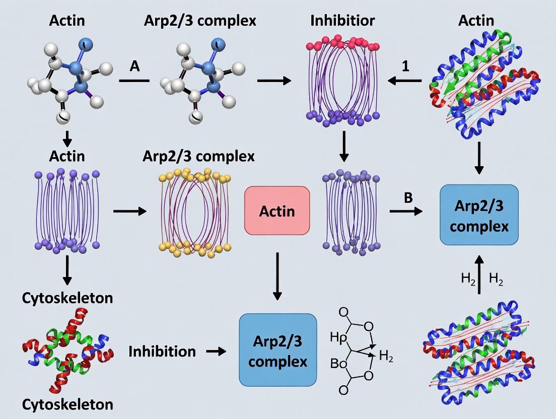
Targeting Cellular Motility: The Mechanism, Inhibition, and Therapeutic Potential of the Arp2/3 Complex in Actin Polymerization
This article provides a comprehensive analysis of the Arp2/3 complex, a pivotal regulator of branched actin network assembly, and its pharmacological inhibition.
Targeting Cellular Motility: The Mechanism, Inhibition, and Therapeutic Potential of the Arp2/3 Complex in Actin Polymerization
Abstract
This article provides a comprehensive analysis of the Arp2/3 complex, a pivotal regulator of branched actin network assembly, and its pharmacological inhibition. Designed for researchers and drug development professionals, it explores the structural biology and nucleation mechanism of Arp2/3 (Intent 1), details experimental methods, assays, and emerging inhibitor classes (Intent 2), addresses common challenges in inhibition studies and strategies for improving inhibitor specificity (Intent 3), and evaluates validation techniques while comparing Arp2/3 inhibition to alternative cytoskeletal targets (Intent 4). The synthesis highlights the complex's role as a promising but challenging target in cancer metastasis, immunology, and other pathologies.
The Arp2/3 Complex Unveiled: Structural Basis and Core Mechanism in Actin Network Assembly
Actin dynamics, the regulated assembly and disassembly of actin filaments (F-actin), are fundamental to cellular processes such as motility, division, and vesicular trafficking. This guide details the core mechanisms within the specific context of research on Arp2/3 complex inhibitors as a therapeutic strategy, providing a technical resource for drug development professionals.
Actin Polymerization: The Monomer-to-Filament Transition
Actin polymerization is a non-equilibrium, ATP-driven process. Globular actin (G-actin) monomers bind ATP and assemble head-to-tail to form polarized filaments with structurally distinct barbed (plus) and pointed (minus) ends. The critical concentration (C~c~) for polymerization differs at each end, creating a steady-state "treadmilling" flux.
Key Quantitative Parameters of Actin Polymerization
| Parameter | Barbed End Value | Pointed End Value | Measurement Conditions (Typical) |
|---|---|---|---|
| Critical Concentration (C~c~) | ~0.1 µM | ~0.6 µM | 1 mM MgATP, 50 mM KCl, pH 7.0, 25°C |
| Elongation Rate Constant (k~+~) | ~11.6 µM⁻¹s⁻¹ | ~1.3 µM⁻¹s⁻¹ | Pyrenyl-actin assay, as above |
| Depolymerization Rate Constant (k~-~) | ~1.4 s⁻¹ | ~0.8 s⁻¹ | As above |
| Treadmilling Rate | ~0.2 - 0.5 µm/min | (Mathematically derived) | Varies with monomer pool & regulatory proteins |
Protocol: Pyrenyl-Actin Polymerization Assay
- Purpose: To quantify the kinetics of actin filament assembly in real-time.
- Materials: Purified G-actin, pyrenyl-labeled actin (Cytoskeleton, Inc. cat # AP-05), polymerization buffer (5 mM Tris-HCl pH 8.0, 0.2 mM CaCl₂, 50 mM KCl, 2 mM MgCl₂, 1 mM ATP), fluorescence spectrophotometer.
- Procedure:
- Prepare a 2 µM G-actin solution (10% pyrenyl-actin, 90% unlabeled) in G-buffer (low salt, 0.2 mM ATP, 0.1 mM CaCl₂).
- Load into a quartz cuvette in a thermostatted fluorometer (25°C, excitation 365 nm, emission 407 nm).
- Initiate polymerization by rapid addition of 1/10 volume 10X polymerization buffer (final: 50 mM KCl, 2 mM MgCl₂, 1 mM ATP).
- Record fluorescence increase over 20-30 minutes. Convert fluorescence to F-actin concentration using a standard curve.
- Fit data to derive elongation rates and lag phase (nucleation).
The Arp2/3 Complex and Branched Network Nucleation
The Actin-Related Protein 2/3 (Arp2/3) complex is the central nucleator of branched actin networks. It binds to the side of a pre-existing "mother" filament and nucleates a new "daughter" filament at a characteristic 70° angle, enabling rapid force generation.
Mechanism of Action & Inhibition: The complex is activated by Nucleation-Promoting Factors (NPFs) like WASP/WAVE. Activated Arp2/3 mimics an actin dimer to initiate a new filament. Inhibitors (e.g., CK-666, CK-869, Arpin) bind to distinct sites, locking the complex in an inactive conformation or preventing branch formation.
Protocol: In Vitro Total Internal Reflection Fluorescence (TIRF) Microscopy of Branched Networks
- Purpose: To visualize and quantify Arp2/3-mediated actin branch formation in real-time.
- Materials: Flow chamber (PEG-silanized coverslip), 1% BSA in buffer, 0.2 µM N-WASH (activated), 50 nM Arp2/3 complex, 2 µM spectrin-actin seeds, 1.5 µM G-actin (20% Alexa Fluor 488-labeled), 100 nM CP, 50 nM GFP-Arp2/3 (for complex localization), TIRF microscope.
- Procedure:
- Assemble flow chamber. Passivate with 1% BSA for 5 min.
- Introduce spectrin-actin seeds and incubate for 2 min to immobilize.
- Rinse with polymerization buffer.
- Flow in the reaction mix containing G-actin (labeled/unlabeled), CP, Arp2/3, N-WASH, and an oxygen scavenging system.
- Immediately acquire time-lapse TIRF images (1-2 sec intervals, 5-10 min).
- Analyze branch density, growth velocity, and lifetime using software (e.g., FIJI, KymoAnalyzer).
Cellular Functions and Therapeutic Targeting
Dysregulated Arp2/3-mediated actin dynamics drive pathological processes, including cancer metastasis (invadopodia formation) and bacterial infection (actin-based motility). This establishes the Arp2/3 complex as a high-value drug target.
Research Reagent Solutions Toolkit
| Reagent / Material | Function / Purpose | Example Vendor (Catalog) |
|---|---|---|
| Purified Skeletal Muscle Actin | Core protein for in vitro assays. Can be labeled. | Cytoskeleton, Inc. (AKL99) |
| Recombinant Arp2/3 Complex | Purified nucleator for mechanistic studies. | Cytoskeleton, Inc. (RP01P) |
| CK-666 / CK-869 | Small-molecule allosteric inhibitors of Arp2/3 complex. | Sigma-Aldrich (SML0006 / 5383410001) |
| Wiskostatin | NPF (N-WASP) inhibitor; indirectly inhibits Arp2/3 activation. | Tocris Bioscience (2979) |
| SMIFH2 | Formin inhibitor; used to isolate Arp2/3-specific effects. | Sigma-Aldrich (S4826) |
| Latrunculin A/B | G-actin sequestering agent; negative control for actin polymerization. | Cayman Chemical (10010630) |
| Jasplakinolide | Actin filament stabilizer; promotes polymerization. | Cayman Chemical (11705) |
| Pyrenyl-labeled Actin | Fluorophore-conjugated actin for kinetic polymerization assays. | Cytoskeleton, Inc. (AP-05) |
| Alexa Fluor Phalloidin | High-affinity F-actin stain for fixed-cell imaging. | Thermo Fisher Scientific (A12379) |
| SiR-Actin Kit | Live-cell compatible, fluorogenic actin probe for microscopy. | Cytoskeleton, Inc. (CY-SC001) |
Diagrams
Title: Mechanism of Arp2/3-Mediated Branching and Inhibition
Title: Pyrenyl-Actin Polymerization Assay Workflow
Within the broader thesis on Arp2/3 complex inhibitors and actin polymerization mechanism research, understanding the precise molecular composition and structure of the Arp2/3 complex is foundational. This nucleator is a central regulator of branched actin filament networks, driving cell motility, endocytosis, and cancer metastasis. Inhibiting its function is a prime therapeutic strategy, necessitating a deep structural knowledge for rational drug design.
Composition and Subunit Architecture
The Arp2/3 complex is a stable, evolutionarily conserved assembly of seven subunits. Its composition is summarized below.
Table 1: Subunit Composition of the Arp2/3 Complex
| Subunit | Gene Name (Human) | Molecular Weight (kDa) | Primary Function/Characteristic |
|---|---|---|---|
| ARPC1 (p41) | ARPC1A/B | ~41 | Scaffolding; binds activating factors (NPFs, WASP) |
| ARPC2 (p34) | ARPC2 | ~34 | Structural core; nucleates branch junction stability |
| ARPC3 (p21) | ARPC3 | ~21 | Bridges ARPC2 and ARPC4; stabilizes complex |
| ARPC4 (p20) | ARPC4 | ~20 | Structural core with ARPC2; essential for complex integrity |
| ARPC5 (p16) | ARPC5 | ~16 | Binds ARPC2 and ARPC4; implicated in branch stabilization |
| ARP2 | ACTR2 | ~44 | Actin-related protein; mimics actin monomer in filament |
| ARP3 | ACTR3 | ~47 | Actin-related protein; ATP-binding site for nucleation |
The complex can be divided into two structural modules:
- The Actin-Related Protein Module: Contains ARP2 and ARP3, which structurally mimic two actin monomers to serve as the nucleation seed.
- The Structural Core Module: Composed of ARPC1-5, which stabilizes the complex and provides binding sites for activators and the mother filament.
Three-Dimensional Structure
High-resolution structural studies (cryo-EM, X-ray crystallography) reveal the complex's architecture in inactive and active states.
Table 2: Key Structural Features and Dimensions
| Feature | Measurement / Description | Method & Resolution (Example) |
|---|---|---|
| Overall Dimensions (Inactive) | ~15 nm x 10 nm x 10 nm | Cryo-EM, ~2.3 Å (PDB: 6WYF) |
| ARP2-ARP3 Separation (Inactive) | ~3.5 nm (too far to mimic actin dimer) | Cryo-EM, ~2.3 Å |
| ARP2-ARP3 Separation (Active) | ~1.2 nm (closes to mimic short-pitch actin dimer) | Cryo-EM, ~4.0 Å (Branch) |
| Mother Filament Binding Angle | ~70° branch angle between mother and daughter filaments | Cryo-EM of branch junctions |
| Key Binding Sites | NPF (WASP/V) binding: ARP2, ARPC1, ARPC3. Mother filament binding: ARP2, ARP3, ARPC2. | Mutagenesis & Cryo-EM |
The transition from an inactive to an active, branch-nucleating conformation involves a major conformational change: ARP2 rotates into a position adjacent to ARP3, creating a template that mimics the barbed end of an actin filament. This movement is triggered by simultaneous binding to a Nucleation-Promoting Factor (NPF, e.g., WASP) and a pre-existing "mother" actin filament.
Diagram 1: Arp2/3 Activation and Branch Nucleation Pathway
Experimental Protocols for Structural & Functional Analysis
Cryo-EM Workflow for Branch Junction Determination
This protocol outlines the process for determining the high-resolution structure of the Arp2/3 complex bound to a branch junction.
1. Sample Preparation:
- Purify recombinant human Arp2/3 complex (e.g., from baculovirus system) and rabbit muscle actin.
- Polymerize mother filaments from actin (2 µM) in F-buffer (2 mM MgCl₂, 100 mM KCl, 1 mM ATP, 1 mM EGTA, 10 mM imidazole pH 7.0).
- Activate Arp2/3 (50 nM) with a WASP-VCA domain fragment (200 nM) in the presence of pre-formed mother filaments.
- Add actin monomers (2 µM) to initiate daughter filament growth. Quench after 60s.
- Apply 3.5 µL of sample to a glow-discharged cryo-EM grid, blot, and plunge-freeze in liquid ethane.
2. Data Collection & Processing:
- Acquire ~5,000 micrograph movies on a 300 keV cryo-EM detector (e.g., K3). Dose: ~50 e⁻/Ų.
- Motion correct and dose-weight micrographs (e.g., MotionCor2).
- Perform template-based particle picking to isolate branch junctions (~1 million particles).
- Execute iterative rounds of 2D classification, 3D classification, and 3D auto-refinement (e.g., in Relion or cryoSPARC).
- Apply CTF refinement and Bayesian polishing. Final map resolution: ~4.0 Å.
3. Model Building & Refinement:
- Dock existing crystal structures of Arp2/3 and F-actin into the cryo-EM density map.
- Manually rebuild and adjust models in Coot to fit density.
- Perform real-space refinement in Phenix.
Pyrene-Actin Polymerization Assay (Standard Kinetic Readout)
Objective: Quantify the effect of an Arp2/3 inhibitor on nucleation activity.
Protocol:
- Reagent Setup: Prepare G-actin (10% pyrene-labeled) in G-buffer (2 mM Tris-HCl pH 8.0, 0.2 mM CaCl₂, 0.2 mM ATP, 0.5 mM DTT). Pre-mix Arp2/3 complex (10 nM final) with/without inhibitor (varying concentrations) in a black 96-well plate.
- Initiation: Rapidly inject a master mix containing actin (2 µM final), WASP-VCA (50 nM final), and 1X F-buffer to initiate polymerization. Final volume: 100 µL.
- Data Acquisition: Immediately monitor pyrene fluorescence (Ex: 365 nm, Em: 407 nm) every 5-10 seconds for 1 hour in a plate reader at 25°C.
- Analysis: Plot fluorescence vs. time. Calculate the maximum polymerization rate (slope) and final extent of polymerization. Fit data to derive IC₅₀ values for inhibitors.
Diagram 2: Pyrene-Actin Assay Workflow
The Scientist's Toolkit: Key Research Reagents & Materials
Table 3: Essential Reagents for Arp2/3 Mechanistic Research
| Reagent/Material | Supplier Examples | Function in Research |
|---|---|---|
| Recombinant Human Arp2/3 Complex | Cytoskeleton, Inc.; in-house purification | The core target for structural and inhibition studies. |
| Pyrene-labeled Actin (10% label) | Cytoskeleton, Inc. | Fluorescent probe for real-time, quantitative measurement of actin polymerization kinetics. |
| WASP/VCA Domain Peptides | GenScript, Peptide 2.0 | Defined NPFs to consistently activate the Arp2/3 complex in assays. |
| CK-666 / CK-869 Inhibitors | Sigma-Aldrich, Tocris | Well-characterized, cell-permeable small molecule inhibitors; used as experimental controls. |
| Latrunculin A/B | Sigma-Aldrich | Actin monomer sequestering agent; negative control for actin-dependent assays. |
| Cryo-EM Grids (Quantifoil R1.2/1.3) | Electron Microscopy Sciences | Sample support for high-resolution structural analysis by cryo-electron microscopy. |
| Size-Exclusion Chromatography Columns (Superdex 200) | Cytiva | Essential for polishing protein complexes to homogeneity for biochemical/structural work. |
| Anti-Arp2/3 Subunit Antibodies (e.g., ARPC2) | Cell Signaling, Abcam | Validation of complex integrity, localization (IF), and expression levels (Western). |
This whitepaper details the Nucleation-Promoting Factor (NPF) paradigm, focusing on the canonical WASP and WAVE family proteins. This discussion is framed within a critical research context: the investigation of Arp2/3 complex inhibitors and their therapeutic potential. As dysregulated actin polymerization drives cancer metastasis, immune dysfunction, and other pathologies, the Arp2/3 complex—the central actin nucleator—is a prime drug target. NPFs are the essential, rate-limiting activators of the Arp2/3 complex. Therefore, a mechanistic understanding of WASP/WAVE regulation and their activation triggers is foundational for rational drug design. Inhibitors may function by blocking Arp2/3 directly, or, more selectively, by disrupting the activation signals or interactions of specific NPFs.
Core NPF Families: WASP and WAVE
WASP (Wiskott-Aldrich Syndrome protein) and WAVE (WASP-family Verprolin-homologous protein) are the two major classes of NPFs. They share a common C-terminal VCA domain (Verprolin homology, Cofilin homology, Acidic region) that binds actin monomers (G-actin) and the Arp2/3 complex to catalyze branched network formation.
Table 1: Key Characteristics of Canonical NPFs
| Feature | WASP (WAS, N-WASP) | WAVE (WAVE1/SCAR, WAVE2, WAVE3) |
|---|---|---|
| Primary Expression | Hematopoietic cells (WAS), ubiquitous (N-WASP) | Ubiquitous (all isoforms) |
| Regulatory State (Basal) | Auto-inhibited (VCA domain blocked by intramolecular interactions) | Inactive within multi-subunit WAVE Regulatory Complex (WRC) |
| Core Activation Trigger | Small GTPases (Cdc42, Rac1) + PIP2 (phosphatidylinositol 4,5-bisphosphate) | Small GTPase (Rac1) exclusively, in conjunction with specific lipids (PIP3, acidic phospholipids) |
| Key Allosteric Activators | Phosphorylation (e.g., by Src kinases), SH3 domain proteins (e.g., Nck, Grb2) | Specific kinases (e.g., Abl, ERK), membrane lipids |
| Primary Cellular Role | Endocytosis, podosome/invadopodium formation, immune synapse assembly | Lamellipodium protrusion, cell migration, membrane ruffling |
| Disease Association | Wiskott-Aldrich Syndrome (immunodeficiency), cancer invasion | Cancer metastasis, neural developmental disorders |
Activation Triggers and Molecular Mechanisms
WASP/N-WASP Activation: Relief of Auto-inhibition
The WASP homology 1 (WH1) domain binds regulatory partners, while a GTPase-binding domain (GBD) interacts with Cdc42/Rac1. The central region and the VCA are connected via a linker. In the auto-inhibited state, the GBD and linker region bind the VCA, blocking its activity.
Activation Mechanism: Cooperative binding of Cdc42•GTP and PIP2 to the GBD and basic region, respectively, induces a conformational change that releases the VCA domain. This is often potentiated by phosphorylation of the linker region (e.g., Y291 on N-WASP) and by SH3 domain-containing adaptors (e.g., Nck) that bind proline-rich regions (PRR), further stabilizing the active conformation.
Detailed Protocol: In Vitro Actin Polymerization Pyrene Assay with N-WASP Activation
- Objective: Quantify actin assembly kinetics triggered by N-WASP under different activation conditions.
- Reagents:
- Pyrene-labeled actin (10%): Actin conjugated with pyrene iodoacetamide. Pyrene fluorescence increases >20-fold upon polymerization, serving as a real-time readout.
- Unlabeled G-actin: Purified rabbit skeletal muscle actin in G-buffer (2 mM Tris-HCl pH 8.0, 0.2 mM CaCl2, 0.2 mM ATP, 0.5 mM DTT).
- Proteins: Purified Arp2/3 complex, N-WASP (full-length, auto-inhibited).
- Activators: Recombinant Cdc42 (loaded with GTPγS, a non-hydrolyzable GTP analog), PIP2-containing liposomes, Src kinase (with ATP for phosphorylation).
- Procedure:
- Prepare reaction mixtures (50 µL final) in polymerization buffer (10 mM imidazole pH 7.0, 50 mM KCl, 1 mM MgCl2, 1 mM EGTA, 0.2 mM ATP) containing: 2 µM G-actin (10% pyrene-labeled), 25 nM Arp2/3 complex, 10 nM N-WASP.
- Set up activation conditions:
- Condition A: N-WASP only (basal).
- Condition B: + 100 nM Cdc42•GTPγS.
- Condition C: + 20 µM PIP2 liposomes.
- Condition D: + Cdc42•GTPγS + PIP2 liposomes.
- Condition E: Pre-phosphorylated N-WASP (incubated with Src kinase + ATP for 30 min at 30°C prior) + Cdc42•GTPγS + PIP2.
- Load mixtures (minus actin) into a quartz cuvette in a fluorometer. Initiate polymerization by adding pre-cleared G-actin. Mix rapidly.
- Monitor pyrene fluorescence (excitation 365 nm, emission 407 nm) every 2 seconds for 600-1200 seconds at 25°C.
- Analyze the fluorescence versus time curves. Key metrics: lag phase (duration before rapid increase), initial polymerization rate (slope during exponential phase), and final plateau (total F-actin).
WAVE Activation: Dissociation of the WAVE Regulatory Complex (WRC)
WAVE isoforms are constitutively incorporated into a stable, ~400 kDa WRC composed of WAVE, Cyfip, Nap1, Abi, and HSPC300. The WRC sterically occludes the VCA domain.
Activation Mechanism: The primary trigger is Rac1•GTP, which binds directly to the Cyfip subunit. This, combined with interaction with acidic phospholipids (PIP3, PIP2) via a basic surface on the WRC, induces a conformational change that partially releases the VCA. Additional inputs, like phosphorylation of WAVE or Abi subunits by kinases such as ERK or Abl, modulate the sensitivity and localization of the WRC.
Detailed Protocol: Co-sedimentation Assay for WRC Activation and Membrane Recruitment
- Objective: Assess the membrane recruitment and activation of the WRC by Rac1 and PIP3 using liposome co-sedimentation.
- Reagents:
- Purified Proteins: WRC (reconstituted from recombinant subunits), Rac1 (loaded with GTPγS or GDP).
- Liposomes: Prepared by extrusion, containing:
- Neutral: 70% PC (phosphatidylcholine), 30% PS (phosphatidylserine).
- PIP3-containing: 67% PC, 30% PS, 3% PIP3.
- Ultracentrifugation equipment.
- Procedure:
- Prepare liposomes in assay buffer (20 mM HEPES pH 7.4, 100 mM NaCl, 1 mM MgCl2).
- Mix 100 nM WRC with 1 µM Rac1•GTPγS (or Rac1•GDP) and 200 µM liposomes (final lipid concentration) in 100 µL total volume. Incubate 30 min at 25°C.
- Load samples onto a 200 µL sucrose cushion (20% sucrose in assay buffer) in a thick-walled polycarbonate ultracentrifuge tube.
- Centrifuge at 100,000 x g for 30 minutes at 4°C. This pellets liposomes and any bound protein.
- Carefully aspirate the top supernatant (unbound protein). Wash the pellet surface gently with buffer. Resuspend the pellet in SDS-PAGE loading buffer.
- Analyze equal proportions of supernatant (S) and pellet (P) fractions by SDS-PAGE and Coomassie or immunoblotting.
- Quantification: The amount of WRC in the pellet fraction indicates membrane recruitment. Co-sedimentation is expected only with Rac1•GTPγS + PIP3-containing liposomes, demonstrating cooperative activation.
Diagrams of Signaling Pathways and Experimental Workflows
Diagram 1: WASP/N-WASP Activation Pathway in Invadopodia
Diagram 2: WAVE Regulatory Complex (WRC) Activation at Lamellipodia
Diagram 3: Pyrene Actin Polymerization Assay Workflow
The Scientist's Toolkit: Essential Research Reagents
Table 2: Key Reagent Solutions for NPF/Arp2/3 Mechanistic Research
| Reagent Category | Specific Example(s) | Function & Application |
|---|---|---|
| Actin Proteins | Purified monomeric (G-) actin (rabbit muscle, non-muscle isoforms), Pyrene-labeled actin (10-30% labeling ratio) | Core substrate for polymerization. Pyrene-actin provides a sensitive fluorescent readout for assembly kinetics in in vitro assays. |
| Effector Proteins | Recombinant, purified Arp2/3 complex (from bovine, human, or insect cell expression), Full-length WASP/N-WASP, Reconstituted WAVE Regulatory Complex (WRC) | Essential reaction components. Full-length, properly regulated NPFs are required for activation studies. |
| Activation Reagents | Small GTPases (Cdc42, Rac1) pre-loaded with GTPɣS or GDP, PIP2/PIP3 lipids (as liposomes or micelles), Active kinases (e.g., Src, Abl, ERK) with ATP. | Used to trigger specific NPF activation pathways in controlled in vitro or cellular assays. |
| Inhibitors (Tool Compounds) | CK-666 / CK-869 (allosteric Arp2/3 inhibitors), Wiskostatin (stabilizes N-WASP auto-inhibition), PIR121-derived peptide (blocks Rac-WRC interaction). | Pharmacological tools to dissect pathway necessity. Serve as prototypes for therapeutic development. |
| Cellular Probes | Fluorescent protein-tagged NPFs (GFP-WASP, GFP-WAVE2), FRET biosensors (for Rac/Cdc42 activity), LifeAct or Utrophin F-actin probes. | For live-cell imaging of NPF localization, activation dynamics, and actin network formation. |
| Antibodies | Phospho-specific antibodies (e.g., anti-N-WASP pY291), Conformation-sensitive antibodies (distinguishing open/closed WASP), Isoform-specific WAVE antibodies. | Detect activation states, protein localization, and expression levels in immunoblotting, immunofluorescence, and flow cytometry. |
Within the research framework of developing Arp2/3 complex inhibitors to modulate actin cytoskeleton dynamics, understanding the precise nucleation mechanism is paramount. The Arp2/3 complex is the central cellular machine that nucleates new "daughter" actin filaments from the sides of pre-existing "mother" filaments, creating the branched networks essential for cell motility, endocytosis, and pathogen invasion. This whitepaper details the structural and kinetic journey from the inactive Arp2/3 complex to the formation of a stabilized branched junction, providing the mechanistic foundation necessary for rational inhibitor design.
Structural Transitions: From Inactive State to Nucleation-Competent Branch
The Arp2/3 complex exists in an inactive, auto-inhibited conformation. Activation requires both a nucleating promoting factor (NPF) and a mother filament. Recent cryo-EM structures have elucidated this transition.
Key Structural States and Data
Table 1: Structural States of the Arp2/3 Complex
| State | Key Features | Stabilizing Factors | Resolution (Approx.) | PDB ID (Example) |
|---|---|---|---|---|
| Inactive | Arp2 & Arp3 separated; blocked nucleation face. | Auto-inhibitory domains. | 4.5 Å | 7KQ9 |
| NPF-Bound | Partial opening; Arp2/3 closer, but not actin-like. | VCA domains (WASP/N-WASP). | 3.8 Å | 7KQA |
| Mother Filament-Bound | Complex anchored to mother filament via Arp2/3 subunits. | ATP-actin in mother filament. | 3.6 Å | 7KQB |
| "Short-Pitch" Daughter Nucleus | Arp2 & Ar3 mimic barbed end of actin dimer; first daughter actin monomers incorporated. | ATP, NPF, mother filament. | 4.0 Å | 8FOE |
Visualization of the Activation Pathway
Title: Arp2/3 Activation and Branch Nucleation Pathway
Kinetic Mechanism and Experimental Quantification
The nucleation process follows a multi-step kinetic pathway. Key rates determine the efficiency of branch formation.
Table 2: Kinetic Parameters for Arp2/3-Mediated Branch Formation
| Kinetic Step | Rate Constant (Approx.) | Method of Determination | Impact of CK-666 (Inhibitor) |
|---|---|---|---|
| NPF (VCA) Binding | Kd ~ 0.1 - 1 µM | Fluorescence Anisotropy | No direct effect. |
| Mother Filament Binding | Kd ~ 10-100 nM | TIRF Microscopy / FRET | Increases Kd (weakens binding). |
| Nucleation (Dimer Stabilization) | k_nuc ~ 0.1 - 1 s⁻¹ | Pyrene-Actin Assembly | Drastically reduces rate. |
| Branch Stability (Debranching) | k_off ~ 0.01 s⁻¹ | TIRF Microscopy (single filament) | Can increase debranching rate. |
Detailed Experimental Protocol: TIRF Microscopy for Single-Filament Branch Kinetics
Objective: Quantify the rate of branch nucleation and debranching from individual mother filaments.
Materials: See "Scientist's Toolkit" below. Procedure:
- Flow Cell Preparation: Passivate a glass flow chamber with methoxy-PEG-silane. Functionalize with biotin-PEG-silane in defined lanes.
- Mother Filament Tethering: Introduce 0.5 µM streptavidin for 2 min, wash. Introduce 1-10 nM biotinylated, spectrin-actin seeds (or pre-formed rhodamine-phalloidin-stabilized filaments) in G-buffer for 5 min. Wash thoroughly.
- Reaction Mix Introduction: Prepare a mix containing:
- 1.5 µM monomeric actin (10-30% Oregon Green 488-labeled)
- 50 nM Arp2/3 complex
- 50 nM full-length NPF (e.g., WASP) or saturating VCA fragment
- 1 mM ATP
- TIRF imaging buffer (10 mM Imidazole pH 7.4, 50 mM KCl, 1 mM MgCl2, 1 mM EGTA, 50 mM DTT, 0.2% methyl cellulose, 100 µg/ml glucose oxidase, 20 µg/ml catalase, 5 mg/ml glucose).
- Image Acquisition: Immediately introduce mix to flow cell. Image using a 488 nm laser on a TIRF microscope with EMCCD/sCMOS camera at 1-5 second intervals for 10-20 minutes.
- Analysis: Use software (e.g., FIJI, KymographClear) to track mother filaments. Count the appearance of new, growing filaments originating from the side of a mother filament as a nucleation event. Plot nucleation events vs. time. Measure the lifetime of branches from initiation to dissociation (debranching).
The "Mother-Daughter" Model: Geometry and Stabilization
The mature branch exhibits a characteristic 70° angle between the mother and daughter filaments. Stabilization involves multiple contacts.
Title: Mother-Daughter Branch Geometry and Contacts
Table 3: Critical Interfaces in the Branched Junction
| Interface | Contributing Subunits/Proteins | Function | Targeted by Inhibitor? |
|---|---|---|---|
| Arp2/3 - Mother Filament | Arp2, Arp3, ARPC1/2/3 | Anchoring & activation. | Yes (CK-666, Arpin) |
| Arp2/3 - Daughter D1 Actin | Arp2, Arp3 | Mimics actin-actin bond. | Yes (CK-869) |
| NPF - Arp2/3 | VCA linker | Releases auto-inhibition. | Potential target |
| D1 - Mother Filament | D1 actin & mother filament actin | Stabilizes branch angle. | -- |
The Scientist's Toolkit: Key Research Reagents
Table 4: Essential Reagents for Arp2/3 Nucleation Studies
| Reagent | Function & Description | Example Supplier/Cat # |
|---|---|---|
| Purified Arp2/3 Complex | Core heptameric complex from bovine thymus, human platelets, or recombinant expression (Sf9 cells). Essential substrate. | Cytoskeleton Inc. (RP01), in-house purification. |
| NPF Fragments (VCA) | Minimal active domain (WASP, N-WASP, WAVE). Used to activate Arp2/3. Often GST- or His-tagged. | Custom peptide synthesis, Cytoskeleton Inc. (AP09). |
| Pyrene-Labeled Actin | Actin conjugated with pyrene fluorophore. Polymerization increases fluorescence >10x. For bulk nucleation kinetics. | Cytoskeleton Inc. (AP05). |
| Fluorophore-Labeled Actin | Actin labeled with Oregon Green 488, Alexa 568, etc., for TIRF/fluorescence microscopy. | Thermo Fisher (A12373), Cytoskeleton Inc. (AB05). |
| Biotinylated Actin | Actin conjugated with biotin for tethering to streptavidin-coated surfaces in single-filament assays. | Cytoskeleton Inc. (AB03). |
| Phalloidin (Rhodamine/ATTO) | Fungal toxin that stabilizes F-actin. Used to label and stabilize mother filaments. | Sigma-Aldrich (P1951), ATTO-TEC. |
| Arp2/3 Inhibitors (CK-666/869) | Small molecule allosteric inhibitors. CK-666 locks inactive state; CK-869 binds Arp3-D1 interface. Tool compounds for mechanism. | Sigma-Aldrich (SML0006 / SML1660). |
| TIRF Imaging Buffer System | Oxygen scavenging (GlOx/Cat) and anti-photobleaching (methyl cellulose) system for prolonged single-molecule imaging. | Home-made or commercial kits. |
The Arp2/3 complex is a conserved, seven-subunit actin nucleator that is fundamental to the creation of branched actin networks. Its activation by Nucleation-Promoting Factors (NPFs) such as the WASP/WAVE family is a central regulatory node in eukaryotic cell physiology and pathogenesis. Research into Arp2/3 complex inhibitors has become a critical pathway for dissecting its precise mechanistic contributions and for developing therapeutic strategies against pathologies driven by aberrant actin dynamics, including metastatic cancer and bacterial infection. This whitepaper details the biological roles of Arp2/3-mediated actin assembly within three key cellular processes, framed by insights gained from pharmacological inhibition.
The following tables consolidate key quantitative findings related to Arp2/3 function and the impact of its inhibition.
Table 1: Arp2/3 in Core Cellular Processes
| Process | Key NPF Activator | Primary Actin Structure | Measurable Impact of Arp2/3 Knockdown/Inhibition |
|---|---|---|---|
| Lamellipodia Protrusion | WAVE Regulatory Complex (WRC) | Dense, branched network at leading edge | ~70-80% reduction in protrusion velocity (from ~2-3 µm/min to ~0.5 µm/min). Loss of persistent directional migration. |
| Clathrin-Mediated Endocytosis | N-WASP & WASH | Patches of branched actin at endocytic sites | ~60% decrease in successful vesicle internalization rate. Prolonged pit maturation time (from ~30s to >60s). |
| Pathogen Propulsion (Listeria) | Bacterial ActA protein | "Comet tail" of branched actin | Complete cessation of intracellular motility. Tail disintegration within minutes of inhibitor addition. |
Table 2: Characterized Small-Molecule Arp2/3 Inhibitors
| Inhibitor Name | Proposed Target / Mechanism | Reported IC₅₀ (In Vitro) | Key Phenotypic Effect in Cells |
|---|---|---|---|
| CK-666 | Binds Arp2/3 complex, stabilizes inactive state. | 5-25 µM (pyrene-actin assay) | Inhibits lamellipodia, endocytosis, and pathogen motility. Reversible. |
| CK-869 | Binds Arp2/3 complex, alternative inhibitory conformation. | ~10 µM (pyrene-actin assay) | Similar to CK-666 but with distinct structural effects. |
| Arpin (natural protein) | Competes with NPFs for binding to Arp2/3 complex. | N/A (endogenous regulator) | Negatively regulates lamellipodial persistence. |
Experimental Protocols for Assessing Arp2/3 Function
These protocols are foundational for research utilizing Arp2/3 inhibitors.
Protocol 1: In Vitro Pyrene-Actin Polymerization Assay
- Purpose: To quantitatively measure the effect of inhibitors on Arp2/3-mediated actin nucleation and branching.
- Materials: Purified actin (with ~5% pyrene-labeled actin), purified Arp2/3 complex, purified NPF (e.g., WASP-VCA domain), inhibitor (e.g., CK-666), polymerization buffer (1X KMEI: 50 mM KCl, 1 mM MgCl₂, 1 mM EGTA, 10 mM Imidazole pH 7.0).
- Procedure:
- Prepare a master mix of G-actin (2 µM final) in polymerization buffer.
- In a 96-well plate, mix Arp2/3 (10-50 nM), NPF (100-200 nM), and varying concentrations of inhibitor. Include controls lacking Arp2/3, NPF, or inhibitor.
- Initiate polymerization by adding the actin master mix to each well.
- Immediately monitor fluorescence (ex: 365 nm, em: 407 nm) in a plate reader at 25-30°C for 30-60 minutes.
- Analyze the initial polymerization rate and final steady-state fluorescence. Calculate IC₅₀ for the inhibitor.
Protocol 2: Live-Cell Imaging of Lamellipodia Dynamics Post-Inhibition
- Purpose: To assess the real-time impact of Arp2/3 inhibition on cell edge protrusion.
- Materials: Migratory cell line (e.g., B16F1 melanoma, MEF), fluorescent actin marker (LifeAct-GFP or similar), cell culture medium, spinning-disk confocal microscope, inhibitor (e.g., CK-666 in DMSO).
- Procedure:
- Plate cells expressing LifeAct-GFP on a glass-bottom dish to ~70% confluency.
- Acquire time-lapse images (1 frame/5-10s) of a lamellipodial edge for 5 minutes to establish baseline dynamics.
- Without moving the field of view, carefully add inhibitor to the medium (final CK-666 concentration ~100 µM).
- Continue time-lapse imaging for 20-30 minutes.
- Use kymograph analysis (drawing a line perpendicular to the cell edge) to quantify protrusion velocity and persistence before and after inhibitor addition.
Signaling Pathways and Experimental Workflows
The Scientist's Toolkit: Key Research Reagents
| Reagent / Material | Provider Examples | Primary Function in Arp2/3 Research |
|---|---|---|
| Recombinant Arp2/3 Complex | Cytoskeleton Inc., custom purification | Essential substrate for in vitro biochemical assays of nucleation and inhibitor screening. |
| CK-666 & CK-869 | Sigma-Aldrich, Tocris Bioscience | Bench-standard small-molecule inhibitors used to probe Arp2/3 function in live cells and in vitro. |
| WAVE2 / N-WASP VCA Domain | Cytoskeleton Inc., custom purification | Minimalist, constitutively active NPF domains used to activate Arp2/3 in simplified in vitro systems. |
| Pyrene-Labeled Actin | Cytoskeleton Inc. | Fluorescent actin derivative used to monitor polymerization kinetics in real-time via fluorescence increase. |
| LifeAct-EGFP/RFP | Addgene, commercial vectors | Genetically encoded peptide tag for non-invasive, high-contrast visualization of F-actin in live cells. |
| siRNA against Arp2/3 Subunits | Dharmacon, Qiagen | For genetic knockdown to validate pharmacological inhibition phenotypes and study long-term adaptation. |
| Total Internal Reflection Fluorescence (TIRF) Microscope | N/A (Core Facility) | Enables high-resolution imaging of actin dynamics at the cell membrane (e.g., endocytosis, lamellipodia). |
Tools and Techniques: From In Vitro Assays to Emerging Arp2/3 Inhibitor Chemotypes
Within the research framework aimed at elucidating the mechanism of action of Arp2/3 complex inhibitors, a multi-faceted technical approach is indispensable. This guide details three cornerstone assays—pyrene-actin polymerization, TIRF microscopy, and electron microscopy—that together provide complementary, quantitative data on actin dynamics and network architecture. These techniques are critical for characterizing how inhibitory compounds affect the nucleation, elongation, and ultrastructure of actin filaments.
Pyrene-Actin Polymerization Assay
This fluorometric assay is the biochemical workhorse for quantifying actin polymerization kinetics in real-time. Pyrene-labeled actin incorporates into filaments, causing a dramatic increase in fluorescence intensity, allowing monitoring of nucleation and elongation phases.
Experimental Protocol
- Reagent Preparation: Prepare G-actin buffer (2 mM Tris-HCl pH 8.0, 0.2 mM CaCl₂, 0.2 mM ATP, 0.5 mM DTT). Thaw rabbit skeletal muscle G-actin (≥99% pure) and pyrene-labeled G-actin (typically 10% labeled) on ice. Pre-complex unlabeled G-actin with 0.2 mM MgCl₂ and 50 μM EGTA for 2 minutes to exchange Ca²⁺ for Mg²⁺.
- Master Mix: In a black 96-well plate, mix components to final volumes of 50-100 μL. A standard reaction contains: 1-4 μM total G-actin (5% pyrene-labeled), polymerizing buffer (final: 1 mM MgCl₂, 50 mM KCl, 1 mM EGTA, 10 mM imidazole pH 7.0). For Arp2/3 studies, include purified Arp2/3 complex (5-50 nM) and a nucleation promoting factor (e.g., 10-100 nM VCA domain of N-WASP) with or without the inhibitor of interest.
- Initiation & Data Acquisition: Use a plate reader pre-heated to 25°C or 30°C. Initiate polymerization by adding the salt-containing polymerizing buffer. Immediately monitor fluorescence (ex: 365 nm, em: 407 nm) every 5-10 seconds for 30-60 minutes.
- Data Analysis: Normalize fluorescence to the maximum and minimum values. Calculate key parameters: lag time (time to 10% max fluorescence), maximum slope (polymerization rate), and final steady-state fluorescence.
Table 1: Quantitative Parameters from Pyrene-Actin Assay for Arp2/3 Inhibition
| Parameter | Control (No Inhibitor) | With Inhibitor A (100 nM) | With Inhibitor B (500 nM) | Interpretation |
|---|---|---|---|---|
| Lag Time (s) | 120 ± 15 | 300 ± 25 | 450 ± 40 | Increased lag indicates suppressed nucleation efficiency. |
| Max Slope (RFU/s) | 55 ± 5 | 20 ± 3 | 8 ± 2 | Reduced slope indicates slower filament elongation or fewer barbed ends. |
| Final RFU (%) | 100 ± 2 | 75 ± 5 | 50 ± 6 | Lower plateau suggests reduced total F-actin mass or altered critical concentration. |
Diagram 1: Inhibitor Impact on Polymerization Phases (83 chars)
TIRF Microscopy for Single-Filament Dynamics
Total Internal Reflection Fluorescence (TIRF) microscopy visualizes real-time dynamics of individual actin filaments near the coverslip surface, providing direct insight into filament nucleation, growth, severing, and depolymerization.
Experimental Protocol
- Flow Chamber Preparation: Create a passivated flow chamber using a glass coverslip and slide separated by double-sided tape. Sequentially flow in: (i) 0.2 mg/mL Biotin-PEG for 5 min, (ii) Blocking solution (1% BSA, 1% pluronic F-127) for 5 min, (iii) 0.5 mg/mL NeutrAvidin for 2 min, (iv) Biotinylated anti-His antibody (for His-tagged nucleation factors).
- Surface Tethering: Introduce His-tagged Arp2/3 complex (or nucleation promoting factor) in TIRF buffer (10 mM Imidazole pH 7.4, 50 mM KCl, 1 mM MgCl₂, 1 mM EGTA, 0.2 mM ATP, 50 mM DTT, 0.5% Methyl Cellulose).
- Imaging Mixture: Prepare the actin mix: 1 μM G-actin (30% labeled with Alexa Fluor 488/568), 0.2 μM profilin (optional), oxygen scavenging system (0.2 mg/mL glucose oxidase, 0.035 mg/mL catalase, 2.5 mM glucose), and 2.5 mM Trolox to reduce photobleaching. Add the inhibitor at the desired concentration.
- Image Acquisition: Flow the imaging mixture into the chamber. Use a 100x or 60x TIRF objective (NA ≥ 1.45) on an inverted microscope. Acquire time-lapse images (100-500 ms intervals) for 10-20 minutes using appropriate laser power and EMCCD/sCMOS camera settings.
- Analysis: Use software (e.g., FIJI/ImageJ with plugins, or KymoToolBox) to generate kymographs from regions of interest. Measure filament nucleation frequency, elongation rate (from kymograph slope), lifetime, and filament length distribution.
Table 2: TIRF Microscopy Quantification of Filament Dynamics with Arp2/3 Inhibition
| Parameter | Control (Arp2/3 + VCA) | + CK-666 (100 μM) | Interpretation |
|---|---|---|---|
| Nucleation Events / FOV / min | 15.2 ± 2.1 | 2.1 ± 0.8 | Direct measure of inhibited Arp2/3 nucleation activity. |
| Average Elongation Rate (subunits/s) | 8.5 ± 0.7 | 8.3 ± 0.9 | Confirms inhibitor does not directly cap barbed ends. |
| Average Filament Lifetime (s) | 180 ± 25 | 250 ± 40 | Longer lifetime may indicate reduced branch turnover/network disassembly. |
| Branch Angle (degrees) | 70 ± 5 | N/A (no branches) | Loss of characteristic Arp2/3-mediated 70° branching. |
Diagram 2: TIRF Assay Workflow for Actin Dynamics (60 chars)
Electron Microscopy for Network Ultrastructure
Electron microscopy (EM), particularly negative staining and vitrification (cryo-EM), provides high-resolution snapshots of actin network architecture and the morphology of individual branches.
Experimental Protocol: Negative Stain EM for Actin Branches
- Sample Preparation: Polymerize 4 μM actin with 50 nM Arp2/3 complex and 100 nM VCA domain in polymerization buffer for 5-10 minutes at room temperature. Include inhibitor in relevant samples.
- Grid Preparation: Apply 3-5 μL of sample to a glow-discharged carbon-coated EM grid for 60 seconds.
- Staining: Blot excess liquid with filter paper. Immediately apply 3-5 μL of 1% uranyl acetate solution for 30 seconds. Blot and air dry.
- Imaging: Examine grids using a transmission electron microscope (TEM) operated at 80-100 kV. Collect images at 20,000x – 60,000x magnification.
- Image Analysis: Identify and count branch junctions. Measure branch angles using image analysis software (e.g., ImageJ). For cryo-EM, samples are vitrified and imaged under cryo-conditions, allowing for 3D reconstruction of the Arp2/3 complex at the branch junction.
Table 3: Electron Microscopy Analysis of Network Architecture
| Structural Feature | Control Network | Network + Inhibitor | Method |
|---|---|---|---|
| Branch Density (per μm²) | 42 ± 8 | < 5 | Negative Stain |
| Average Branch Angle (°) | 70 ± 7 | N/A | Negative Stain |
| Arp2/3 Conformation at Junction | "Active" Short Pitch | "Inactive" Open State | Cryo-EM Single Particle Analysis |
Diagram 3: Inhibitor Block of Arp2/3-Mediated Branching (78 chars)
The Scientist's Toolkit: Key Research Reagent Solutions
Table 4: Essential Materials for Actin Polymerization Mechanism Research
| Reagent / Material | Function & Rationale | Key Considerations |
|---|---|---|
| Pyrene-labeled G-actin | Fluorescent reporter for polymerization kinetics. Pyrene excimer formation upon incorporation increases fluorescence ~25-fold. | Labeling ratio typically 5-10%. Ensure low free pyrene content to avoid background. |
| Purified Arp2/3 Complex | Core nucleation machinery from bovine brain, human platelets, or recombinant expression. | Activity varies by source/prep. Check nucleation activity in pyrene assay vs. known standards. |
| Nucleation Promoting Factor (NPF) | Activates Arp2/3 complex. Commonly used: VCA domain of N-WASP or WAVE2. | Use truncated, purified domains for consistent, high-affinity activation. |
| TIRF-compatible Fluorescent Actin | G-actin conjugated to bright, photostable dyes (e.g., Alexa Fluor 488, 568). | Degree of labeling critical. High labeling inhibits polymerization; low labeling yields dim filaments. |
| Profilin | Binds G-actin, prevents spontaneous nucleation, promotes elongation at barbed ends. | Essential for clean TIRF assays to observe regulated nucleation, not background assembly. |
| CK-666 / CK-869 | Well-characterized, cell-permeable allosteric inhibitors of Arp2/3 complex. | CK-666 stabilizes inactive state. Use as positive control for biochemical and cellular inhibition. |
| Uranyl Acetate (EM Grade) | High-contrast negative stain for visualizing actin filaments and branches by TEM. | Light-sensitive, mildly radioactive. Prepare fresh solutions and dispose of properly. |
| ATP (Magnesium Salt) | Essential cofactor for actin monomer stability and polymerization. | Use Mg²⁺-ATP. Always include in all buffers (0.1-2 mM) to maintain actin health. |
| Oxygen Scavenging System | Reduces photobleaching and free radical damage in fluorescence microscopy. | Glucose oxidase/catalase + Trolox is standard for TIRF. Protects fluorophores and actin. |
| Methyl Cellulose | Added to TIRF buffer to reduce filament diffusion and tumbling, keeping them in focal plane. | Viscous agent. Use low concentration (0.1-0.5%) to minimize artifacts. |
This technical guide details functional cell-based assays critical for evaluating the effects of Arp2/3 complex inhibitors, such as CK-666, CK-869, and Arp2/3-targeting compounds. Inhibition of the Arp2/3 complex prevents nucleation of branched actin networks, a primary mechanism driving cell motility, protrusion, and invasion. These assays measure downstream phenotypic consequences of disrupted actin polymerization, directly linking molecular mechanism to cellular function.
Lamellipodia Formation Assay
Lamellipodia are broad, sheet-like membrane protrusions driven by Arp2/3-mediated branched actin networks. This assay quantitatively assesses the impact of inhibitors on protrusion dynamics.
Detailed Protocol
- Cell Plating: Plate serum-starved fibroblasts (e.g., NIH/3T3, MEF) or cancer cells (e.g., MDA-MB-231) on fibronectin-coated (5 µg/mL) glass-bottom dishes at low density.
- Inhibitor Treatment: Pre-treat cells with Arp2/3 inhibitor (e.g., 50-200 µM CK-666) or DMSO control in serum-free medium for 1-2 hours.
- Stimulation & Fixation: Stimulate lamellipodia formation with 10-20 ng/mL EGF or 10% FBS for 5-15 minutes. Immediately fix with 4% paraformaldehyde (PFA) for 15 minutes.
- Staining: Permeabilize with 0.1% Triton X-100, block with 1% BSA, and stain for F-actin using Alexa Fluor 488- or 594-conjugated phalloidin (1:500) for 1 hour.
- Imaging & Quantification: Acquire high-resolution images using a 60x or 63x oil objective on a confocal microscope. Quantify:
- Lamellipodial area: Threshold-based measurement of peripheral F-actin-rich protrusions.
- Protrusion width/height: Using line-scan analysis.
- Intensity of peripheral F-actin: Mean fluorescence intensity at the cell edge.
Table 1: Representative Effects of Arp2/3 Inhibitors on Lamellipodia Formation
| Cell Line | Arp2/3 Inhibitor | Concentration | Stimulus | Reduction in Lamellipodial Area | Key Citation |
|---|---|---|---|---|---|
| Mouse Embryonic Fibroblast (MEF) | CK-666 | 100 µM | 10% FBS | 70-80% | Nolen et al., Nature, 2009 |
| MDA-MB-231 (Breast Cancer) | CK-869 | 50 µM | 10 ng/mL EGF | 60-75% | Yang et al., JCB, 2020 |
| U2OS (Osteosarcoma) | Arpin overexpression | N/A | 10% FBS | ~50% | Dang et al., Nature, 2013 |
Title: Signaling to Lamellipodia via Arp2/3 Complex
Invadopodia Formation & Degradation Assay
Invadopodia are actin-rich protrusions that degrade the extracellular matrix (ECM), crucial for invasion. Their formation is Arp2/3-dependent.
Detailed Protocol (Fluorescent Gelatin Degradation Assay)
- Substrate Preparation: Coat glass coverslips with a thin layer of fluorescein-conjugated gelatin (0.2% gelatin, 0.2% sucrose) and cross-link with 0.5% glutaraldehyde. Quench with 5 mg/mL sodium borohydride, then sterilize and coat with 5 µg/mL fibronectin.
- Cell Plating & Treatment: Plate invasive cancer cells (e.g., MDA-MB-231, SCC-61) on the coated coverslips. Allow to adhere, then treat with inhibitor or vehicle in complete medium for 4-24 hours.
- Fixation & Staining: Fix with 4% PFA, permeabilize, and block. Stain for F-actin (phalloidin, 1:500) and cortactin (anti-cortactin antibody, 1:200) as an invadopodia marker. Use DAPI for nuclei.
- Imaging & Quantification: Image using a confocal microscope (63x oil). Identify invadopodia as F-actin/cortactin puncta colocalizing with dark areas of degraded gelatin. Quantify:
- % of cells with invadopodia: Cells with ≥3 degradation spots.
- Number of invadopodia per cell.
- Total degradation area per cell: Area of black (degraded) spots relative to cell area.
Table 2: Effects of Arp2/3 Inhibition on Invadopodia Activity
| Cell Line | Arp2/3 Inhibitor | Concentration | Incubation Time | Reduction in Degradation Area | Key Citation |
|---|---|---|---|---|---|
| MDA-MB-231 | CK-666 | 100 µM | 18 hours | ~85% | Clark et al., Cancer Res, 2021 |
| SCC-61 (HNSCC) | CK-666 | 200 µM | 6 hours | 70-80% | Hoppe et al., Mol Biol Cell, 2022 |
| PC-3 (Prostate Cancer) | siRNA Arp3 | N/A | 48 hours | >90% | Gligorijevic et al., Nat Protoc, 2014 |
Transwell Migration & Invasion Assay
This assay measures directed cell movement through porous membranes, with or without an ECM coating, to model chemotaxis and invasion.
Detailed Protocol
- Chamber Preparation: For invasion assays, coat the upper side of a Transwell insert (8 µm pore) with 50-100 µL of Matrigel (1-2 mg/mL). Allow to polymerize for 2 hours at 37°C. For migration assays, use uncoated inserts.
- Cell Preparation: Serum-starve cells for 12-24 hours. Harvest, resuspend in serum-free medium containing inhibitor or DMSO.
- Assay Setup: Place 500-750 µL of chemoattractant medium (with 10% FBS or specific factor) in the lower chamber. Seed 50,000-100,000 cells in serum-free medium into the upper chamber.
- Incubation & Treatment: Incubate at 37°C for 6-48 hours (time varies by cell line).
- Quantification: Remove non-migrated cells from the upper chamber with a cotton swab. Fix migrated/invaded cells on the lower membrane with methanol or 4% PFA. Stain with 0.1% crystal violet or DAPI. Count cells in 5-10 random fields per insert under a 20x objective. Alternatively, dissolve crystal violet in 10% acetic acid and measure absorbance at 590 nm.
Table 3: Impact of Arp2/3 Inhibition on Transwell Migration/Invasion
| Cell Line | Assay Type | Arp2/3 Inhibitor | Concentration | % Inhibition of Migration/Invasion | Key Citation |
|---|---|---|---|---|---|
| MDA-MB-231 | Invasion (Matrigel) | CK-666 | 100 µM | 60-70% | Wong et al., Cell Rep, 2021 |
| HeLa | Migration | CK-869 | 25 µM | ~50% | Rizvi et al., JCS, 2022 |
| HT-1080 (Fibrosarcoma) | Invasion (Collagen I) | siRNA Arp2 | N/A | 75-85% | Steffen et al., J Cell Sci, 2013 |
Title: Transwell Migration Assay Experimental Workflow
The Scientist's Toolkit: Research Reagent Solutions
Table 4: Essential Reagents for Arp2/3 Functional Assays
| Reagent/Material | Supplier Examples | Function in Assay |
|---|---|---|
| CK-666 & CK-869 | Sigma-Aldrich, Tocris, Cayman Chemical | Small-molecule allosteric inhibitors of the Arp2/3 complex; used to block branched actin nucleation. |
| Fluorescent Phalloidin | Thermo Fisher (e.g., Alexa Fluor conjugates), Cytoskeleton Inc. | High-affinity probe for staining F-actin to visualize lamellipodia, invadopodia, and cytoskeleton. |
| Matrigel / GFR Matrigel | Corning | Basement membrane extract used to coat Transwell inserts for invasion assays. |
| Fluorescein-Gelatin | Thermo Fisher, prepared from pig skin gelatin | Fluorescently-labeled substrate for quantifying invadopodia-mediated ECM degradation. |
| Cortactin Antibody | Cell Signaling Tech., Abcam, Santa Cruz | Common marker for invadopodia; used in immunofluorescence to confirm invadopodia identity. |
| Transwell Permeable Supports | Corning, Falcon, Millicell (Merck) | Polycarbonate membrane inserts with defined pores (e.g., 8 µm) for migration/invasion assays. |
| Fibronectin, Human Plasma | Sigma-Aldrich, Corning, R&D Systems | Coating protein for plates/coverslips to promote cell adhesion and standardized spreading. |
| Recombinant EGF / PDGF | PeproTech, R&D Systems | Growth factor stimuli to induce lamellipodia formation and chemotaxis in migration assays. |
This whitepaper details the canonical small-molecule inhibitors CK-666 and CK-869 within the broader thesis of Arp2/3 complex inhibition as a cornerstone for actin polymerization mechanism research. The Arp2/3 complex is a seven-subunit protein machinery that nucleates new actin filaments from the sides of pre-existing filaments, creating branched networks essential for cell motility, endocytosis, and vesicular trafficking. Selective pharmacological inhibition of this complex is paramount for dissecting its precise role in cellular dynamics and for validating it as a therapeutic target in pathological processes involving aberrant cell migration, such as cancer metastasis and inflammatory diseases. CK-666 and CK-869 represent the foundational chemical tools for this endeavor, offering distinct yet complementary mechanisms of action.
Chemical Profiles and Direct Mechanisms of Action
CK-666 and CK-869 are structurally related compounds identified through high-throughput screening for inhibitors of actin polymerization driven by the Arp2/3 complex.
- CK-666 (1-(1,3,5-Triazin-2-yl)piperidine-4-carboxylic acid): This inhibitor functions as a stabilizer of the inactive state. It binds to a hydrophobic cleft at the interface of the Arp2 and Arp3 subunits, preventing their movement into the active, "short-pitch" conformation that mimics an actin dimer nucleation seed. CK-666 does not dissociate the complex but locks it in an auto-inhibited state, thereby inhibiting nucleation.
- CK-869 (2-(4-Fluorobenzamido)-N-[4-(4-morpholinyl)phenyl]benzamide): This analog acts as a promoter of complex dissociation. While its binding site overlaps with CK-666, its interaction induces conformational changes that weaken the integrity of the entire Arp2/3 complex, leading to its partial disassembly and loss of nucleation activity.
Table 1: Comparative Profile of Canonical Arp2/3 Inhibitors
| Parameter | CK-666 | CK-869 |
|---|---|---|
| Chemical Name | 1-(1,3,5-Triazin-2-yl)piperidine-4-carboxylic acid | 2-(4-Fluorobenzamido)-N-[4-(4-morpholinyl)phenyl]benzamide |
| Primary Mechanism | Allosteric inhibition; stabilizes inactive complex | Promotes dissociation of the complex |
| IC₅₀ (In Vitro Pyrene-Actin Assay) | ~20-40 µM | ~10-20 µM |
| Cellular Working Concentration | 50-200 µM | 25-100 µM |
| Reversibility | Reversible upon washout | Largely reversible |
| Key Structural Effect | Blocks Arp2/3 movement to active state | Induces subunit dissociation |
| Selectivity | High for Arp2/3 complex; no direct effect on formins or profilin. | High for Arp2/3 complex. |
Detailed Experimental Protocols
Core Protocol: Pyrene-Actin Polymerization Assay
This fluorometric assay is the standard for quantifying Arp2/3-mediated nucleation and inhibitor efficacy.
Materials:
- Purified rabbit skeletal muscle G-actin (10% pyrene-labeled)
- Purified Arp2/3 complex (from bovine brain or recombinant)
- Purified nucleation-promoting factor (e.g., GST-VCA domain of N-WASP)
- CK-666 or CK-869 stock solution (in DMSO)
- Assay Buffer: 10 mM Tris-HCl (pH 7.5), 50 mM KCl, 1 mM MgCl₂, 0.1 mM CaCl₂, 0.2 mM ATP, 0.5 mM DTT.
- Fluorometer with thermostatic control.
Methodology:
- Prepare a master mix of G-actin (2 µM final, 10% pyrene-labeled) in assay buffer on ice.
- Pre-incubate the Arp2/3 complex (10-50 nM final) with varying concentrations of inhibitor (or DMSO vehicle) for 10 minutes at room temperature.
- Add the nucleation-promoting factor (VCA, 50-200 nM final) to the Arp2/3-inhibitor mix.
- Rapidly mix the actin master mix with the Arp2/3/VCA/inhibitor solution in a fluorometer cuvette to initiate polymerization.
- Monitor fluorescence (excitation 365 nm, emission 407 nm) every 5-10 seconds for 30-60 minutes at 25°C.
- Calculate the initial polymerization rate (slope of the early linear phase) for each condition. Normalize rates to the DMSO control to determine percent inhibition and calculate IC₅₀ values.
Protocol for Cellular Lamellipodia Inhibition Assay
This assay visualizes the functional consequence of Arp2/3 inhibition in live cells.
Materials:
- Cell line (e.g., B16-F1 melanoma, MEFs)
- Serum-containing growth medium
- CK-666 or CK-869 stock solution
- Live-cell imaging medium
- Cell membrane dye (e.g., CellMask Deep Red)
- Confocal or TIRF microscope.
Methodology:
- Plate cells on glass-bottom dishes and culture until ~70% confluent.
- Serum-starve cells for 4-6 hours to suppress constitutive motility.
- Replace medium with live-cell imaging medium containing a cell membrane dye.
- Acquire a baseline time-lapse series (1 frame/10 sec for 5 min).
- Gently add inhibitor (or DMSO) directly to the dish to the desired final concentration without moving it.
- Continue time-lapse imaging for 30-60 minutes.
- Analyze lamellipodial dynamics (area, protrusion/retraction rates) before and after treatment using image analysis software (e.g., Fiji/ImageJ). CK-666 treatment typically causes rapid cessation of lamellipodial protrusion and a "curling" of the cell edge within 2-5 minutes.
Signaling Pathway and Experimental Logic
Title: Arp2/3 Activation Pathway & Inhibitor Mechanism
Title: Workflow for Validating Arp2/3 Inhibitors
The Scientist's Toolkit: Research Reagent Solutions
Table 2: Essential Materials for Arp2/3 Inhibition Studies
| Item | Function/Description | Key Consideration |
|---|---|---|
| Purified Arp2/3 Complex | Core protein target. Source can be bovine brain (native) or recombinant (e.g., from Sf9 insect cells). | Recombinant complexes allow for mutagenesis studies to map inhibitor binding sites. |
| Pyrene-Labeled Actin | Fluorometric probe for polymerization kinetics. Typically a 10% labeled:unlabeled mix. | Ensure high labeling efficiency and avoid freeze-thaw cycles to maintain reproducibility. |
| Nucleation-Promoting Factor (NPF) | Activator to stimulate Arp2/3. Commonly used: purified VCA domain of N-WASP or WAVE2. | Use at saturating concentrations in biochemical assays to isolate inhibitor effect on Arp2/3 itself. |
| CK-666 & CK-869 (Powder) | Primary inhibitors. Prepare high-concentration stocks (e.g., 100 mM) in DMSO. Aliquot and store at -20°C. | DMSO concentration must be matched in all controls (typically ≤1% final). |
| Inactive Control Compound (CK-689) | Structural analog of CK-666 with no inhibitory activity. Essential negative control for cellular experiments. | Rules out off-target effects caused by the chemical scaffold. |
| Live-Cell Imaging Chamber | Environmentally controlled chamber for microscopy. Maintains temperature and CO₂. | Critical for observing rapid, dynamic lamellipodial responses over time. |
| Cell-Permeant Actin Dyes (e.g., SiR-Actin) | Fluorogenic probes for visualizing actin dynamics in live cells with low toxicity. | Useful for confirming loss of branched network without fixation artifacts. |
The Arp2/3 complex is a central actin nucleation factor that catalyzes the formation of branched actin networks, essential for processes like cell motility, endocytosis, and cancer cell invasion. Dysregulation of this pathway is implicated in metastatic disease and immune disorders. Inhibition of the Arp2/3 complex presents a promising therapeutic strategy. This review focuses on natural product and peptide-based inhibitors, highlighting their unique binding modes distinct from small-molecule ATP-competitive inhibitors. Their structural complexity often allows them to target protein-protein interfaces crucial for Arp nucleation, offering high specificity.
Natural Product Inhibitors: Examples and Mechanisms
Natural products provide privileged scaffolds that bind to complex biological targets.
Latrunculins (LatA and LatB)
- Source: Sponges of the genus Latrunculia.
- Target & Binding Mode: Bind with high affinity to monomeric G-actin (Kd ~ 0.1-0.4 µM) in a 1:1 molar ratio. They sequester G-actin by inserting their macrocyclic ring into the nucleotide-binding cleft, stabilizing a non-polymerizable conformation. This depletes the pool of actin available for both Arp2/3-mediated and formin-mediated polymerization.
- Key Quantitative Data:
Table 1: Characteristics of Latrunculin Inhibitors
| Inhibitor | Source | Primary Target | Reported Kd/IC50 (Actin) | Effect on Arp2/3 |
|---|---|---|---|---|
| Latrunculin A | Latrunculia magnifica | G-actin | 0.1 - 0.2 µM | Indirect inhibition via monomer sequestration |
| Latrunculin B | Latrunculia spp. | G-actin | 0.4 µM | Indirect inhibition via monomer sequestration |
CK-666 and Its Natural Product Analogs
- CK-666 (Synthetic): A well-characterized, allosteric inhibitor that binds at the interface of Arp2 and Arp3, locking the complex in an inactive conformation.
- Natural Product Context: While CK-666 itself is synthetic, its design principle—targeting the complex interface—is inspired by natural product mechanisms. Recent screening campaigns from natural product libraries have identified compounds with similar binding pockets.
Pectenotoxins (PTX-2)
- Source: Dinoflagellates.
- Target & Binding Mode: While primarily known as actin-depolymerizing agents, recent evidence suggests they may also influence branch stability. They bind at the inter-strange cleft of F-actin, inducing severing and potentially preventing Arp2/3 complex stabilization on filament branches.
Peptide-Based Inhibitors: Examples and Mechanisms
Peptides offer high specificity for disrupting protein-protein interactions (PPIs) critical for Arp2/3 activation.
CA-Derived Peptides: Targeting the Nucleation-Promoting Factor (NPF) Interface
The central event in Arp2/3 activation is its binding to the "CA" (Central and Acidic) region of NPFs like WASP/N-WASP.
- Example Peptide: The VCA-derived peptide (e.g., residues 392-502 of human N-WASP).
- Mechanism & Binding Mode: This peptide contains the Verprolin homology (V), Connector (C), and Acidic (A) domains. The A region directly binds to the Arp2/3 complex. Competitive inhibitors have been developed using just the CA or A sequence to occupy the NPF-binding site on Arp2/3, preventing endogenous activator binding.
- Key Quantitative Data:
Table 2: Characteristics of Peptide-Based Arp2/3 Inhibitors
| Inhibitor | Sequence/Origin | Target Site | Reported IC50 | Binding Mode |
|---|---|---|---|---|
| CA Peptide | C-terminal CA region of N-WASP | Arp2/3 complex (NPF site) | ~2-5 µM | Competitive inhibition of NPF binding |
| Arpin-derived peptide | C-terminal region of Arpin protein | Arp2/3 complex | ~10 µM | Mimics inhibitory tail, binds surface of Arp2 |
| PPI Inhibitor (e.g., UPN peptides) | Engineered α-helical peptides | WCA/Arp2/3 interface | Sub-µM range | Disrupts the α-helical V-domain interaction |
Arpin Mimetic Peptides
- Source: The endogenous inhibitory protein Arpin.
- Target & Binding Mode: Arpin's C-terminal peptide mimics the CA region of NPFs but delivers an inhibitory signal. Synthetic peptides based on this sequence (e.g., residues 1-22 of Arpin's C-terminus) bind to a site on Arp2, competitively inhibiting NPF binding and potentially inducing a conformational change.
Engineered α-Helical PPI Inhibitors
- Design: Rational design of stabilized α-helical peptides based on the V-domain of WASP, which binds actin. These peptides disrupt the ternary complex formation between actin, the V-domain of an NPF, and the Arp2/3 complex.
Experimental Protocols for Key Assays
Protocol: Pyrene-Actin Polymerization Assay for Inhibitor Screening
Purpose: To measure the kinetics of actin polymerization and the inhibitory effect of compounds on Arp2/3-mediated branching. Reagents: G-actin (from rabbit muscle, >99% pure), pyrene-labeled G-actin, Arp2/3 complex (purified from bovine thymus or recombinant), NPF (e.g., GST-VCA), assay buffer (10 mM imidazole pH 7.0, 50 mM KCl, 1 mM MgCl2, 1 mM EGTA, 0.2 mM ATP, 0.2 mM DTT). Procedure:
- Prepare a master mix of G-actin (2 µM final, containing 5% pyrene-labeled actin) in G-buffer (2 mM Tris pH 8.0, 0.2 mM CaCl2, 0.2 mM ATP, 0.5 mM DTT).
- In a black 96-well plate, mix 45 µL of the actin master mix with inhibitor at varying concentrations. Include DMSO-only controls.
- Initiate polymerization by adding 5 µL of a 10X initiation mix containing Arp2/3 complex (10-50 nM final) and VCA (50-100 nM final) in 1X assay buffer.
- Immediately measure fluorescence (excitation 365 nm, emission 407 nm) in a plate reader every 10-30 seconds for 30-60 minutes at 25°C.
- Analysis: Plot fluorescence vs. time. Calculate the initial polymerization rate (slope of the initial linear phase) and the final steady-state fluorescence. Express inhibition as % reduction in initial rate relative to control.
Protocol: Co-sedimentation Assay for Binding Affinity
Purpose: To assess direct binding of an inhibitor to F-actin or the Arp2/3 complex. Reagents: Target protein (F-actin or Arp2/3), inhibitor, ultracentrifuge. Procedure:
- Incubate a fixed concentration of the target protein with varying concentrations of the inhibitor (e.g., peptide) in appropriate buffer for 30 min at room temperature.
- For F-actin binding, polymerize actin by adding 1X polymerization buffer (final 50 mM KCl, 2 mM MgCl2) during incubation.
- Ultracentrifuge samples at 100,000 x g for 30 min (F-actin) or 1 hr (Arp2/3 complex) at 4°C to pellet the protein and any bound ligand.
- Carefully separate supernatant (S) and pellet (P). Resuspend the pellet in an equal volume of buffer.
- Analyze S and P fractions by SDS-PAGE or quantify ligand concentration via fluorescence/absorbance.
- Analysis: Plot fraction of ligand pelleted vs. total target protein concentration. Fit data to a binding isotherm to derive Kd.
Visualizations
Diagram 1: Arp2/3 Activation Pathway & Inhibitor Sites (100 chars)
Diagram 2: Inhibitor Characterization Workflow (79 chars)
The Scientist's Toolkit: Research Reagent Solutions
Table 3: Essential Research Reagents for Arp2/3 Inhibition Studies
| Reagent / Material | Supplier Examples | Function in Research |
|---|---|---|
| Purified G-Actin (non-muscle or muscle) | Cytoskeleton Inc., Hypermol | The fundamental monomeric subunit for all in vitro polymerization assays. Often labeled (pyrene, rhodamine) for detection. |
| Recombinant Arp2/3 Complex | Cytoskeleton Inc., custom expression in Sf9/baculovirus systems | The direct target of inhibition studies. Purity and activity are critical for reliable results. |
| GST- or His-tagged VCA Protein (N-WASP/WASP) | MilliporeSigma, custom recombinant production | The standard NPF activator used to stimulate Arp2/3 complex activity in in vitro assays. |
| Pyrene-Labeled G-Actin | Cytoskeleton Inc., Hypermol | Enables real-time, fluorescence-based kinetic measurement of actin polymerization in plate readers. |
| Latrunculin A/B (Control Inhibitor) | Tocris, Cayman Chemical | Well-characterized natural product control that sequesters G-actin, providing a benchmark for inhibition. |
| CK-666 / CK-869 (Control Inhibitor) | MilliporeSigma, Tocris | Direct, allosteric Arp2/3 complex inhibitors used as positive controls in mechanistic studies. |
| Fluorescent Phalloidin (e.g., Alexa Fluor 488-phalloidin) | Thermo Fisher, Cytoskeleton Inc | Stains and stabilizes F-actin for fluorescence microscopy visualization of cellular actin structures post-inhibition. |
| Ultracentrifuge & Rotors | Beckman Coulter | Essential for co-sedimentation assays to separate bound vs. unbound inhibitor and protein complexes. |
The Arp2/3 complex, a conserved actin nucleation factor, is a master regulator of cell motility and cytoskeletal remodeling. Its activity drives the formation of branched actin networks, which are fundamental to processes such as cell invasion, phagocytosis, and vesicular trafficking. This whitepaper frames recent advances in therapeutic targeting of cancer metastasis, inflammation, and infectious disease within the broader mechanistic thesis of Arp2/3 complex inhibition. Disrupting pathological actin polymerization presents a unifying strategy across these diverse indications, as each involves aberrant cellular motility or shape change dependent on Arp2/3 activity.
Core Mechanisms and Therapeutic Rationale
Cancer Metastasis: The Arp2/3 complex is a critical effector downstream of oncogenic signaling pathways (e.g., Rac, N-WASP, WAVE). It drives the formation of invadopodia and lamellipodia, enabling cancer cell invasion through extracellular matrices and intravasation into blood vessels. Inhibiting the Arp2/3 complex halts this motility at a convergent point.
Inflammation: In immune cells, Arp2/3-mediated actin polymerization is essential for chemotaxis toward sites of inflammation, phagocytic cup formation during pathogen engulfment, and immunological synapse formation. Excessive or chronic activation contributes to inflammatory tissue damage in conditions like rheumatoid arthritis and atherosclerosis.
Infectious Disease: Intracellular pathogens such as Listeria monocytogenes and Shigella flexneri hijack host actin polymerization machinery, including the Arp2/3 complex, to propel themselves through the cytoplasm and spread cell-to-cell. Blocking this hijacking mechanism can contain infection.
Quantitative Data on Arp2/3 Inhibition Effects
Table 1: In Vitro Efficacy of Select Arp2/3 Complex Inhibitors Across Disease Models
| Compound / Target | Cancer Cell Invasion (% Reduction vs. Control) | Immune Cell Chemotaxis (% Inhibition) | Intracellular Pathogen Spread (% Reduction) | Key Model System | Reference (Year) |
|---|---|---|---|---|---|
| CK-666 (Arp2/3 allosteric) | 75-85% | 70% | 90% (Listeria) | MDA-MB-231, Neutrophils, Macrophages | PMID: 33927415 (2021) |
| Arp2 siRNA (Genetic Knockdown) | 60-70% | 65% | 95% (Shigella) | HeLa, Primary T-cells | PMID: 35021084 (2022) |
| CAMKII Inhibitor (Upstream) | 50% | 55% | N/A | Breast Cancer Spheroids, Monocytes | PMID: 35273102 (2022) |
| Compound A (N-WASP VCA disruptor) | 80% | 40% | 85% (Rickettsia) | Pancreatic Cancer Cells, Dendritic Cells | PMID: 36224333 (2022) |
Table 2: In Vivo Efficacy of Arp2/3-Targeting Strategies
| Strategy | Disease Model | Key Metric (Improvement vs. Control) | Dosage/Route | Study Duration |
|---|---|---|---|---|
| CK-869 (prodrug of CK-666) | Murine Breast Cancer Metastasis (4T1) | Lung Nodules: 60% reduction | 25 mg/kg, i.p. | 4 weeks |
| Arp3 shRNA Lentivirus | Rheumatoid Arthritis (Collagen-Induced) | Clinical Arthritis Score: 55% lower | Intra-articular | 14 days |
| Wiskostatin (N-WASP inhibitor) | Listeria Systemic Infection | Spleen Bacterial Load: 2-log decrease | 5 mg/kg, i.v. | 3 days |
Detailed Experimental Protocols
Protocol 1: Assessing Invadopodia Formation and Matrix Degradation (In Vitro Metastasis Assay)
- Objective: Quantify the effect of Arp2/3 inhibitors on cancer cell invasive structures.
- Materials: Fluorescently labelled gelatin (e.g., Oregon Green 488 gelatin), Matrigel, MDA-MB-231 cells, CK-666, DMSO, confocal microscope.
- Method:
- Coat glass-bottom dishes with a thin layer of fluorescent gelatin and cross-link.
- Plate cells on coated dishes in serum-free media containing inhibitor (e.g., 100 µM CK-666) or DMSO control.
- Incubate for 16-24 hours at 37°C, 5% CO₂.
- Fix cells, stain for actin (Phalloidin) and nuclei (DAPI), and mount.
- Image using confocal microscopy. Invadopodia appear as actin-rich puncta colocalized with areas of degraded (dark) gelatin.
- Quantify: % of cells with invadopodia, total invadopodia per cell, and total degradation area per field.
Protocol 2: Transwell Chemotaxis Assay for Immune Cell Migration
- Objective: Measure inhibition of directional migration of immune cells toward a chemoattractant.
- Materials: Transwell inserts (5.0 µm pore), fMLP (for neutrophils) or CCL19 (for T-cells), CK-666, HBSS + 0.1% BSA, cell counter.
- Method:
- Pre-treat isolated human neutrophils with inhibitor (50 µM CK-666) or vehicle for 30 min.
- Add chemoattractant to the lower chamber of a 24-well plate.
- Place the transwell insert and add pre-treated cells to the upper chamber.
- Incubate for 1-2 hours at 37°C.
- Carefully remove the insert, collect cells that migrated to the lower chamber, and count using a hemocytometer or automated cell counter.
- Calculate % migration relative to vehicle control with chemoattractant.
Protocol 3: Intracellular Pathogen Cell-to-Cell Spread Assay
- Objective: Evaluate the containment of bacterial infection upon actin polymerization inhibition.
- Materials: HeLa cells, Listeria monocytogenes (wild-type), Gentamicin, CK-666, Cell culture incubator.
- Method:
- Infect a monolayer of HeLa cells with Listeria at an MOI of 0.1 for 1 hour.
- Wash and add media containing gentamicin (5 µg/mL) to kill extracellular bacteria.
- Add Arp2/3 inhibitor (CK-666, 100 µM) or DMSO to the media.
- Incubate for 6-8 hours to allow for intracellular replication and spread.
- Fix and perform immunofluorescence staining for Listeria and actin.
- Score the percentage of infected cells containing >10 bacteria (indicative of successful spread) versus those with 1-3 bacteria (contained infection).
Visualizations: Signaling Pathways and Workflows
Title: Arp2/3 in Cancer Metastasis Signaling (100 chars)
Title: Arp2/3 Inhibitor Screening Workflow (100 chars)
The Scientist's Toolkit: Research Reagent Solutions
Table 3: Essential Research Reagents for Arp2/3-Targeted Research
| Reagent / Material | Function / Application | Key Provider Example |
|---|---|---|
| CK-666 & CK-869 | Small molecule, allosteric inhibitors of Arp2/3 complex. CK-869 is an in vivo prodrug. Used in cellular and animal models. | Merck Millipore, Sigma-Aldrich |
| siRNA Pools (Arp2, Arp3) | For genetic knockdown of specific Arp2/3 subunits to confirm phenotypic effects are on-target. | Dharmacon, Qiagen |
| Pyrene-Actin Polymerization Kit | Gold-standard biochemical assay to directly measure the kinetics of actin filament nucleation and elongation in the presence of Arp2/3 and its inhibitors. | Cytoskeleton, Inc. |
| Fluorescent Gelatin (DQ) | A quenched fluorescent matrix substrate used to visualize and quantify invadopodia-mediated degradation in cancer cells. | Thermo Fisher Scientific |
| Cell-Based Invadopodia Assay Kit | Includes ready-to-use fluorescent gelatin coated plates and staining buffers for standardized invadopodia quantification. | Abcam |
| Recombinant N-WASP/WAVE Proteins | Purified activator proteins for in vitro reconstruction of Arp2/3 activation pathways. | Sino Biological, Proteintech |
| Anti-Arp3 / p34-Arc Antibodies | For immunofluorescence (localization) and Western blot (expression analysis) of the complex. | Cell Signaling Technology |
| Actin Live-Cell Probes (SiR-Actin, LifeAct) | Fluorogenic probes for real-time, low-background imaging of actin dynamics in living cells under inhibitor treatment. | Spirochrome, Ibidi |
Overcoming Hurdles in Arp2/3 Inhibition: Specificity, Toxicity, and Experimental Pitfalls
Common Artifacts and Controls in Actin Polymerization Assays
Within the context of research on Arp2/3 complex inhibitors and actin polymerization mechanisms, reliable assay data is paramount. In vitro actin polymerization assays are fundamental for characterizing inhibitor potency, mechanism of action, and kinetics. However, these assays are susceptible to numerous artifacts that can lead to erroneous conclusions. This guide details common pitfalls, essential controls, and robust methodologies to ensure data integrity in inhibitor discovery and development.
Core Assay Principles and Artifacts
Actin polymerization is typically monitored fluorometrically using pyrene-labeled actin, where fluorescence increases upon filament incorporation. Key artifacts arise from:
- Inner Filter Effects: High fluorophore concentration or turbidity absorbs excitation/emission light.
- Fluorophore Quenching/Enhancement: Test compounds may directly interact with the pyrene label.
- Non-Specific Compound Effects: Compound fluorescence, absorbance, or precipitation.
- Salt and Buffer Artifacts: Impurities or lot-to-lot variability in KCl/MgCl₂.
- Nucleation Seeds: Pre-formed actin oligomers in G-actin stocks.
- Temperature and Mixing Inconsistencies: Critical for reproducible nucleation kinetics.
Essential Controls and Validation Experiments
To mitigate artifacts, the following controls must be integrated into any experimental series investigating Arp2/3 inhibitors.
Table 1: Mandatory Assay Controls for Artifact Identification
| Control Type | Purpose | Experimental Setup | Interpretation of Result |
|---|---|---|---|
| Buffer-Only Control | Detects signal from buffer/compound fluorescence. | Run assay with compound in assay buffer (no actin). | Any signal indicates compound fluorescence/artifact. Subtract from test data. |
| G-Actin Baseline | Establifies baseline fluorescence of unpolymerized actin. | Measure pyrene-actin in G-buffer for entire assay duration. | Flat line confirms no spontaneous nucleation. Upward drift indicates actin stock issues. |
| DMSO/Solvent Control | Accounts for solvent effects on polymerization. | Use matching solvent concentration in polymerization reaction. | Essential for normalizing test wells; corrects for minor solvent inhibition. |
| Light Scattering Control | Identifies compound turbidity or precipitation. | Monitor scattering at a wavelength where pyrene does not emit (e.g., 350 nm). | Increased signal coincident with polymerization suggests particulate interference. |
| Positive & Negative Inhibition Controls | Validates assay sensitivity. | Include known Arp2/3 inhibitor (e.g., CK-666) and inert compound. | Confirms assay can detect inhibition; sets dynamic range for inhibitor screening. |
Detailed Experimental Protocols
Protocol 1: Primary Pyrene-Actin Polymerization Assay with Controls
Objective: Measure the effect of a test compound on Arp2/3-mediated actin assembly.
Reagents:
- G-buffer: 2 mM Tris-HCl pH 8.0, 0.2 mM CaCl₂, 0.2 mM ATP, 0.5 mM DTT.
- Polymerization buffer (10X): 200 mM KCl, 20 mM MgCl₂, 10 mM EGTA, 1 M Tris-HCl pH 7.5.
- Pyrene-labeled G-actin (10% labeled, cytoskeleton.com).
- Purified Arp2/3 complex (from bovine brain or recombinant).
- Nucleation promoting factor (NPF), e.g., GST-VCA.
- Test compound and control inhibitor (CK-666, 100 mM stock in DMSO).
Procedure:
- Thaw and clarify all components by centrifugation (100,000 x g, 30 min, 4°C).
- Prepare master mixes in G-buffer on ice:
- Actin Mix: 2 µM G-actin (10% pyrene-labeled).
- NPF/Arp2/3 Mix: 50 nM Arp2/3 complex, 100 nM NPF.
- Dispense 45 µL of Actin Mix into a black 96-well plate.
- Add 0.5 µL of compound (or DMSO) to appropriate wells. Include wells for all controls in Table 1.
- Initiate polymerization by adding 5 µL of 10X polymerization buffer containing the NPF/Arp2/3 Mix. Use a multichannel pipette for consistency.
- Immediately monitor fluorescence (λex = 365 nm, λem = 407 nm) every 10-30 seconds for 30-60 minutes in a plate reader pre-equilibrated to 25°C.
Protocol 2: Direct Fluorophore Interaction Test (Compound-Pyrene Quenching)
Objective: Determine if the compound directly quenches pyrene fluorescence, independent of actin.
Procedure:
- Prepare a solution of 0.5 µM pyrene-butyric acid (a pyrene analog) in assay buffer.
- In a cuvette or plate, add buffer, pyrene-butyric acid, and compound to match final assay concentrations.
- Measure fluorescence (same wavelengths as main assay) before and after compound addition.
- A decrease >5% indicates direct quenching, necessitating a correction factor or alternative label (e.g., Oregon Green).
Data Analysis and Quantification
Key parameters extracted from polymerization curves include the maximum polymerization rate (V_max, slope at inflection point), final steady-state fluorescence, and lag time. Data must be normalized to the DMSO control (100% polymerization) and buffer-only baseline (0%).
Table 2: Quantitative Parameters for Inhibitor Characterization
| Parameter | Definition | How to Calculate | Relevance to Arp2/3 Inhibition |
|---|---|---|---|
| Lag Time (T_lag) | Time before rapid elongation. | X-intercept of tangent at V_max. | Increased lag suggests impaired nucleation. |
| Maximum Rate (V_max) | Peak polymerization speed. | First derivative maximum (dF/dt_max). | Reduced rate indicates inhibition of branch formation. |
| Half-Time (T₁/₂) | Time to reach 50% of max fluorescence. | Read directly from curve. | Holistic measure of inhibition potency. |
| Final Extent | Total F-actin at steady-state. | Fluorescence at plateau. | Severe inhibition may reduce total polymer. |
| IC₅₀ | Compound conc. for 50% V_max inhibition. | Non-linear fit of V_max vs. [Inhibitor]. | Primary potency metric for drug development. |
The Scientist's Toolkit: Research Reagent Solutions
Table 3: Essential Materials for Actin Polymerization Assays
| Item | Function & Rationale | Example Source/Catalog |
|---|---|---|
| Pyrene-labeled Actin | Fluorescent probe for polymerization. High-quality, lot-consistent labeling is critical. | Cytoskeleton Inc. (AP-05), Hypermol (AKL99). |
| Recombinant Arp2/3 Complex | Purified, active target protein. Recombinant source minimizes variability. | Creative BioMart (ARP2/3-265H), in-house expression. |
| Nucleation Promoting Factors (NPFs) | To specifically activate Arp2/3 (e.g., VCA domain, WASP, WAVE). | Purified GST-VCA domains are standard. |
| Validated Reference Inhibitors | Positive controls for inhibition (CK-666) and inactivity (CK-689). | Sigma-Aldrich (SML1666), Tocris (3950). |
| Ultra-Pure ATP & DTT | Prevents actin denaturation and maintains monomer stability. | Roche (10127523001), Thermo Fisher (R0861). |
| Low-Binding Microplates | Minimizes actin loss to plastic surfaces. | Corning (CLS3991), Greiner (655209). |
| Centrifugal Filters (100kDa MWCO) | For clarifying G-actin stocks and removing oligomers. | Amicon Ultra (UFC510024). |
Visualization of Assay Workflow and Mechanisms
Diagram 1: Assay Workflow & Arp2/3 Mechanism
Diagram 2: Artifact Sources & Detection
This whitepaper addresses a critical challenge emerging from the targeted inhibition of the Arp2/3 complex, a primary driver of branched actin nucleation in cells. The broader thesis posits that Arp2/3 complex inhibitors (e.g., CK-666, CK-869, Arpin) represent a promising therapeutic strategy for pathologies involving aberrant cell motility and invasion, such as metastatic cancer. However, pharmacological inhibition of one actin nucleator creates a biochemical and mechanical void, leading to compensatory upregulation and rewiring of other actin assembly pathways. This document provides an in-depth technical analysis of the off-target effects and specificity challenges encountered when Arp2/3 inhibition inadvertently impacts three key regulators of linear actin filaments: formins, profilin, and capping protein (CP). Understanding these secondary effects is essential for interpreting experimental data and developing effective combination therapies.
Core Mechanisms and Off-Target Interactions
The Actin Polymerization Landscape
Actin dynamics are governed by a delicate equilibrium between nucleation, elongation, and capping. Arp2/3 generates dense, branched networks, while formins processively elongate unbranched filaments. Profilin-actin complexes supply monomers for elongation by both Arp2/3-nucleated branches and formins. CP terminates elongation by binding filament barbed ends.
The Compensation Hypothesis
Inhibition of Arp2/3-mediated branching shifts the cellular actin budget. This can lead to:
- Upregulation of Formin Activity: Cellular compensation through increased mDia1/2 or FMNL formin expression and membrane localization.
- Altered Profilin Dynamics: Changes in the pool of available profilin-actin, affecting nucleation and elongation rates across all pathways.
- CP Redistribution: As branched network density decreases, CP may become more available for formin-elongated filaments, paradoxically suppressing the very compensatory pathway it could enhance.
Table 1: Documented Off-Target Effects of Chronic Arp2/3 Inhibition In Cellulo
| Target Affected | Measured Parameter | Change Post-Arp2/3 Inhibition | Experimental System | Key Implication |
|---|---|---|---|---|
| Formin (mDia1/2) | Cellular protein level | ↑ 40-60% | MDA-MB-231 cells (72h CK-666) | Compensatory transcriptional/translational upregulation. |
| Formin Activity | Filoformaxin lifetime | ↑ 300% | MEFs (24h CK-869) | Increased stability of formin-mediated protrusions. |
| Profilin:Actin Ratio | Free Profilin pool | ↑ ~35% | U2OS cells (siRNA Arp2/3) | Altered monomer sequestration and availability. |
| Capping Protein | Barbed End Availability | Initial ↑, then ↓ | In vitro reconstitution | Dynamic shift in barbed end capping equilibrium. |
| Actin Network Architecture | Filament Orientation | Branched: ↓ 80% Linear: ↑ 220% | HT-1080 cells (Arpin overexpression) | Structural rewiring from dendritic to bundled networks. |
Table 2: Common Research Reagents for Probing Specificity
| Reagent Name | Target | Primary Function | Use in Specificity Studies |
|---|---|---|---|
| CK-666 / CK-869 | Arp2/3 Complex | Allosteric inhibitor of nucleation. | Positive control for Arp2/3 inhibition; baseline for observing compensatory effects. |
| SMIFH2 | Formin FH2 Domain | Inhibits formin-mediated nucleation/elongation. | To block compensatory formin activity post-Arp2/3 inhibition. |
| Profilin I/II Mutants | Profilin-Actin Interaction | e.g., H119E (low actin affinity). | To dissect profilin's role in supplying monomers to different nucleators. |
| CARMIL / V-1 Proteins | Capping Protein (CP) | CP inhibitors that uncap barbed ends. | To test the effect of releasing CP on network recovery after Arp2/3 inhibition. |
| LifeAct / Utrophin | F-actin | Fluorescent F-actin labeling. | To visualize the shift from branched to linear actin structures. |
Detailed Experimental Protocols
Protocol: Quantifying Formin Upregulation via Western Blot
Objective: Measure compensatory increase in formin protein levels after chronic Arp2/3 inhibition.
- Cell Treatment: Seed 5x10^5 cells (e.g., MDA-MB-231) in 6-well plates. At 70% confluency, treat with 50-100 µM CK-666 or DMSO vehicle control for 24-72 hours.
- Lysis: Wash with PBS, lyse in RIPA buffer + protease inhibitors on ice for 30 min. Centrifuge at 16,000g for 15 min at 4°C.
- Protein Quantification: Use BCA assay to normalize protein concentrations.
- Western Blot: Load 20-30 µg protein per lane on 8% SDS-PAGE gel. Transfer to PVDF membrane.
- Detection: Block, then probe with primary antibodies: anti-mDia1 (1:1000), anti-mDia2 (1:1000), anti-GAPDH (loading control, 1:5000). Use HRP-conjugated secondaries and chemiluminescence.
- Analysis: Densitometry to compare formin/GAPDH ratio between treated and control samples.
Protocol: FRAP Assay for Profilin-Actin Turnover
Objective: Assess changes in actin monomer exchange dynamics post-inhibition.
- Cell Transfection: Transfect cells with GFP-Profilin I using standard protocols.
- Inhibition & Imaging: Treat cells with 100 µM CK-666 for 2 hours. Mount in live-cell imaging medium.
- FRAP Execution: On confocal microscope, define a 2µm x 2µm ROI in cytoplasm. Bleach with 100% 488nm laser power. Acquire images every 500ms for 60s post-bleach.
- Data Processing: Normalize fluorescence intensity in bleached ROI to unbleached region. Fit recovery curve to single exponential: f(t) = A(1 - e^(-τt)).
- Output: Compare recovery half-time (τ) and mobile fraction between treated and control cells.
Protocol:In VitroTIRF Microscopy Actin Assembly
Objective: Directly observe the effect of Arp2/3 inhibitors on formin-mediated elongation from immobilized seeds.
- Flow Chamber Preparation: Prepare biotinylated, N-ethylmaleimide (NEM)-myosin coated coverslips in a flow chamber. Sequentially introduce: streptavidin, biotinylated formin (e.g., mDia1 FH1-FH2) or Arp2/3 complex with WCA activator.
- Assembly Reaction: Introduce TIRF imaging buffer containing: 1 µM Mg-ATP G-actin (10% Alexa-488 labeled), 2 µM profilin, oxygen scavengers, and capping protein (as variable). For Arp2/3 condition, include 50 nM WCA.
- Inhibition Test: In separate experiments, pre-mix the reaction buffer with 200 µM CK-666 before introduction.
- Imaging & Analysis: Acquire time-lapse movies (1 frame/s). Use FIJI to track filament elongation rates (formin) or branch density (Arp2/3) in the presence vs. absence of inhibitor.
Visualization: Pathways and Workflows
Diagram Title: Actin Network Rewiring After Arp2/3 Inhibition
Diagram Title: Specificity Challenge Experimental Workflow
Optimizing Cellular Uptake and Pharmacokinetics of Inhibitor Candidates
Within the framework of research on Arp2/3 complex inhibitors and actin polymerization mechanisms, a critical bottleneck in translating potent in vitro inhibitors into effective in vivo therapeutics is suboptimal cellular delivery and systemic pharmacokinetics (PK). This guide details strategies and experimental approaches for optimizing these parameters for inhibitor candidates targeting the Arp2/3 complex.
Key Physicochemical Determinants of Cellular Uptake
Cellular uptake, particularly for cytosolic targets like the Arp2/3 complex, is governed by molecular properties. The following table summarizes target ranges for key parameters.
Table 1: Target Physicochemical Property Ranges for Optimized Uptake & PK
| Property | Optimal Range for Cell Penetration | Impact on Pharmacokinetics | Measurement Method |
|---|---|---|---|
| Molecular Weight (MW) | < 500 Da | Higher MW often reduces volume of distribution (Vd) and oral bioavailability. | LC-MS |
| Calculated Log P (cLogP) | 1 - 3 (or cLogD7.4 1-4) | Extremes can impair solubility (low LogP) or cause precipitation/toxicity (high LogP). | HPLC/Shake Flask |
| Polar Surface Area (tPSA) | < 140 Ų | High tPSA limits passive diffusion across membranes. | Computational (e.g., OSIRIS) |
| H-Bond Donors (HBD) | ≤ 5 | Affects permeability and metabolic clearance. | Computational / pKa |
| Charge at pH 7.4 | Primarily neutral or cationic | Cationic compounds often show enhanced endocytic uptake but may increase toxicity. | Capillary electrophoresis |
| Solubility (PBS) | > 50 µM | Critical for formulation and oral absorption. | Nephelometry/UV |
Strategies to Enhance Cellular Uptake
Passive Diffusion Optimization
Modify scaffold to fall within ranges in Table 1. Key tactics include:
- Prodrugs: Mask polar groups (e.g., phosphates, carboxylic acids) with ester linkages cleaved by intracellular esterases.
- Bioisosteric Replacement: Swap polar heterocycles with less polar counterparts to lower tPSA/HBD count.
- Alkyl Chain Adjustment: Fine-tune LogP by adding/removing methylene groups or incorporating halogen atoms.
Active Transport Mechanisms
- Cationic Cell-Penetrating Peptides (CPPs): Conjugate inhibitors to CPPs (e.g., TAT, penetratin) via cleavable disulfide or protease-sensitive linkers.
- Receptor-Mediated Endocytosis: Attach ligands for overexpressed receptors on target cells (e.g., folate for cancer cells).
Nanocarrier Systems
For highly insoluble or large inhibitor complexes, utilize:
- Liposomes: Phospholipid bilayers encapsulating drug. PEGylation extends circulation.
- Polymeric Nanoparticles: PLGA-based particles allowing for controlled release.
- Antibody-Drug Conjugates (ADCs): Monoclonal antibodies targeting cell-surface antigens linked to potent Arp2/3 inhibitor warheads.
Core Experimental Protocols for Uptake & PK Assessment
Protocol 3.1: Quantitative Cellular Uptake Assay using LC-MS/MS
Objective: Measure intracellular concentration of inhibitor candidate over time. Materials: Candidate compound, cell line (e.g., MDA-MB-231 for cancer research), LC-MS/MS system, Hanks' Balanced Salt Solution (HBSS), lysis buffer (RIPA + 1% SDS). Procedure:
- Seed cells in 12-well plates at 300,000 cells/well. Incubate 24h.
- Dilute compound in pre-warmed serum-free medium at desired concentration (e.g., 1 µM, 10 µM).
- Aspirate medium from cells, add compound-containing medium. Incubate at 37°C for time points (e.g., 15min, 30min, 1h, 4h).
- Termination & Wash: At each time point, rapidly aspirate medium and wash cells 3x with ice-cold PBS.
- Lysis: Add 200 µL ice-cold lysis buffer with internal standard. Scrape cells, transfer lysate to microtube, vortex 10s.
- Sample Prep: Precipitate proteins with 400 µL acetonitrile, vortex, centrifuge at 15,000g for 10min. Transfer supernatant for LC-MS/MS analysis.
- Quantification: Use a standard curve from spiked cell lysates. Normalize intracellular concentration to total cellular protein (BCA assay).
Protocol 3.2: In Vivo Pharmacokinetics in Rodents
Objective: Determine key PK parameters after IV and oral administration. Materials: Inhibitor formulated in suitable vehicle (e.g., 5% DMSO, 10% Solutol HS-15, 85% saline for IV; 0.5% methylcellulose for PO), cannulated rats or mice, LC-MS/MS. Procedure:
- Dosing: Administer compound (e.g., 1 mg/kg IV bolus; 5 mg/kg PO) to groups of animals (n=3 per time point).
- Serial Blood Sampling: Collect blood (e.g., 50 µL) from tail vein or cannula at pre-dose, 2, 5, 15, 30min, 1, 2, 4, 8, 12, 24h post-dose into heparinized tubes.
- Plasma Processing: Centrifuge blood at 4°C, 2000g for 10min. Transfer plasma to new tube.
- Bioanalysis: Protein precipitate plasma samples with acetonitrile containing IS. Analyze via LC-MS/MS.
- PK Analysis: Use non-compartmental analysis (NCA) software (e.g., Phoenix WinNonlin) to calculate: AUC (Area Under the curve), C~max~, T~max~, t~1/2~ (half-life), V~d~, CL (Clearance), and F% (oral bioavailability).
Table 2: Example PK Data for Two Arp2/3 Inhibitor Analogs
| Parameter | Unit | Compound A (IV) | Compound A (PO) | Compound B (IV) | Compound B (PO) |
|---|---|---|---|---|---|
| Dose | mg/kg | 1.0 | 5.0 | 1.0 | 5.0 |
| AUC~0-∞~ | ng·h/mL | 450 | 550 | 1200 | 4800 |
| t~1/2~ | h | 1.5 | - | 6.2 | - |
| C~max~ | ng/mL | - | 120 | - | 850 |
| T~max~ | h | - | 0.5 | - | 1.0 |
| V~d~ | L/kg | 2.1 | - | 0.8 | - |
| CL | mL/min/kg | 25.9 | - | 9.3 | - |
| F% | % | - | 24.4 | - | 80.0 |
Visualization of Pathways and Workflows
Title: Cellular Uptake Pathways for Cytosolic Inhibitors
Title: In Vivo Pharmacokinetic Study Workflow
The Scientist's Toolkit: Research Reagent Solutions
Table 3: Essential Materials for Uptake and PK Studies
| Item/Category | Example Product/Brand | Primary Function |
|---|---|---|
| LC-MS/MS System | Sciex Triple Quad 6500+, Agilent 6470 | Gold-standard for quantitative bioanalysis of inhibitors in biological matrices. |
| Caco-2 Cell Line | ATCC HTB-37 | Model for predicting intestinal permeability and active transport mechanisms. |
| Ready-to-Use PK Assay Kits | BioVision P450-Glo CYP450 Assay | Assess metabolic stability and potential for drug-drug interactions. |
| Cell-Penetrating Peptides | TAT (GRKKRRQRRRPQC), from Bachem or AnaSpec | Conjugation to improve uptake of polar, large inhibitors. |
| Cleavable Linkers | SM(PEG)₂ (Succinimidyl-(PEG)₂), from Thermo Fisher | For reversible conjugation of CPPs or targeting ligands. |
| PLGA Nanoparticles | RESOMER RG 502H, MilliporeSigma | Biodegradable polymer for sustained-release nanoparticle formulation. |
| Pharmacokinetic Software | Phoenix WinNonlin (Certara) | Industry standard for non-compartmental and compartmental PK analysis. |
| Protein Binding Assay | Rapid Equilibrium Dialysis (RED) Device, Thermo Fisher | Determine fraction of inhibitor unbound in plasma (critical for PK/PD). |
Addressing Compensatory Pathways and Adaptive Cellular Responses
Within the pursuit of Arp2/3 complex inhibitors as therapeutic agents (e.g., in oncology), a critical challenge emerges: cellular systems activate compensatory pathways and adaptive responses to bypass the inhibition of branched actin nucleation. This whitepaper details the mechanisms underlying these adaptations and provides a technical guide for their systematic investigation, ensuring robust therapeutic development.
Core Compensatory Mechanisms in Actin Cytoskeleton Regulation
Upon pharmacological inhibition of the Arp2/3 complex, cells frequently upregulate alternative actin nucleators and modify upstream signaling to maintain essential motility and survival functions.
Table 1: Major Compensatory Pathways in Response to Arp2/3 Inhibition
| Compensatory Mechanism | Key Molecular Players | Quantitative Change (Example) | Functional Outcome |
|---|---|---|---|
| Upregulation of Formins | mDia1, mDia2, FHOD1 | mRNA ↑ 2.5-4 fold (qPCR) | Linear actin filament assembly, filopodia formation |
| Enhanced Ena/VASP Activity | VASP, Mena | Phosphorylation ↑ ~3 fold (Western blot) | Anticapping activity, elongation of unbranched filaments |
| Differential Cofilin Activity | Cofilin, LIMK, SSH1 | Cofilin activity ↑ 40% (FRET assay) | Increased actin severing & turnover for new nucleation |
| Activation of Alternative Nucleators | SPIRE, Cordon-bleu | Protein level ↑ ~2 fold (mass spectrometry) | Generation of actin seeds independent of Arp2/3 |
| Re-wiring of RTK Signaling | EGFR, PDGFR, FAK | Phospho-FAK ↑ 2-3 fold (Luminex assay) | Altered integrin signaling, increased membrane protrusion |
Experimental Protocols for Detection and Quantification
Protocol 3.1: Multiplexed Quantification of Actin Regulator Phosphorylation Objective: To simultaneously measure activity-dependent phosphorylation changes in key compensatory proteins (e.g., VASP, FAK, LIMK). Methodology:
- Cell Treatment & Lysis: Seed MDA-MB-231 cells in 6-well plates. Treat with Arp2/3 inhibitor (e.g., CK-666, 100 µM) or DMSO for 6, 24, and 48 hours. Lyse in RIPA buffer with phosphatase/protease inhibitors.
- MagPlex Assay: Use a customized Luminex xMAP bead-based immunoassay.
- Couple antibodies against total protein targets to distinct magnetic bead regions.
- Incubate cell lysates (25 µg protein) with bead mix for 18h at 4°C.
- Detect with biotinylated phospho-specific antibodies followed by streptavidin-PE.
- Data Acquisition & Analysis: Read on a Luminex MAGPIX. Report Median Fluorescence Intensity (MFI). Normalize phospho-signal to total protein signal per target. Express as fold-change vs. DMSO control.
Protocol 3.2: Fixed-Cell Phalloidin Co-staining for Actin Architecture Objective: To visualize and quantify shifts in actin filament subtypes. Methodology:
- Fixation & Staining: Seed cells on glass coverslips. After treatment, fix with 4% PFA for 15 min, permeabilize (0.1% Triton X-100), and block.
- Dual Staining: Incubate with:
- Alexa Fluor 488-conjugated phalloidin (1:500, 1 hr, RT) to label all F-actin.
- Recombinant LifeAct-TagRFP (1 µg/mL, 1 hr, RT). LifeAct preferentially labels linear over branched actin in fixed cells.
- Imaging & Analysis: Acquire confocal z-stacks with identical settings. Use Fiji/ImageJ to calculate a "Linear Actin Index": (LifeAct intensity at cell edge) / (Phalloidin intensity at cell edge). Compare indices between treated and control cells.
The Scientist's Toolkit: Research Reagent Solutions
Table 2: Essential Reagents for Studying Compensatory Pathways
| Reagent / Material | Supplier Examples | Function in Experimental Context |
|---|---|---|
| CK-666 (Arp2/3 Inhibitor) | Sigma-Aldrich, Tocris | Selective, reversible allosteric inhibitor used to induce compensatory responses. |
| SMIFH2 (Formin Inhibitor) | MilliporeSigma | Pan-formin inhibitor used in combination studies to block a major compensatory route. |
| Luminex xMAP Custom Kit | R&D Systems, Millipore | Multiplexed bead-based immunoassay for quantifying phospho-protein signaling dynamics. |
| Cell-Based N-WASP Activity Assay | Cytoskeleton, Inc. | Pull-down assay using GST-VCA domain to measure GTPase-driven Arp2/3 activation state. |
| siRNA Library (Actin Nucleators) | Dharmacon, Qiagen | For systematic knockdown of compensatory genes (e.g., DIAPH1, SPIRE1/2) in synergy studies. |
| Live-Cell Actin Probe (SiR-Actin) | Cytoskeleton, Inc. | Far-red fluorescent probe for longitudinal imaging of actin dynamics in living cells. |
| Matrigel Invasion Chambers | Corning | To assess functional recovery of invasive capability post-inhibition in 3D. |
Strategic Workflow for Pathway Dissection
A systematic approach is required to deconvolute complex adaptive networks.
Addressing compensatory pathways is not a peripheral concern but a central pillar in the development of robust Arp2/3-targeted therapies. A combination of longitudinal phenotypic screening, multi-omics profiling, and rational combinatorial targeting, as outlined in this guide, will be essential to overcome adaptive resistance and achieve durable therapeutic efficacy. Future research must integrate computational modeling of these redundant networks to predict emergent resistance pathways in silico.
Strategies for Isoform-Selective Inhibition and Targeted Delivery
Within the broader thesis on Arp2/3 complex inhibition and actin polymerization mechanisms, a central challenge is translating fundamental biochemical inhibition into effective in vivo therapeutics. This requires strategies for isoform-selective inhibition of Arp2/3 complex subunits and targeted delivery to specific cell populations. This guide details current technical approaches.
Isoform-Selective Inhibition of the Arp2/3 Complex
The Arp2/3 complex comprises seven subunits: ARPC1-5, ARP2, and ARP3. Isoforms exist, most notably ARPC1 (ARPC1A and ARPC1B) and ARPC5 (ARPC5 and ARPC5L). Selective inhibition aims to modulate specific cellular functions (e.g., cancer cell invasion vs. immune cell function) and minimize off-target effects.
Targeting Allosteric Isoform-Specific Pockets
Recent structural studies (cryo-EM) reveal conformational differences between isoforms, particularly in the ARPC1 and ARPC5 subunits. Small molecules or macrocyclic peptides can be designed to bind regions distant from the active site but unique to a specific isoform, allosterically disrupting complex nucleation.
Protocol:Surface Plasmon Resonance (SPR) for Allosteric Inhibitor Screening
- Immobilization: Purified individual Arp2/3 complex isoforms (e.g., containing ARPC1A or ARPC1B) are immobilized on a CM5 sensor chip via amine coupling.
- Ligand Injection: Candidate small-molecule libraries are injected over the chip surface at a flow rate of 30 µL/min in running buffer (20 mM HEPES, 150 mM NaCl, 1 mM TCEP, pH 7.4).
- Binding Kinetics: The association phase is monitored for 120 seconds, followed by a 300-second dissociation phase. Sensograms are generated in real-time.
- Data Analysis: Equilibrium dissociation constants (KD) are calculated using a 1:1 Langmuir binding model. Hits showing >10-fold selectivity for one isoform are advanced.
Protocol:Cell-Based Lamellipodia Inhibition Assay
- Cell Seeding: Plate MDA-MB-231 cells (highly invasive, Arp2/3-dependent) on fibronectin-coated glass-bottom dishes.
- Transfection: Transfect cells with siRNA targeting a specific isoform (e.g., ARPC5) or pre-treat with the candidate isoform-selective inhibitor for 4 hours.
- Stimulation & Fixation: Stimulate with 50 ng/mL EGF for 5 minutes to induce lamellipodia. Fix immediately with 4% paraformaldehyde for 15 min.
- Staining & Imaging: Stain for F-actin (Phalloidin-647) and a lamellipodia marker (e.g., GFP-LifeAct). Image using confocal microscopy (63x oil objective).
- Quantification: Lamellipodia area per cell is quantified using ImageJ. % Inhibition is calculated relative to vehicle control.
PROTACs for Isoform-Specific Degradation
Proteolysis-Targeting Chimeras (PROTACs) offer a powerful alternative to inhibition. A bifunctional molecule links an isoform-binding ligand to an E3 ubiquitin ligase recruiter, inducing selective ubiquitination and proteasomal degradation of the target isoform.
Protocol:PROTAC Efficiency & Selectivity Validation (Western Blot)
- Treatment: Treat cells (e.g., HeLa and primary T-cells) with increasing concentrations (0-10 µM) of the Arp2/3 isoform-targeting PROTAC for 18 hours.
- Lysis & Quantification: Lyse cells in RIPA buffer, quantify total protein via BCA assay.
- Electrophoresis: Load 20 µg of protein per lane on a 4-12% Bis-Tris gel for SDS-PAGE.
- Blotting & Detection: Transfer to PVDF membrane. Probe with isoform-specific primary antibodies (e.g., anti-ARPC1A mAb and anti-ARPC1B mAb). Use β-actin as a loading control.
- Analysis: Quantify band intensity. DC50 (degradation concentration 50%) and selectivity ratio (ARPC1A DC50 / ARPC1B DC50) are calculated.
Table 1: Comparison of Isoform-Selective Inhibition Strategies
| Strategy | Molecular Target | Example Agent (Hypothetical) | Selectivity Metric | Key Advantage | Key Challenge |
|---|---|---|---|---|---|
| Allosteric Small Molecule | ARPC5L-specific surface pocket | ARC-5Li | 15-fold KD preference for ARPC5L over ARPC5 | Reversible; fine-tuned pharmacology | High-throughput screening for cryptic pockets required |
| Macrocyclic Peptide | ARPC1A-Binding Interface | MacArp-1A | Inhibits ARPC1A-complex nucleation (IC50= 80 nM); no effect on ARPC1B at 1 µM | High specificity & affinity | Poor cell permeability often requires delivery vector |
| PROTAC | ARPC1B + VHL E3 Ligase | ProTab-1B | DC50 = 50 nM for ARPC1B; >200 nM for ARPC1A | Catalytic; removes scaffolding function | Potential on-target, off-tissue toxicity |
| siRNA / ASO | ARPC5 mRNA | siRNA-ARPC5 | >80% mRNA knockdown in hepatocytes in vivo | Ultimate specificity at genetic level | Requires robust delivery system to target cells |
Targeted Delivery Systems
Effective delivery must overcome systemic distribution, cellular uptake, and endosomal escape barriers. This is critical for macromolecular inhibitors (peptides, siRNA, PROTACs).
Ligand-Conjugated Lipid Nanoparticles (LNPs)
LNPs can be functionalized with antibodies or ligands to target cell-specific surface receptors (e.g., EGFR on carcinoma cells, CD4 on T-cells).
Protocol:Formulation of Targeted LNPs for siRNA Delivery
- Lipid Mixture: Prepare an ethanolic lipid mixture of ionizable lipid (DLin-MC3-DMA), cholesterol, DSPC, and PEG-lipid at a 50:38.5:10:1.5 molar ratio. Incorporate 1 mol% of a maleimide-headgroup lipid for conjugation.
- Aqueous Phase: Prepare an siRNA solution (against ARPC3) in citrate buffer (pH 4.0).
- Microfluidic Mixing: Use a staggered herringbone micromixer to combine ethanolic and aqueous phases at a 1:3 volumetric flow rate ratio (total flow rate 12 mL/min).
- Conjugation: React freshly formed LNPs with thiol-functionalized targeting ligand (e.g., an EGFR-binding nanobody) via maleimide-thiol chemistry for 1 hour at room temperature.
- Purification & Characterization: Dialyze against PBS. Characterize by DLS (size, PDI) and measure siRNA encapsulation efficiency (RiboGreen assay).
Cell-Penetrating Peptides (CPPs) for Cytosolic Delivery
CPPs like TAT or designed sequences can be fused to inhibitory peptides (e.g., CA-derived Arp2/3 inhibitors) to facilitate cytosolic entry.
Protocol:Evaluating CPP-Inhibitor Conjugate Uptake & Efficacy
- Synthesis: Synthesize conjugate via solid-phase peptide synthesis: [CPP sequence]-[GSG linker]-[CA-derived inhibitory peptide]. Label with TAMRA fluorophore at N-terminus.
- Uptake Kinetics: Treat cells with 5 µM conjugate. At time points (15, 30, 60, 120 min), trypsinize cells to remove surface-bound peptide, and analyze mean fluorescence intensity via flow cytometry.
- Functional Assay: Use the in vitro pyrene-actin polymerization assay. Compare inhibition curves of the conjugated vs. unconjugated inhibitory peptide.
- In Vivo Distribution: Inject TAMRA-labeled conjugate intravenously into a tumor xenograft model. Image organ and tumor fluorescence ex vivo at 24 hours.
The Scientist's Toolkit: Research Reagent Solutions
Table 2: Essential Reagents for Isoform-Selective & Delivery Research
| Reagent / Material | Supplier Examples | Function in Research |
|---|---|---|
| Recombinant Human Arp2/3 Complex Isoforms | Cytoskeleton Inc., custom expression (Sf9/baculovirus) | Biochemical screening and structural studies for selectivity. |
| Isoform-Specific siRNA Pools | Dharmacon, Sigma-Aldrich | Genetic validation of isoform-specific phenotypic outcomes. |
| Pyrene-Actin Polymerization Assay Kit | Cytoskeleton Inc. (BK003) | Gold-standard in vitro quantification of Arp2/3 complex activity and inhibition. |
| Ionizable Cationic Lipid (e.g., SM-102, DLin-MC3-DMA) | Avanti Polar Lipids, MedKoo | Critical component of LNPs for encapsulating nucleic acid-based inhibitors (siRNA, mRNA). |
| Microfluidic Mixer (NanoAssemblr, SHM) | Precision NanoSystems, Dolomite | Enables reproducible, scalable formation of uniform LNPs. |
| Maleimide-Functionalized PEG-Lipid | Avanti Polar Lipids (DSPE-PEG(2000)-Maleimide) | Enables post-formation conjugation of targeting ligands (antibodies, peptides) to LNP surface. |
| Cell-Penetrating Peptide (TAT, polyarginine) | Genscript, AnaSpec | Facilitates cytosolic delivery of impermeable peptide inhibitors and probes. |
| RiboGreen Quantitation Assay | Thermo Fisher Scientific | Accurately measures encapsulation efficiency of nucleic acids in delivery vehicles. |
Visualization of Strategies and Workflows
Diagram 1: Strategy Overview for Isoform Inhibition & Delivery
Diagram 2: Typical R&D Workflow for an Inhibitor
Diagram 3: Actin Pathway & Selective Inhibition Node
Validating Inhibition and Strategic Alternatives: Comparing Arp2/3 to Other Cytoskeletal Targets
Within the study of actin cytoskeleton dynamics, particularly in the evaluation of Arp2/3 complex inhibitors, rigorous validation of mechanism and efficacy is paramount. This whitepaper details three critical, complementary validation techniques: FRET-based biosensors for real-time quantification of actin polymerization states, super-resolution imaging for nanoscale visualization of filament architecture, and in vivo models for holistic physiological assessment. Together, these methods form a cornerstone for robust research and drug development targeting pathological actin remodeling.
FRET-Based Biosensors for Real-Time Arp2/3 Activity Monitoring
FRET (Förster Resonance Energy Transfer) biosensors enable live-cell, spatiotemporal quantification of molecular activity. For Arp2/3 research, biosensors typically consist of an actin-binding module (e.g., the calponin homology domain) flanked by a FRET donor (e.g., CFP) and acceptor (e.g., YFP). Upon actin polymerization, conformational changes alter FRET efficiency.
Key Experimental Protocol: FRET Ratio Imaging of Actin Polymerization
- Cell Culture & Transfection: Plate appropriate cells (e.g., MEFs, cancer cell lines) on glass-bottom dishes. Transfect with a validated actin FRET biosensor (e.g., F-actin biosensor FAB) using lipid-based methods.
- Imaging Setup: Use a confocal or widefield microscope with environmental control (37°C, 5% CO₂). Configure filters for CFP excitation (∼433 nm) and simultaneous emission collection for donor (CFP, ∼475 nm) and acceptor (YFP, ∼527 nm).
- Acquisition: Acquire time-lapse images before and after treatment with an Arp2/3 inhibitor (e.g., CK-666) or a stimulant (e.g., EGF). Maintain low laser power to minimize photobleaching.
- Image Analysis: Calculate the FRET ratio (YFP emission/CFP emission) for each pixel/time point. Correct for background, bleed-through, and cross-excitation. Normalize the ratio to the pre-treatment baseline (R/R₀). A decrease indicates reduced F-actin assembly.
Table 1: Representative FRET ratio changes in response to CK-666 (100 µM) in serum-starved MEFs stimulated with EGF (50 ng/mL).
| Cell Type | Basal FRET Ratio (R₀) | Peak FRET Ratio Post-EGF (R) | FRET Ratio after CK-666 (R) | % Inhibition of EGF Response |
|---|---|---|---|---|
| Wild-type MEF | 1.00 ± 0.05 | 1.35 ± 0.08 | 1.05 ± 0.06 | 86.2 ± 5.1 |
| Arp2/3 KD MEF | 0.95 ± 0.06 | 1.10 ± 0.07 | 1.02 ± 0.05 | 53.3 ± 8.7 |
| Cancer Line A | 1.12 ± 0.10 | 1.60 ± 0.12 | 1.15 ± 0.09 | 93.8 ± 4.5 |
The Scientist's Toolkit: FRET Imaging Essentials
Table 2: Key Reagents and Materials for FRET Biosensor Experiments.
| Item | Function & Explanation |
|---|---|
| Actin FRET Biosensor Plasmid (e.g., FAB) | Genetically encoded sensor for reporting F-/G-actin equilibrium via FRET efficiency. |
| Lipid Transfection Reagent | For efficient, low-toxicity delivery of biosensor plasmid into mammalian cells. |
| Glass-Bottom Culture Dishes | Provide optimal optical clarity for high-resolution live-cell imaging. |
| CK-666 (Arp2/3 Inhibitor) | Small molecule allosteric inhibitor used to block Arp2/3-mediated nucleation (positive control). |
| Jasplakinolide (F-actin Stabilizer) | Used as a positive control to increase FRET ratio by promoting F-actin. |
| Latrunculin B (G-actin Sequesterer) | Used as a negative control to decrease FRET ratio by depolymerizing F-actin. |
Diagram 1: FRET biosensor states and response to stimuli.
Super-Resolution Imaging of Actin Networks
Techniques like STORM (Stochastic Optical Reconstruction Microscopy) or STED (Stimulated Emission Depletion) microscopy bypass the diffraction limit, resolving actin filament ultrastructure at ~20 nm resolution. This is critical for visualizing the dense, branched networks nucleated by the Arp2/3 complex.
Key Experimental Protocol: STORM Imaging of Cortical Actin
- Sample Preparation: Fix cells (treated with inhibitor/vehicle) with 4% PFA + 0.1% glutaraldehyde for 1 min. Permeabilize (0.1% Triton X-100), quench autofluorescence, and block.
- Staining: Incubate with primary antibody against actin (or Arp3) followed by a photoswitchable dye-conjugated secondary antibody (e.g., Alexa Fluor 647). Alternatively, use phalloidin conjugated to a suitable dye.
- STORM Imaging: Image in a photoswitching buffer (containing thiols and oxygen scavengers). Acquire 10,000-60,000 frames. Use high laser power (640 nm) to switch molecules to a dark state; a weak activation laser (405 nm) stochastically reactivates individual fluorophores for precise localization.
- Analysis: Reconstruct a super-resolution image from all localized molecules. Quantify network mesh size, filament length, branch point density, and Arp2/3 colocalization using software like ThunderSTORM or Fiji plugins.
Table 3: STORM-derived metrics of cortical actin in cells treated with Arp2/3 inhibitor CK-869 (10 µM, 1 hr).
| Metric | Control (Vehicle) | CK-869 Treated | p-value |
|---|---|---|---|
| Filament Density (filaments/µm²) | 12.5 ± 1.8 | 6.2 ± 1.1 | <0.001 |
| Branch Point Density (points/µm²) | 8.1 ± 0.9 | 2.5 ± 0.7 | <0.001 |
| Mean Branch Angle (degrees) | 77.2 ± 5.1 | N/A (linear filaments) | N/A |
| Arp3 Cluster Colocalization (%) | 85.3 ± 4.2 | 22.7 ± 8.5 | <0.001 |
Diagram 2: STORM imaging workflow for actin.
In Vivo Models for Physiological Validation
In vivo models provide the indispensable context of tissue architecture, immune response, and systemic pharmacology for evaluating Arp2/3 inhibitors.
Key Experimental Protocol: Orthotopic Tumor Model for Metastasis Assessment
- Model Generation: Implant syngeneic cancer cells or patient-derived xenografts (PDX) into the physiologically relevant organ (e.g., mammary fat pad for breast cancer) of immunocompromised or immunocompetent mice.
- Treatment Regimen: Randomize mice into vehicle and treatment groups once tumors are palpable. Administer Arp2/3 inhibitor (e.g., via oral gavage or IP injection) at the maximum tolerated dose (MTD) established in prior studies.
- Endpoint Analysis: Primary tumor growth is monitored by caliper. For metastasis, sacrifice at endpoint and perform ex vivo bioluminescent imaging (if cells are luciferase-tagged) or histological examination of lungs/liver/lymph nodes. Quantify metastatic burden.
- Tissue Analysis: Fix primary tumors and metastatic foci. Section and stain for H&E, F-actin (phalloidin), Arp3, and markers of invasion (e.g., invadopodia marker TKS5).
Table 4: Efficacy data for inhibitor "ARPi-01" in an orthotopic MDA-MB-231 breast cancer model (n=10/group).
| Parameter | Vehicle Control | ARPi-01 (50 mg/kg, QD) | p-value |
|---|---|---|---|
| Primary Tumor Volume (Day 21, mm³) | 785 ± 145 | 420 ± 98 | <0.01 |
| Lung Metastatic Nodules (count) | 22.5 ± 6.8 | 5.2 ± 3.1 | <0.001 |
| Intratumoral F-actin Intensity (a.u.) | 1.00 ± 0.15 | 0.62 ± 0.11 | <0.05 |
| Animal Body Weight Change (%) | +3.2 ± 1.5 | -1.8 ± 2.1 | NS |
The Scientist's Toolkit: In Vivo Model Essentials
Table 5: Key Resources for In Vivo Validation of Arp2/3 Inhibitors.
| Item | Function & Explanation |
|---|---|
| Immunodeficient Mice (e.g., NSG) | Host for human xenograft studies, enabling assessment of human-specific drug effects. |
| Syngeneic Cancer Cell Lines | For immunocompetent models, allowing evaluation of drug effects in the context of an intact immune system. |
| In Vivo Imaging System (IVIS) | For non-invasive, longitudinal tracking of tumor growth/metastasis via bioluminescence. |
| CK-666 / CK-869 (In Vivo Formulation) | Tool compounds for proof-of-concept studies; often formulated in DMSO/PEG/saline. |
| Phalloidin-Alexa Conjugates | High-affinity probe for F-actin staining in fixed tumor tissue sections. |
Diagram 3: In vivo validation workflow for Arp2/3 inhibitors.
The integration of FRET-based biosensors, super-resolution imaging, and in vivo models creates a powerful, multi-scale validation framework for Arp2/3 complex inhibitor research. FRET provides dynamic, mechanistic insight in living cells; super-resolution microscopy reveals the nanoscale architectural consequences of inhibition; and in vivo models confirm therapeutic potential and physiological relevance. Employing these techniques in concert is essential for advancing targeted therapeutics aimed at modulating actin polymerization in cancer, inflammation, and other diseases.
Within the field of actin cytoskeleton research, the Arp2/3 complex is a critical nucleator of branched actin networks, driving processes such as cell migration, endocytosis, and vesicle trafficking. This whitepaper, framed within a broader thesis on Arp2/3 complex inhibition mechanisms, provides a technical comparison of two well-characterized inhibitors: CK-666 and CK-869. While both target the Arp2/3 complex, their distinct modes of action, efficacies, and resultant cellular phenotypes necessitate a detailed, data-driven analysis for researchers and drug development professionals.
CK-666 and CK-869 are structurally distinct small molecules that inhibit the Arp2/3 complex via different mechanisms.
- CK-666 is considered a "classical" inhibitor that binds to the complex and prevents it from adopting the active, filament-like conformation. It essentially locks the complex in an inactive state.
- CK-869 acts as a "sequestering" agent. It binds to the Arp2/3 complex and promotes its dissociation into inactive subcomplexes (Arp2/3*CK-869), effectively reducing the pool of available nucleators.
This fundamental difference underpins the variation in their dose-response profiles and cellular effects.
Table 1: Core Pharmacological Properties
| Property | CK-666 | CK-869 |
|---|---|---|
| Primary Mechanism | Stabilizes inactive conformation | Promotes complex dissociation |
| Reported IC₅₀ (In Vitro Pyrene-Actin Assay) | 10 - 25 µM | 5 - 15 µM |
| Cellular Working Concentration | 50 - 200 µM | 10 - 50 µM |
| Solubility | DMSO, limited aqueous solubility | DMSO, limited aqueous solubility |
| Key Target Site | Interface of Arp2 and Arp3 subunits | Likely distinct site inducing disassembly |
Quantitative Efficacy Data in Cellular Systems
The following table summarizes key experimental outcomes from peer-reviewed studies comparing the two inhibitors across various cellular assays.
Table 2: Comparative Cellular Efficacy & Phenotypes
| Assay / Readout | CK-666 Treatment (Typical Dose) | CK-869 Treatment (Typical Dose) | Notes & References |
|---|---|---|---|
| Lamellipodial Dynamics | ~70-80% reduction in protrusion rate (100 µM) | ~85-90% reduction in protrusion rate (25 µM) | CK-869 often shows more complete inhibition at lower doses. |
| F-Actin Content (Phalloidin Stain) | Moderate decrease (~30%) | Significant decrease (~50-60%) | Correlates with sequestering mechanism depleting nucleation sites. |
| Podosome/Invadopodia Turnover | Effective disruption; delayed disassembly. | Rapid and complete disassembly. | CK-869's action is often more immediate. |
| Endocytic Rate (Clathrin-Mediated) | Partial inhibition (~40-50%) | Strong inhibition (~70-80%) | Consistent with greater efficacy of complex sequestration. |
| Cytotoxicity (24h treatment) | Low up to 200 µM | Moderate observed at >50 µM | CK-869's destabilizing mechanism may have broader off-target effects at high doses. |
Experimental Protocols for Key Assays
1. Pyrene-Actin Polymerization Assay (In Vitro Efficacy)
- Objective: Quantify inhibition of Arp2/3-mediated actin nucleation.
- Reagents: Purified Arp2/3 complex, actin (10% pyrene-labeled), VCA domain of a nucleation-promoting factor (e.g., N-WASP), CK-666/CK-869 in DMSO, control DMSO.
- Protocol:
- Prepare actin monomer mix (2 µM final, 10% pyrene-labeled) in G-buffer (5 mM Tris HCl pH 8.0, 0.2 mM CaCl₂, 0.2 mM ATP, 0.5 mM DTT).
- In a 96-well plate, mix Arp2/3 complex (10-50 nM), VCA domain (50-200 nM), and inhibitor at varying concentrations.
- Initiate polymerization by adding the actin monomer mix and 10X initiation buffer (final: 1 mM MgCl₂, 50 mM KCl).
- Immediately monitor pyrene fluorescence (ex: 365 nm, em: 407 nm) in a plate reader for 300-600 seconds.
- Calculate IC₅₀ from the initial polymerization rates normalized to DMSO control.
2. Lamellipodia Dynamics Analysis (Live-Cell Imaging)
- Objective: Assess impact on leading-edge protrusion.
- Reagents: Serum-starved, motile cells (e.g., B16-F1 melanoma, MEFs), inhibitor stocks, fluorescent membrane dye (e.g., CellMask Deep Red), imaging medium.
- Protocol:
- Plate cells on fibronectin-coated glass-bottom dishes. Serum-starve for 2-4 hours.
- Stain with membrane dye (1:1000) for 10 min, wash.
- Acquire a 5-minute baseline time-lapse series (1 frame/3 sec) using TIRF or widefield microscopy.
- Gently add pre-warmed medium containing inhibitor (or DMSO) directly to the dish without moving it.
- Continue imaging for 30-60 minutes.
- Analyze kymographs along the leading edge to quantify protrusion rate and persistence before and after treatment.
Visualization of Mechanisms and Workflows
Title: Comparison of CK-666 and CK-869 Inhibition Mechanisms
Title: Lamellipodia Dynamics Live-Cell Assay Workflow
The Scientist's Toolkit: Research Reagent Solutions
Table 3: Essential Materials for Arp2/3 Inhibition Studies
| Reagent / Material | Function & Rationale |
|---|---|
| CK-666 (CAS 442633-00-3) | The standard allosteric Arp2/3 inhibitor. Used to probe the role of complex activation without disassembly. Requires DMSO vehicle control. |
| CK-869 (CAS 388592-44-7) | The dissociating Arp2/3 inhibitor. Used for experiments requiring rapid and potent depletion of functional complex. |
| Purified Arp2/3 Complex | Essential for in vitro mechanistic studies (e.g., pyrene-actin assays) to determine direct inhibitory constants (IC₅₀). |
| N-WASP or WAVE2 VCA Domain | Nucleation-promoting factor (NPF) fragment required to activate the Arp2/3 complex in in vitro reconstitution assays. |
| Pyrene-Labeled Actin | Fluorescent actin derivative whose fluorescence increases upon polymerization, enabling real-time kinetic measurements. |
| Cell-Permeant F-Actin Dyes (e.g., SiR-Actin, LifeAct) | Allow visualization of actin cytoskeleton dynamics in live cells with minimal perturbation. |
| Fibronectin or Poly-L-Lysine | For coating imaging dishes to promote cell adhesion and standardized lamellipodial protrusion. |
| Low-Serum or Serum-Free Media | For synchronizing and stimulating cell motility prior to imaging experiments to elicit lamellipodia. |
The choice between CK-666 and CK-869 is not merely a matter of potency but of mechanistic objective. CK-666 serves as a precise tool to inhibit the activated state of the Arp2/3 complex, while CK-869 acts as a potent depleting agent. This analysis, contributing to the broader thesis on actin polymerization mechanisms, underscores that the selection of inhibitor must be guided by the specific experimental question—whether studying the kinetics of nucleation itself or the cellular consequences of acute Arp2/3 complex removal. The provided protocols and data tables offer a foundational guide for rigorous, comparative investigation in this field.
This technical guide, framed within a broader thesis on Arp2/3 complex inhibitors and actin polymerization mechanism research, provides a comparative analysis of two central classes of actin nucleators—the Arp2/3 complex and Formins (specifically mDia and FMNL isoforms)—as prospective therapeutic targets. Dysregulation of actin dynamics underpins pathologies such as cancer metastasis, immunological disorders, and invasive infections, making these molecular machines compelling for pharmacological intervention.
Biological Functions and Mechanisms
Arp2/3 Complex
The Arp2/3 (Actin-Related Protein 2/3) complex is a stable assembly of seven subunits (ARP2, ARP3, and ARPC1-5). It nucleates new actin filaments as branched networks off the sides of existing "mother" filaments. This activity is tightly regulated by Nucleation-Promoting Factors (NPFs) like WASP/WAVE proteins. The complex is essential for lamellipodia formation, endocytosis, and the pathogen actin-based motility.
Formins
Formins are a large family of multidomain proteins (e.g., mDia1/2/3, FMNL1/2/3, DAAM1) that processively nucleate and elongate unbranched, linear actin filaments. They act as dimers, binding the barbed end via their Formin Homology 2 (FH2) domains while recruiting profilin-actin via FH1 domains for rapid elongation. They drive filopodia formation, cytokinesis, and adhesion dynamics.
Quantitative Comparison of Key Properties
Table 1: Core Biochemical and Functional Properties
| Property | Arp2/3 Complex | Formins (mDia/FMNL) |
|---|---|---|
| Nucleation Trigger | Activated by NPFs (WASP/N-WASP) & pre-existing filament. | Autoinhibition relieved by Rho GTPase binding (e.g., RhoA→mDia; Cdc42→FMNL2). |
| Filament Architecture | Creates branched, Y-shaped networks (∼70° angle). | Nucleates linear, unbranched filaments. |
| Polymerization Rate | Moderate; depends on NPF and actin monomer availability. | Very high; mDia1 can elongate at ∼100 subunits/sec with profilin-actin. |
| Processivity | Non-processive; remains at branch junction. | Highly processive; remains associated with barbed end during elongation. |
| Key Regulatory Input | Signals: PI(4,5)P2, Cdc42/Rac. | Signals: Rho GTPases (RhoA, Cdc42), PIP2. |
| Cellular Structures | Lamellipodia, phagocytic cups, endocytic sites. | Filopodia, stress fibers, contractile rings. |
| Disease Association | Cancer invasion, immunodeficiency (WAS), Listeria/Shigella motility. | Cancer metastasis, inflammation, cardiovascular defects. |
Table 2: Pharmacological Landscape (Representative Examples)
| Target Class | Compound Name | Mechanism/Target | IC50/Kd | Developmental Stage |
|---|---|---|---|---|
| Arp2/3 | CK-666 / CK-869 | Allosteric inhibitor; stabilizes inactive state. | CK-666: IC50 ∼5-30 µM (cellular) | Research tool |
| Arp2/3 | Arpin-derived peptides | Competitive inhibitor; blocks NPF binding site. | Kd ∼0.2 µM (for Arp2/3) | Preclinical |
| Formins | SMIFH2 | Pan-formin inhibitor; blocks FH2 domain. | IC50 ∼10-40 µM (varies by formin) | Research tool (low specificity) |
| Formins (mDia) | mDia inhibitor (e.g., BDP-13176) | Binds FH2 domain; inhibits nucleation. | IC50 ∼3 µM (in vitro) | Early preclinical |
| Formins (FMNL) | Secramine analogs | Inhibits Cdc42-FMNL interaction in filopodia. | - | Research tool |
Experimental Protocols for Target Validation
Protocol: Pyrene-Actin Polymerization Assay (In Vitro Nucleation)
Purpose: Quantify nucleation and elongation kinetics of Arp2/3 vs. Formins.
- Reagents: Purified actin (10% pyrene-labeled), Arp2/3 complex (with activating NPF, e.g., VCA domain), formin (e.g., mDia1 FH1-FH2), Profilin, ATP, polymerization buffer (10 mM imidazole pH 7.0, 50 mM KCl, 1 mM MgCl2, 0.2 mM CaCl2, 1 mM ATP, 0.5 mM DTT).
- Procedure:
- Pre-incubate actin in G-buffer (low salt) on ice.
- In a fluorescence cuvette, mix 2 µM actin (10% pyrene) with or without nucleator (50 nM Arp2/3 + 100 nM VCA; or 20 nM formin) in polymerization buffer.
- For formin assays, include 2 µM profilin if studying profilin-enhanced elongation.
- Rapidly transfer to a thermostatted spectrofluorometer at 25°C.
- Monitor pyrene fluorescence (ex: 365 nm, em: 407 nm) every 2 sec for 1 hour.
- Analysis: Plot fluorescence vs. time. Calculate nucleation efficiency from the lag phase and initial polymerization rate from the slope.
Protocol: Immunofluorescence-Based Filopodia/Lamellipodia Quantification
Purpose: Assess cellular phenotypic consequences of target inhibition.
- Reagents: Cells (e.g., MDA-MB-231), target inhibitor (e.g., CK-666, SMIFH2), Phalloidin (Alexa Fluor 488/568), transfection reagent, GFP-tagged actin or marker plasmids.
- Procedure:
- Plate cells on glass coverslips. Treat with inhibitor or DMSO for 4-24 hrs.
- Fix with 4% PFA, permeabilize with 0.1% Triton X-100, stain F-actin with Phalloidin.
- For Arp2/3 inhibition, stain for cortactin (lamellipodia marker). For formin inhibition, stain for VASP or myosin X (filopodia tips).
- Image using confocal microscopy (63x/100x oil objective).
- Analysis: Use ImageJ/Fiji. For lamellipodia: measure peripheral actin ruffling intensity. For filopodia: count the number of linear, phalloidin-positive protrusions >2 µm per cell edge.
Visualization of Pathways and Workflows
Diagram Title: Arp2/3 Complex Activation Pathway (78 chars)
Diagram Title: Formin (mDia) Activation and Polymerization (76 chars)
Diagram Title: In Vitro Pyrene-Actin Polymerization Workflow (78 chars)
The Scientist's Toolkit: Key Research Reagent Solutions
Table 3: Essential Reagents for Actin Polymerization & Target Research
| Reagent / Material | Supplier Examples | Function / Application |
|---|---|---|
| Purified Non-Muscle Actin (Lyophilized, ≥99% pure) | Cytoskeleton Inc., Hypermol. | Core substrate for all in vitro polymerization assays; can be labeled with pyrene, biotin, or fluorophores. |
| Pyrene-Labeled Actin | Cytoskeleton Inc. | Fluorescent reporter for real-time actin polymerization kinetics in fluorometer-based assays. |
| Recombinant Arp2/3 Complex (Human, purified) | Cytoskeleton Inc., Merck. | Direct target protein for biochemical characterization and inhibitor screening. |
| Recombinant Formin FH1-FH2 Domains (e.g., mDia1, FMNL2) | Custom expression (e.g., Baculovirus) often required. | Isolated functional domains for studying nucleation/elongation without full-length regulatory complexity. |
| Profilin I (Human) | Sigma-Aldrich, Cytoskeleton Inc. | Actin-binding protein critical for formin-mediated elongation; used in in vitro assays. |
| WASP/WAVE VCA Domain Peptides | AnaSpec, GenScript. | Minimal NPF to maximally activate Arp2/3 complex in assays. |
| CK-666 / CK-869 (Arp2/3 inhibitors) | Sigma-Aldrich, Tocris. | Standard small-molecule tools to inhibit Arp2/3 complex in cells and in vitro. |
| SMIFH2 (Formin inhibitor) | Sigma-Aldrich, Tocris. | Commonly used (but non-specific) pharmacological inhibitor of formin FH2 domains. |
| Latrunculin A/B | Cayman Chemical, Tocris. | Actin monomer sequestering agent; negative control for actin polymerization assays. |
| Jasplakinolide | Cayman Chemical, Tocris. | Actin filament stabilizing compound; positive control for filament staining; tool to alter dynamics. |
| Rho GTPase Activity Assay Kits (RhoA, Rac1, Cdc42) | Cytoskeleton Inc., Merck. | Measures upstream GTPase activation states to link signaling to Arp2/3 or formin activity. |
| Phalloidin Conjugates (Alexa Fluor, ATTO dyes) | Thermo Fisher, Sigma-Aldrich. | High-affinity F-actin stain for fixed-cell imaging to visualize cytoskeletal architecture. |
| siRNA/miRNA Libraries targeting ARPC genes or Formins | Dharmacon, Qiagen. | For genetic knockdown to validate target-specific phenotypes and probe compensatory mechanisms. |
Within the context of developing novel Arp2/3 complex inhibitors for actin polymerization mechanism research, a comparative analysis of its function relative to other key actin regulatory proteins is essential. The Arp2/3 complex nucleates new actin filaments from the sides of existing filaments, creating branched networks. In contrast, capping protein terminates filament elongation by binding to barbed ends, and cofilin severs existing filaments and promotes depolymerization. This whitepaper provides a technical comparison of these targets, focusing on their molecular mechanisms, quantitative biochemical parameters, and experimental approaches for their study in the context of inhibitor development.
Molecular Mechanisms & Quantitative Comparison
Table 1: Core Functional & Biochemical Properties
| Property | Arp2/3 Complex | Capping Protein (e.g., CapZ) | Cofilin (ADF/Cofilin) |
|---|---|---|---|
| Primary Action | Nucleates new actin filaments as branches from mother filaments. | Binds barbed ends of actin filaments, preventing subunit addition/loss. | Severs aged ADP-actin filaments and increases pointed-end depolymerization. |
| Structural Composition | 7 subunits (ARP2, ARP3, ARPC1-5). | Heterodimer (α and β subunits). | Small (15-21 kDa) single polypeptide. |
| Key Regulators | NPFs (WASP, WAVE), ATP, pre-existing filament, acidic phospholipids. | PIP2, V-1/myotrophin, phosphorylation. | pH, PIP2, phosphorylation (Ser3), LIM-kinase. |
| Actin Binding Site | Binds pointed end of daughter filament and side of mother filament. | Binds barbed end terminal subunits. | Binds to ADP-actin subunits along filament sides (F-actin). |
| Effect on Polymerization Kinetics | Increases filament number, accelerates polymerization initially. | Reduces filament number by blocking growth, lowers total polymer mass at steady state. | Increases filament number via severing, but reduces polymer mass. |
| Critical Concentration (Cc) Impact | Does not alter Cc; creates new ends. | Does not alter Cc; caps available ends. | Does not alter Cc; increases subunit turnover. |
| Typical In Vitro Kd | ~0.1-1 µM (for NPF-activated binding to actin) | ~0.1-1 nM (for barbed end capping) | ~0.1-1 µM (for F-actin binding) |
| Pharmacological Targeting | Small molecules (e.g., CK-666, CK-869), natural products (e.g., Wiskostatin). | Few specific small-molecule inhibitors (e.g., chemical capping disruptors). | Small molecules (e.g., NSC305787), peptide mimetics. |
Table 2: Phenotypic & Disease Relevance
| Aspect | Arp2/3 Complex | Capping Protein | Cofilin |
|---|---|---|---|
| Cellular Phenotype upon Inhibition/Loss | Loss of lamellipodia, impaired endocytosis, defective phagocytosis, adhesion defects. | Increased filament length, unstable protrusions, altered cell motility. | Reduced filament turnover, stabilized stress fibers, impaired migration. |
| Role in Cancer | Promotes invasion, metastasis, and invadopodia formation. | Can be tumor suppressor (reduces motility) or promoter (context-dependent). | Overexpression linked to invasion, metastasis, and poor prognosis. |
| Role in Immunodeficiency | WASP mutations cause Wiskott-Aldrich Syndrome. | Not directly linked. | Not directly linked. |
| Neurological Role | Synaptic plasticity, dendritic spine morphology. | Neuromuscular junction development, synaptic morphology. | Alzheimer's (cofilin-actin rods), neurodegeneration. |
Experimental Protocols for Mechanistic Analysis
Pyrene-Actin Polymerization Assay (Comparative Application)
Purpose: To distinguish the kinetic effects of each target on actin assembly. Protocol:
- Prepare G-actin (10% pyrene-labeled) in G-buffer (2 mM Tris pH 8.0, 0.2 mM CaCl₂, 0.2 mM ATP, 0.5 mM DTT).
- Initiate polymerization by adding 1/10 volume of 10X KMEI buffer (500 mM KCl, 10 mM MgCl₂, 10 mM EGTA, 100 mM Imidazole pH 7.0) to 2-4 µM actin.
- For Arp2/3: Pre-incubate Arp2/3 complex (50-100 nM) with a Nucleation Promoting Factor (e.g., WASP-VCA domain, 200 nM) and pre-formed actin seeds (0.5-1 nM filaments) for 2 min before initiation.
- For Capping Protein: Add CP (0.1-10 nM) to actin simultaneously with KMEI.
- For Cofilin: Add cofilin (0.5-5 µM) to actin simultaneously with or after initial polymerization.
- Monitor fluorescence (Ex: 365 nm, Em: 407 nm) in a plate reader or fluorometer for 30-60 min.
- Analyze initial slope (polymerization rate), final steady-state level, and lag phase.
Total Internal Reflection Fluorescence (TIRF) Microscopy Filament Analysis
Purpose: To directly visualize filament nucleation, branching, capping, and severing events. Protocol:
- Prepare flow chambers using PEG-silane and biotin-PEG-silane coated coverslips.
- Bind neutravidin (0.2 mg/mL) to the chamber, then biotinylated anti-His antibodies (for His-tagged proteins) or directly biotinylated actin seeds.
- Attach stabilized, biotinylated actin filaments (rhodamine-labeled) to the surface.
- Flush in TIRF imaging buffer (1% glucose, 0.1 mg/mL glucose oxidase, 0.02 mg/mL catalase, 10 mM DTT in KMEI) containing:
- For Arp2/3: 1-2 µM G-actin (20% Alexa-488 labeled), 50-100 nM Arp2/3, 100-200 nM NPF.
- For Capping Protein: 1-2 µM G-actin (20% Alexa-488 labeled), 0.1-1 nM CP.
- For Cofilin: Pre-assemble 1-2 µM ADP-actin filaments (50% Alexa-488 labeled), then add 0.5-2 µM cofilin.
- Image at 1-10 sec intervals for 10-30 min. Quantify branch formation per µm/min (Arp2/3), free barbed end disappearance rate (CP), or filament severing frequency (cofilin).
Co-sedimentation (Pull-down) Assay for Binding Affinity
Purpose: To measure the affinity of each regulator for F-actin. Protocol:
- Polymerize 4 µM actin (10% pyrene-labeled for quantification) in KMEI for 1 hr at room temperature.
- Incubate F-actin (2 µM final) with varying concentrations of the target protein (Arp2/3: 0-2 µM; CP: 0-100 nM; Cofilin: 0-5 µM) in KMEI buffer for 30 min at 25°C.
- Ultracentrifuge samples at 150,000 x g for 20 min at 24°C to pellet F-actin and bound proteins.
- Separate supernatant (unbound) and pellet fractions. Resuspend pellet in equal volume to supernatant.
- Analyze both fractions by SDS-PAGE (Coomassie or Western blot) and quantify band intensity.
- Plot fraction bound vs. regulator concentration and fit data to a hyperbolic or quadratic binding equation to determine Kd.
Visualization of Pathways and Workflows
Title: Arp2/3 Complex Activation and Inhibition Pathway
Title: Comparative Effects on Actin Networks
Title: Actin Polymerization Assay Workflow
The Scientist's Toolkit: Research Reagent Solutions
Table 3: Essential Reagents for Comparative Actin Target Research
| Reagent / Material | Supplier Examples | Function & Brief Explanation |
|---|---|---|
| Purified Non-muscle Actin | Cytoskeleton Inc., Hypermol, custom purification. | Core substrate for all polymerization assays. Often prepared from rabbit muscle or human platelets. |
| Pyrene-Iodoacetamide Labeled Actin | Cytoskeleton Inc., Hypermol. | Fluorophore-labeled actin for kinetic pyrene assays (polymerization increases fluorescence ~25-fold). |
| Recombinant Arp2/3 Complex | Cytoskeleton Inc., custom expression (Sf9/baculovirus). | The heptameric target complex. Requires co-expression and multi-step purification. |
| Recombinant NPF Domains (VCA) | GenScript, custom peptide synthesis. | Essential activator for Arp2/3 complex in assays (e.g., WASP-VCA, N-WASP-VCA). |
| Recombinant Capping Protein (CapZ/β2) | Thermo Fisher, custom expression. | Heterodimeric protein to study barbed-end capping. Often expressed with affinity tags in E. coli. |
| Recombinant Cofilin/ADF | R&D Systems, Abcam, custom expression. | Single polypeptide for severing/depolymerization assays. Sensitive to phosphorylation state (use S3E mutant as inactive control). |
| CK-666 & CK-869 | MilliporeSigma, Tocris. | Well-characterized, cell-permeable small-molecule inhibitors of Arp2/3 complex nucleation. CK-666 stabilizes inactive state. |
| NSC305787 | MilliporeSigma. | Small-molecule inhibitor of cofilin, blocks its interaction with actin. |
| TIRF Microscope System | Nikon, Olympus, GE/Amersham. | Essential for single-filament visualization of branching, capping, and severing dynamics. Requires high NA objective, lasers. |
| Anti-ARP2/3 (p34-Arc) Antibody | Cell Signaling, Abcam. | For Western blot quantification of complex levels in cellular lysates or pull-down assays. |
| Phalloidin Derivatives (Rhodamine, Alexa Fluor conjugates) | Thermo Fisher, Cytoskeleton Inc. | Binds and stabilizes F-actin for fluorescence microscopy (fixed cells) or creating immobilized seeds in TIRF. |
| LIM Kinase 1 | SignalChem, custom. | Kinase that phosphorylates and inactivates cofilin on Ser3. Critical for studying regulatory circuits. |
Within the broader thesis on Arp2/3 complex inhibitors and actin polymerization mechanism research, this guide examines the strategic integration of complementary inhibitory agents to disrupt cancer cell migration. Metastasis, driven by aberrant cell motility, remains a primary challenge in oncology. The rationale for combination strategies stems from the inherent redundancy and adaptability of migratory signaling pathways. While Arp2/3 complex inhibitors (e.g., CK-666, CK-869) effectively block branched actin nucleation, cells often engage alternative motility mechanisms, such as formin-mediated linear actin polymerization or adhesion-based propulsion. This document provides a technical framework for designing, executing, and analyzing experiments that combine Arp2/3 inhibitors with other targeted agents to achieve synergistic, sustained anti-migratory effects.
Core Signaling Pathways and Rationale for Combination
The migratory apparatus of a cell is governed by interconnected pathways. The diagram below illustrates the primary targets and their crosstalk.
Title: Migratory Signaling Pathways and Inhibitor Targets
Key Research Reagent Solutions
| Reagent / Material | Function in Anti-Migratory Research | Example Product / Cat. No. |
|---|---|---|
| Arp2/3 Complex Inhibitors | Specifically blocks nucleation-promoting factor (NPF)-mediated activation of Arp2/3, halting branched actin assembly. | CK-666 (Sigma-Aldrich, SML0006); CK-869 (Tocris, 3950) |
| ROCK Inhibitors | Inhibits Rho-associated protein kinase (ROCK), reducing actomyosin contractility and stress fiber formation. | Y-27632 (Sigma-Aldrich, Y0503); Fasudil (HCl) (Selleckchem, S1573) |
| Formin Inhibitors | Targets the FH2 domain of formins, inhibiting linear actin polymerization and filopodia formation. | SMIFH2 (Sigma-Aldrich, S4826) |
| Live-Cell Actin Probes | Fluorescent tags for real-time visualization of actin dynamics. | SiR-Actin (Cytoskeleton, Inc., CY-SC001); LifeAct-GFP |
| Boyden Chamber / Transwell | Polycarbonate membrane inserts for quantitative 2D migration and invasion assays. | Corning Transwell (8.0 µm pores, CLS3422) |
| Matrigel | Basement membrane extract for simulating extracellular matrix in invasion assays. | Corning Matrigel Matrix (356234) |
| Time-Lapse Microscopy System | Automated imaging system for capturing cell movement and morphology over time. | Incucyte S3 or equivalent |
Experimental Protocols for Combined Targeting
Protocol: Combinatorial Dose-Response & Synergy Analysis
Objective: To determine synergistic concentrations of Arp2/3 inhibitor (CK-666) and a ROCK inhibitor (Y-27632) that inhibit cell migration.
- Cell Seeding: Seed metastatic MDA-MB-231 cells in 96-well plates at 5,000 cells/well in complete medium. Incubate for 24h.
- Drug Preparation: Prepare serial dilutions of CK-666 (0, 25, 50, 100 µM) and Y-27632 (0, 2.5, 5, 10 µM) in serum-free medium.
- Combination Treatment: Apply drug combinations in a matrix format (4x4), with each condition in sextuplicate. Include single-agent and vehicle (DMSO) controls.
- Wound Healing Assay: After 1h pre-incubation with drugs, create a uniform scratch using a 96-well wound maker. Wash twice to remove debris.
- Live-Cell Imaging: Place plate in an Incucyte or similar system. Image every 2h for 24h under 10x objective.
- Analysis: Use integrated software to calculate relative wound density. Calculate combination index (CI) using the Chou-Talalay method via CompuSyn software. A CI < 1 indicates synergy.
Protocol: Integrated 3D Invasion Assay with Pharmacological Inhibition
Objective: To assess the combined effect of inhibitors on cell invasion through a basement membrane matrix.
- Matrigel Coating: On ice, dilute Matrigel 1:5 in cold serum-free medium. Add 50 µL to the top chamber of a 24-well Transwell insert (8.0 µm pores). Incubate at 37°C for 4h to gel.
- Cell Preparation: Serum-starve MDA-MB-231 cells for 6h. Pretreat with vehicle, 50 µM CK-666, 5 µM Y-27632, or the combination for 1h. Trypsinize and resuspend in serum-free medium with inhibitors at 2.5 x 10^5 cells/mL.
- Invasion Setup: Add 500 µL of complete medium with 10% FBS (chemoattractant) to the lower chamber. Add 200 µL of cell suspension to the top chamber.
- Incubation: Incubate at 37°C, 5% CO2 for 22h.
- Fix & Stain: Remove non-invading cells from the top membrane with a cotton swab. Fix cells on the bottom with 4% PFA for 15 min. Stain with 0.1% crystal violet for 20 min.
- Quantification: Wash, air dry, and image 5 random fields per insert at 20x magnification. Count cells manually or using ImageJ. Express data as percentage invasion relative to vehicle control.
Table 1: Efficacy of Single vs. Combined Inhibitors in 2D Wound Healing (MDA-MB-231 cells, 18h post-wound).
| Treatment Condition | Concentration | % Wound Closure (Mean ± SD) | p-value (vs. Vehicle) | Combination Index (CI) |
|---|---|---|---|---|
| Vehicle (0.1% DMSO) | - | 92.5 ± 4.1 | - | - |
| CK-666 (Arp2/3i) | 50 µM | 68.2 ± 6.7 | <0.01 | - |
| Y-27632 (ROCKi) | 5 µM | 59.8 ± 5.9 | <0.001 | - |
| CK-666 + Y-27632 | 50 µM + 5 µM | 28.4 ± 4.3 | <0.0001 | 0.47 |
Table 2: Impact on 3D Matrigel Invasion (MDA-MB-231 cells, 22h).
| Treatment Condition | Number of Invaded Cells (per Field, Mean ± SD) | % Inhibition vs. Vehicle | p-value (vs. Vehicle) |
|---|---|---|---|
| Vehicle (0.1% DMSO) | 145.3 ± 18.2 | 0% | - |
| CK-666 (Arp2/3i) | 89.6 ± 12.4 | 38.3% | <0.01 |
| Y-27632 (ROCKi) | 71.5 ± 9.7 | 50.8% | <0.001 |
| CK-666 + Y-27632 | 31.2 ± 7.1 | 78.5% | <0.0001 |
Advanced Mechanistic Workflow
The following diagram outlines a comprehensive experimental strategy to validate the mechanism of combined targeting.
Title: Workflow for Validating Combined Inhibitor Mechanism
This guide details a systematic approach for integrating Arp2/3 complex inhibitors with complementary agents, such as ROCK inhibitors, to achieve enhanced and sustained suppression of cell migration. The presented data and protocols underscore the synergistic potential of simultaneously targeting branched actin nucleation and actomyosin contractility. This combined targeting strategy, rooted in a deep understanding of actin polymerization mechanisms, presents a promising avenue for developing more effective anti-metastatic therapeutics. Future work should explore in vivo efficacy and the integration of these strategies with immunotherapies or traditional chemotherapies.
Conclusion
The Arp2/3 complex stands as a master regulator of branched actin dynamics, with its inhibition offering a potent strategy to modulate cell motility in disease. This review synthesizes key insights: the complex's precise activation mechanism (Intent 1) provides a blueprint for rational inhibitor design; a growing toolkit of assays and chemotypes (Intent 2) enables robust discovery; however, challenges in specificity and cellular adaptation (Intent 3) demand careful optimization. When validated against alternative cytoskeletal targets (Intent 4), Arp2/3 inhibitors show unique promise but may be most powerful in combination therapies. Future directions must focus on developing isoform-specific inhibitors, high-resolution in vivo imaging of inhibition, and advancing candidates into clinical trials for metastatic cancers and autoimmune disorders, ultimately translating cytoskeletal biology into novel therapeutics.