PRIMO Contactless Micropatterning: Revolutionizing Cytoskeletal Analysis for Drug Discovery and Cell Biology
This article provides a comprehensive guide to PRIMO contactless micropatterning for advanced cytoskeletal analysis.
PRIMO Contactless Micropatterning: Revolutionizing Cytoskeletal Analysis for Drug Discovery and Cell Biology
Abstract
This article provides a comprehensive guide to PRIMO contactless micropatterning for advanced cytoskeletal analysis. Aimed at researchers, scientists, and drug development professionals, it explores the foundational principles of this innovative light-based lithography system, details step-by-step protocols for creating precise adhesive patterns to guide cell architecture and probe cytoskeletal dynamics, and offers expert troubleshooting advice for optimization. Furthermore, it validates the technique by comparing its performance, cost, and throughput against alternative micropatterning methods like microcontact printing and stencils, establishing PRIMO as a versatile, high-resolution tool for mechanobiology research, compound screening, and disease modeling.
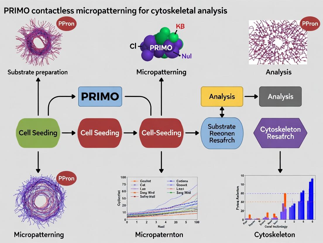
What is PRIMO? The Core Principles of Contactless Photopatterning for Cell Biology
PRIMO (Protein Micropatterning via Light-Induced Optoelectronic Device) is a contactless, UV-free photopatterning technology enabling high-resolution spatial control of protein immobilization on biocompatible surfaces. Within cytoskeletal analysis research, it provides a powerful tool for dissecting cell-biomaterial interactions, guiding cell adhesion, and studying mechanotransduction pathways in precisely engineered microenvironments.
This application note details the use of PRIMO contactless micropatterning as a cornerstone methodology for a broader thesis focused on cytoskeletal dynamics. The core thesis posits that precise spatial control of extracellular matrix (ECM) cues, enabled by PRIMO, is critical for unraveling the causative relationships between adhesion geometry, actomyosin contractility, and nuclear mechanotransduction. By creating defined patterns of adhesive proteins (e.g., fibronectin) surrounded by non-adhesive regions, researchers can standardize cell shape and force distribution, enabling quantitative analysis of cytoskeletal organization and downstream signaling.
Key Principles and Quantitative Performance Data
Table 1: PRIMO System Specifications and Performance Metrics
| Parameter | Specification / Value | Implication for Cytoskeletal Research |
|---|---|---|
| Light Source | 385 nm LED array (UV-free) | Enables long-duration patterning without cell-damaging UV radiation. |
| Spatial Resolution | < 1 µm (theoretical), ~2 µm (practical) | Sufficient to define sub-cellular adhesion sites, controlling focal adhesion size and spacing. |
| Patterning Speed | Up to 10 mm²/s (varies with resolution) | Enables rapid prototyping of multiple pattern designs on a single chip. |
| Substrate Compatibility | Glass, PDMS, plastic, with appropriate coating (e.g., PLPP) | Versatile for various assay formats (microscopy, traction force, etc.). |
| Protein Compatibility | Fibronectin, Collagen I, Laminin, vitronectin, custom peptides | Direct patterning of key ECM proteins relevant to adhesion and signaling. |
| Cell Seeding Post-Patterning | Immediate (no required wash-out step) | Streamlined workflow, maintains protein bioactivity. |
Table 2: Typical Pattern Geometries for Cytoskeletal Studies
| Pattern Geometry | Typical Dimensions | Cytoskeletal Analysis Application |
|---|---|---|
| Micropillars / Dots | 1-5 µm diameter, 5-20 µm center-center spacing | Study of discrete focal adhesion formation and maturation. |
| Lines | 5-20 µm width | Guidance of actin stress fiber alignment and polarization. |
| Square / Rectangle | 20x20 µm to 50x50 µm | Control of cell spreading area to investigate spread area vs. contractility relationships. |
| "Bowtie" or Anisotropic Shapes | Varying aspect ratios | Induce and measure polarized tension and directional traction forces. |
Detailed Experimental Protocols
Protocol 3.1: Substrate Preparation and Patterning with PRIMO
Objective: Create a glass substrate with defined fibronectin micropatterns for cell shape control.
Materials (Research Reagent Solutions):
- PLPP Copolymer Solution: A solution of PLL(20)-g[3.5]-PEG(2)/PEG(3.4)-biotin(50%) in PBS. Functions as a non-fouling, passivating layer that prevents non-specific protein adsorption and cell adhesion.
- Streptavidin Solution: Recombinant streptavidin in PBS. Acts as a molecular bridge, binding to biotin on the PLPP layer and providing a binding site for biotinylated proteins.
- Biotinylated Fibronectin Solution: Human plasma fibronectin, biotinylated, in PBS. The key ECM protein ligand that will be patterned to promote specific integrin-mediated cell adhesion.
- PRIMO-Compatible Microscope and Chip: Inverted epifluorescence microscope integrated with the PRIMO LED module and a digital micromirror device (DMD). The core tool for maskless photopatterning.
- Alvéole Lab or PhiSTOP Software: For designing patterns and controlling the photopatterning process.
Procedure:
- Substrate Cleaning: Plasma treat a glass-bottom dish or coverslip for 1 minute.
- Passivation: Incubate with PLPP Copolymer Solution (150 µL/cm²) for 30 minutes at room temperature (RT) in a humid chamber. Rinse 3x with sterile PBS.
- Streptavidin Coating: Incubate with Streptavidin Solution (50 µg/mL in PBS, 150 µL/cm²) for 10 minutes at RT. Rinse 3x with PBS.
- Priming with Protein: Incubate with Biotinylated Fibronectin Solution (5-25 µg/mL in PBS, 150 µL/cm²) for 5 minutes. DO NOT RINSE. The solution must remain during patterning.
- Patterning: Place the dish on the PRIMO microscope stage. In the control software, load the desired pattern design file (e.g., a grid of 20 µm squares). Initiate the projection sequence. The 385 nm light locally inactivates the streptavidin in the illuminated areas, preventing fibronectin binding. In the dark areas, fibronectin binds stably.
- Completion: After patterning, gently rinse the substrate 3x with PBS to remove unbound fibronectin. The substrate is now ready for cell seeding. Store in PBS at 4°C for up to 48 hours if not used immediately.
Protocol 3.2: Cell Seeding and Immunostaining for F-actin and Focal Adhesions
Objective: Seed cells on patterned substrates and visualize the resulting cytoskeletal organization.
Procedure:
- Cell Seeding: Trypsinize, count, and resuspend cells (e.g., NIH/3T3 fibroblasts, hMSCs) in serum-free medium. Seed onto the patterned substrate at a density of 5,000 - 15,000 cells/cm² to achieve isolated, single cells on patterns. Allow to adhere for 15-30 minutes before carefully adding complete growth medium.
- Incubation: Culture cells for 4-24 hours (time-dependent on the process under study).
- Fixation: Rinse with warm PBS and fix with 4% paraformaldehyde in PBS for 15 minutes at 37°C.
- Permeabilization & Blocking: Rinse with PBS, permeabilize with 0.1% Triton X-100 in PBS for 5 minutes, and block with 3% BSA in PBS for 30 minutes.
- Immunostaining:
- Focal Adhesions: Incubate with primary antibody against vinculin or paxillin (1:200 in 1% BSA/PBS) for 1 hour. Rinse 3x, then incubate with appropriate Alexa Fluor 568-conjugated secondary antibody (1:500) for 45 minutes.
- Actin Cytoskeleton: Incubate with Alexa Fluor 488-conjugated phalloidin (1:200) during the secondary antibody step.
- Nucleus: Incubate with DAPI (1 µg/mL) for 5 minutes.
- Imaging: Rinse and mount. Image using a high-resolution confocal or epifluorescence microscope.
Visualization of Workflows and Pathways
Title: PRIMO Patterning and Cellular Response Workflow
Title: Cytoskeletal Mechanotransduction Pathway on PRIMO Patterns
The Scientist's Toolkit: Research Reagent Solutions
Table 3: Essential Materials for PRIMO-based Cytoskeletal Patterning
| Item | Function in PRIMO Protocol | Key Consideration |
|---|---|---|
| PLPP Copolymer | Creates a universal, non-adhesive background by presenting biotin and PEG. | Batch consistency is critical for reproducible passivation. |
| Recombinant Streptavidin | High-affinity linker protein; its localized photo-inactivation defines the pattern. | Use a clean, azide-free preparation for optimal light response. |
| Biotinylated Fibronectin | The primary cell-adhesive ligand that is spatially organized. | Degree of biotinylation affects patterning efficiency and bioactivity. |
| PRIMO Patterning Buffer | Optimized buffer (often PBS-based) for the patterning reaction. | Maintains protein stability and photo-inactivation kinetics. |
| Phase Guide or Cell Rinsing Buffer | Assists in confining cell suspension during seeding on specific areas. | Reduces waste of precious cells and reagents on large substrates. |
| Validated Cell Line | Cells with robust integrin-mediated adhesion (e.g., fibroblasts, epithelial). | Low passage number and consistent culture conditions are vital. |
| Anti-Vinculin/Paxillin Antibody | Key biomarker for validating focal adhesion localization to patterns. | Validate for immunofluorescence after fixation/permeabilization. |
| Fluorescent Phalloidin | High-affinity probe for staining F-actin stress fibers. | Choose a conjugate color compatible with your microscope filters. |
Within the broader thesis on utilizing PRIMO contactless micropatterning for advanced cytoskeletal analysis, this application note details the core photophysical mechanism enabling sub-micron precision. The synergy between Dynamic Illumination (DI) and Astigmatic Local Phase Adjustment (ALPA) photoactivation allows for unprecedented spatial control in protein patterning, facilitating high-resolution studies of cytoskeletal dynamics, cell mechanics, and polarization in response to defined geometric cues.
Core Photophysical Mechanism
The PRIMO system's precision is achieved through a two-tiered optical and computational process. A digital micromirror device (DMD) generates a dynamic illumination pattern projected into the sample plane. This pattern is not static; it is dynamically modulated in intensity and shape over millisecond timescales to control the photobleaching or photoactivation kinetics of the photoreleasable molecules (e.g., NVOC-caged compounds, PA-GFP). Concurrently, the ALPA algorithm pre-corrects the projected wavefront for optical aberrations inherent to the microscope and sample chamber. By applying a calculated astigmatic phase mask, ALPA ensures the illumination pattern maintains sub-diffraction-limited fidelity at the focal plane, crucial for generating sharp, sub-micron features.
Quantitative Performance Data
Table 1: PRIMO System Performance Specifications
| Parameter | Specification | Impact on Cytoskeletal Patterning |
|---|---|---|
| Minimum Feature Size | 0.5 µm (theoretical), 0.8 µm (practical) | Enables patterning of single adhesion sites, mimicking physiological scale. |
| Pattern Positioning Accuracy | ± 50 nm | Allows precise alignment of patterns relative to existing cellular structures. |
| Photoactivation Wavelength | 375 nm or 405 nm | Compatible with common caged compounds (e.g., RGD, glutamate) and photoactivatable fluorophores. |
| Pattern Write Speed | Up to 10 mm²/s | Enables rapid patterning of large arrays for high-throughput statistical analysis. |
| Lateral (XY) Precision | < 100 nm (with ALPA correction) | Critical for defining precise boundaries to study cytoskeletal confinement. |
Table 2: Comparison of Patterning Outcomes With and Without ALPA
| Condition | Linewidth (FWHM) | Edge Sharpness (10-90% rise) | Pattern Fidelity (vs. Target) |
|---|---|---|---|
| Dynamic Illumination Only | 1.2 ± 0.3 µm | 0.7 µm | 85% |
| DI + ALPA Correction | 0.8 ± 0.1 µm | 0.4 µm | 98% |
Detailed Protocols
Protocol 1: Sub-Micron Patterning of Fibronectin Lines for F-Actin Alignment Studies
Objective: Create 1 µm wide adhesive lines to guide and analyze actin stress fiber formation.
Materials:
- PRIMO-equipped epifluorescence microscope (e.g., Nikon Ti2-E, Olympus IX83).
- Microscope slides with 35 mm µ-dish, glass bottom.
- Phosphate-buffered saline (PBS), pH 7.4.
- Recombinant human fibronectin, conjugated with NVOC-caging group (e.g., K₂⁰⁸NVOC-FN).
- Pluronic F-127 solution (0.2% w/v in PBS).
- Target cells (e.g., NIH/3T3 fibroblasts, U2OS osteosarcoma).
- Cell culture media.
Procedure:
- Surface Preparation: Coat the glass-bottom dish with 150 µL of caged fibronectin solution (25 µg/mL in PBS). Incubate for 1 hour at 37°C or overnight at 4°C.
- Quenching & Washing: Aspirate the solution. Add 2 mL of 0.2% Pluronic F-127 to passivate non-patterned areas. Incubate for 30 minutes at room temperature. Wash 3x with 2 mL PBS.
- System Setup:
- Mount the dish on the microscope stage.
- In the PRIMO control software (e.g., Mosaic), load the target pattern (e.g., array of 1 µm lines with 5 µm spacing).
- Select the 375 nm illumination laser and set power to 80% of maximum (calibrated to ~15 mW/cm² at sample plane).
- Enable the ALPA correction module and load the pre-calibrated correction mask for the 40x oil immersion objective and dish geometry.
- Photoactivation Patterning: Define the exposure time (typically 500-1000 ms per pattern field). Initiate the automated patterning sequence. The DI+ALPA system will project the dynamic, aberration-corrected UV pattern, uncaging fibronectin exclusively in the illuminated zones.
- Cell Seeding: Immediately after patterning, wash the dish once with serum-free media. Seed cells at a low density (e.g., 5,000 cells/dish) in full growth media to allow for attachment and spreading primarily on the patterned lines.
- Analysis: After 4-24 hours, fix and stain cells for F-actin (e.g., phalloidin) and nuclei. Image using confocal microscopy to quantify actin fiber alignment relative to the patterned lines.
Protocol 2: Multi-Protein Patterning for Studying Protein Recruitment
Objective: Create adjacent, sub-micron zones of two different proteins (e.g., an adhesive protein and a repellent cue).
Materials:
- All materials from Protocol 1.
- Second caged protein (e.g., NVOC-caged bovine serum albumin, BSA).
- Fluorescently-tagged secondary antibodies or direct fusion proteins for validation.
Procedure:
- Perform steps 1-3 from Protocol 1 using a mixture of the two caged proteins (e.g., NVOC-FN and NVOC-BSA).
- Sequential Patterning:
- First, expose the dish to the DI+ALPA pattern for Protein A (e.g., FN grid). Use standard 375 nm exposure.
- Without moving the dish, switch the pattern file in the software to the design for Protein B (e.g., BSA dots at grid intersections).
- Change the illumination wavelength to 405 nm (if the second protein cage is optimized for this wavelength) or adjust the intensity/dose to selectively activate the second cage. This sequential, wavelength- or dose-specific uncaging enables multi-component patterning.
- Validate the pattern by immunostaining before cell seeding.
Visualization of Workflows and Pathways
Title: PRIMO DI+ALPA Patterning Workflow
Title: Cytoskeletal Signaling from PRIMO Pattern
The Scientist's Toolkit
Table 3: Key Research Reagent Solutions for PRIMO Cytoskeletal Patterning
| Item | Function & Relevance |
|---|---|
| NVOC-Caged Fibronectin or RGD Peptide | Photoactivatable adhesive ligand. UV uncaging creates defined adhesion sites to study integrin clustering and downstream actin dynamics. |
| Caged Bioactive Molecules (e.g., cAMP, LPA) | Enables sub-cellular, temporally controlled release of signaling molecules to probe their localized effect on cytoskeleton remodeling. |
| Photoactivatable Fluorescent Proteins (e.g., PA-GFP-actin) | Allows precise marking and tracking of actin polymerization dynamics within patterned regions with high spatial resolution. |
| Inhibitors & Activators (e.g., Y-27632 (ROCKi), CN01 (Rho Activator)) | Pharmacological tools used in conjunction with patterns to dissect specific pathway contributions to observed cytoskeletal organization. |
| Anti-FAK pY397 & Anti-Paxillin Antibodies | Key markers for focal adhesion maturation, used to correlate pattern geometry with adhesion signaling strength. |
| SiR-Actin / LifeAct-TagGFP2 | Live-cell, high-contrast probes for visualizing F-actin dynamics over time on patterned substrates without UV activation. |
Application Notes: The PRIMO Platform for Cytoskeletal Analysis
Controlled cell adhesion via protein micropatterning is a foundational technique for cytoskeleton research. It standardizes cell shape and spreading, reducing variability and enabling direct correlation between adhesion geometry, cytoskeletal architecture, and downstream signaling. The PRIMO contactless digital micromirror device (DMD) system allows for rapid, mask-free patterning of any 2D protein design on standard culture surfaces, facilitating high-throughput, reproducible studies.
Table 1: Impact of Adhesion Geometry on Cytoskeletal Organization
| Adhesion Pattern Shape | Actin Stress Fibers | Microtubule Organizing Center (MTOC) | Vimentin Intermediate Filament Network | Primary Cellular Readout |
|---|---|---|---|---|
| Large Square (≥50µm) | Dense, crisscrossing bundles across the cell body. | Centrally located, random orientation. | Perinuclear cage with radial extensions. | Maximum spreading, baseline polarization. |
| Small Circle (20µm) | Concentric cortical ring, few central fibers. | Centrally located. | Tight perinuclear organization. | Restricted spreading, minimal polarization. |
| Asymmetric "Teardrop" or "Polarized" Pattern | Aligned bundles along the long axis. | Polarized towards the wider/adhesive front. | Asymmetrically extended towards the narrow "rear." | Induced polarity, directed intracellular trafficking. |
| Dual "Bowtie" Adhesion Pads (Separated 40µm) | Bundles spanning between pads, tension-generated. | Localized between pads along the axis. | Extended network connecting the two nuclei. | Model for cell-cell tension or bimucleated states. |
| Microlines (5µm wide) | Highly aligned, parallel bundles along the line. | Aligned along the line axis. | Aligned along the long axis of the cell. | Guidance, neurite modeling, migration studies. |
Detailed Protocols
Protocol 1: PRIMO-mediated Patterning of Fibronectin on Glass for Actin/FA Analysis Objective: Create 20µm circular fibronectin islands to study confined adhesion effects on actin stress fiber formation.
- Surface Preparation: Clean 35mm glass-bottom dishes with O₂ plasma for 5 min. Incubate with 0.01% Poly-L-Lysine-PEG (PLL-PEG) in HEPES buffer (pH 7.4) for 30 min at RT to create a non-fouling background.
- PRIMO Patterning: Use the PRIMO system with the "Photon" module. Load the dish. In the software, load the design file (circle, 20µm diameter). Set the patterning parameters: 405nm LED at 100% intensity, exposure time of 400ms per pattern position. Use the "Photon" reagent (photosensitive reagent containing fibronectin). Initiate the digital patterning process (~2 min for a field of view).
- Post-Patterning: Rinse the dish 3x with sterile PBS. Block with 1% heat-denatured BSA in PBS for 30 min. Rinse again with PBS.
- Cell Seeding: Seed U2OS or NIH/3T3 cells at low density (5,000 cells/dish) in serum-free medium. Allow attachment for 15-30 min, then add complete medium.
- Fixation & Staining (4h post-seeding): Fix with 4% PFA for 15 min, permeabilize with 0.1% Triton X-100 for 5 min. Stain for Actin (Phalloidin-488), Focal Adhesions (anti-paxillin), and nuclei (DAPI).
- Imaging: Acquire images using a 63x/1.4NA oil objective on a confocal microscope. Quantify actin fiber alignment (using OrientationJ in ImageJ) and focal adhesion count/size.
Protocol 2: Probing Microtubule Polarity in Polarized Cells Objective: Pattern asymmetric adhesive shapes to direct MTOC positioning and analyze microtubule growth.
- Patterning: Follow Protocol 1, but use a "Polarized Triangle" (30µm base, 60µm length) design with Laminin-511.
- Cell Seeding: Seed RPE1 or MDCK cells as in Protocol 1.
- Live-Cell Imaging of EB3 Comets (6h post-seeding): Transfer dish to a live-cell imaging system (37°C, 5% CO₂). Transfer cells with an EB3-GFP plasmid 24h prior to patterning. Acquire time-lapse images (1 frame/2 sec for 2 min) using a 63x objective.
- Analysis: Track EB3 comets using the TrackMate plugin in Fiji. Generate rose plots of comet trajectories to visualize the predominant direction of microtubule growth relative to the adhesion shape and MTOC location.
Protocol 3: Intermediate Filament Network Remodeling Under Geometric Constraint Objective: Assess vimentin network organization in cells confined to "bowtie" adhesion patterns.
- Patterning: Create a "Bowtie" pattern (two 20µm squares separated by a 40µm non-adhesive gap) using Collagen I via PRIMO.
- Cell Seeding & Staining: Seed MCF7 or vimentin-GFP expressing fibroblasts. Culture for 12h. Fix and stain for vimentin (if not GFP) and DAPI.
- Quantitative IF Analysis: Acquire 3D z-stacks. Use the Squassh or similar plugin for deconvolution and 3D reconstruction. Calculate the vimentin network anisotropy index and the fraction of vimentin signal extending into the inter-nuclear bridge.
The Scientist's Toolkit: Research Reagent Solutions
| Item/Catalog Number (Example) | Function in Cytoskeleton-Adhesion Studies |
|---|---|
| PRIMO System & "Photon" Reagent Kit (Alvéole) | Enables mask-free, contactless UV patterning of any protein of choice on any surface. Key for generating precise adhesion geometries. |
| Cytoskeleton Live Cell Imaging Reagents (SiR-Actin/Tubulin, Spirochrome) | Fluorogenic, cell-permeable probes for super-resolution or long-term live imaging of actin and microtubules with minimal phototoxicity. |
| Inhibitors & Activators (Y-27632 (ROCKi), Nocodazole, SMIFH2, Cytochalasin D) | Pharmacologically perturb actin (CytoD, SMIFH2) or microtubule (Nocodazole) dynamics, or actomyosin contractility (Y-27632) to dissect force-related feedback. |
| ECM Proteins (Fibronectin, Laminin-511, Collagen I, Corning Matrigel) | Different ECM proteins engage specific integrin receptors, initiating distinct signaling cascades that remodel all three cytoskeletal networks. |
| Validated Antibodies for Cytoskeletal Markers (e.g., Anti-acetylated Tubulin, Anti-phospho-Vimentin, Anti-Vinculin) | Essential for fixed-cell endpoint analysis of cytoskeletal post-translational modifications and adhesion complex maturation. |
Diagram Title: Cytoskeletal Crosstalk Controlled by Adhesion Geometry
Diagram Title: PRIMO Micropatterning & Cytoskeleton Analysis Workflow
Within the broader thesis investigating PRIMO contactless micropatterning for high-throughput cytoskeletal analysis in drug discovery, a rigorous understanding of the system's core hardware and consumables is paramount. The reliability and reproducibility of experiments correlating patterned adhesion geometry with cytoskeletal dynamics and cellular responses to pharmacological agents depend entirely on the precise function and interplay of these components. These Application Notes detail the technical specifications and protocols for the essential hardware and reactive substrates of the PRIMO system.
PRIMO System Hardware Breakdown
The PRIMO system (Alvéole) is an integrated solution combining dynamic micromirror device (DMD)-based photopatterning with advanced live-cell imaging. Its hardware is designed for subcellular resolution patterning directly within standard cell culture incubators.
Table 1: Core Hardware Components & Quantitative Specifications
| Component | Model / Specification | Key Function | Critical Parameters for Cytoskeletal Patterning |
|---|---|---|---|
| DMD Chip | Texas Instruments DLP6500 | Creates the digital photomask by reflecting UV light through ~2 million micromirrors. | Resolution: 1920 x 1080 (Full HD). Pixel Size: Projected to ~0.5 µm on sample (with 20x objective). |
| UV LED Source | 365 nm wavelength | Provides illumination for activating the reactive coating on slides. | Power Density: Adjustable, typically 50-200 mW/cm² at sample plane. Exposure Control: 1 ms to 10 s precision. |
| Optical Path | Custom integrated lens system | Projects the DMD pattern onto the sample plane with high fidelity. | Magnification: Ensures 1 DMD pixel = desired micron size on substrate (e.g., 0.5-1.0 µm). Homogeneity: >90% illumination uniformity. |
| Motorized Stage | Marzhauser or equivalent | Precisely positions the sample for multi-field patterning and imaging. | Travel Range: 114 x 75 mm. Repositioning Accuracy: <2 µm. |
| Incubator Integration | Customizable enclosure | Maintains physiological conditions (37°C, 5% CO₂, humidity) during live patterning and imaging. | Stability: ±0.5°C, ±0.5% CO₂. Compatible with most microscope incubators. |
| Control Software | Leonardo (Alvéole) | User interface for pattern design, exposure sequencing, and hardware orchestration. | Features: Multi-shape libraries, array generation, time-lapse patterning protocols. |
Diagram 1: PRIMO Hardware & Patterning Workflow
Reactive Slides: PLPP Coating Chemistry & Handling
The PRIMO process relies on proprietary functionalized slides pre-coated with a Photolabile Phenylazide Polyethylene Glycol (PLPP) layer. This non-fouling PEG coating is rendered adhesive upon precise UV photolysis.
Table 2: PRIMO Reactive Slide Specifications & Handling Data
| Parameter | Specification | Importance for Cytoskeletal Research |
|---|---|---|
| Coating Type | PLPP (Photolabile Peg Polymer) | Inert until UV exposure; prevents non-specific cell adhesion. |
| Activation Wavelength | 365 nm | Optimal for minimal cell damage and efficient photolysis. |
| Standard Slide Format | 25 x 75 mm glass, #1.5 thickness | Compatible with high-resolution oil objectives (60x, 100x). |
| Storage | -20°C, desiccated, in the dark. | Preserves photolabile compound reactivity. Shelf life: 6 months. |
| Post-Thaw Stability | 1 week at 4°C in the dark. | Allows for planned experimental timelines. |
| Protein Grafting Density | Tunable via UV dose & protein concentration. | Enables control over adhesion strength, impacting cytoskeletal tension. |
Diagram 2: PLPP Slide Activation & Protein Grafting Chemistry
Detailed Experimental Protocol: Patterning for F-Actin Stress Fiber Analysis
This protocol details the creation of fibronectin lines (5 µm width) to guide and analyze aligned stress fiber formation in fibroblasts, a common assay for cytoskeletal mechanics.
Protocol: Micropatterning of Adhesive Lines for Directed Cytoskeletal Assembly
I. Pre-Patterning Setup
- Equipment & Software: PRIMO system installed on an inverted microscope within a live-cell incubator (37°C, 5% CO₂). Leonardo software running.
- Reactive Slide Preparation: Thaw a PRIMO slide (25 x 75 mm) at room temperature for 15 min, protected from light. Clean with air duster.
- Protein Solution: Prepare a 50 µg/mL solution of purified fibronectin or similar ECM protein in sterile PBS.
- Pattern Design: In Leonardo, design an array of lines (5 µm width, 20 µm spacing, length 100 µm). Set the pattern to cover the desired number of imaging fields.
II. Photopatterning Process
- Mount the reactive slide on the PRIMO motorized stage.
- In Leonardo, navigate to the exposure settings. Set UV intensity to 100% (typically ~150 mW/cm²). Critical: Calibrate exposure time. A starting point is 500 ms for 5 µm features.
- Execute the exposure protocol. The DMD will project the line pattern onto the slide, activating the PLPP coating only in illuminated regions.
- Post-Exposure Processing:
- Immediately pipette 200 µL of the fibronectin solution (50 µg/mL) onto the patterned area.
- Incubate the slide in a humidified dark chamber for 1 hour at room temperature.
- Rinse gently three times with sterile PBS to remove unbound protein.
- Block non-specific sites by incubating with 1% Pluronic F-127 in PBS for 30 min.
- Rinse thoroughly with PBS. The slide is now ready for cell seeding.
III. Cell Seeding & Imaging
- Seed fluorescently-labeled (e.g., LifeAct-GFP) fibroblasts at a low density (e.g., 2,000 cells/cm²) in complete medium.
- Allow cells to adhere for 15-30 min, then gently rinse to remove non-adherent cells.
- Return slide to the incubator and image at 2-4 hours post-seeding using a 60x oil objective to visualize aligned F-actin stress fibers confined to the patterned lines.
The Scientist's Toolkit: Key Reagent Solutions
Table 3: Essential Research Reagents for PRIMO Cytoskeletal Patterning
| Reagent / Material | Function in Experiment | Critical Notes |
|---|---|---|
| PRIMO Reactive Slides (PLPP) | Photoactivatable substrate for high-resolution protein patterning. | Must be stored at -20°C. Avoid freeze-thaw cycles >2x. |
| Extracellular Matrix Proteins | Provide specific adhesive ligands (e.g., Fibronectin, Laminin, Collagen I). | Use purified, carrier-free proteins at 10-100 µg/mL for grafting. |
| Pluronic F-127 | Blocks non-patterned, non-activated PEG regions to ensure perfect confinement. | 1% (w/v) solution in PBS is standard. Essential for low background. |
| LifeAct-GFP/RFP Live Cell Probe | Fluorescent tag for real-time visualization of F-actin dynamics. | Minimally perturbing; allows long-term imaging of cytoskeletal remodeling. |
| Phenotypic Drugs (e.g., Y-27632, Blebbistatin) | Modulators of cytoskeletal tension (ROCK inhibitor, Myosin II inhibitor). | Used to perturb the system and study mechanotransduction pathways on patterns. |
| Fixed-Cell Staining Kits (Phalloidin, Antibodies) | For endpoint, high-resolution analysis of cytoskeleton and associated proteins. | Enables multiplexing after live-cell experiments on patterned cohorts. |
Application Notes
PRIMO contactless micropatterning utilizes a digital micromirror device to project dynamic UV light patterns onto a photosensitive biocompatible surface, enabling precise, reagent-free protein adsorption. This technology is pivotal for cytoskeletal analysis research, allowing unparalleled control over cell shape, adhesion, and subsequent intracellular signaling.
1. Flexibility in Experimental Design
- Dynamic Patterning: Unlike static stamps, PRIMO allows for in-situ pattern changes, enabling studies on cell adaptation, polarization, and migration in response to sudden geometric cues.
- Substrate Independence: Compatible with glass, PDMS, hydrogels, and multi-well plates, facilitating integration with traction force microscopy, FRET biosensors, or high-content screening.
- Custom Geometry Library: Researchers can design and deploy any pattern shape (dots, lines, squares, complex polygons) with a simple change in the digital mask, enabling systematic study of how spatial constraints dictate cytoskeletal organization.
2. High-Resolution for Precise Manipulation
- Single-Cell Patterning: Achieves feature sizes down to 1 µm, permitting the isolation and analysis of individual cells on specific adhesion islands.
- Subcellular Control: Enables patterning of multiple adhesion sites within a single cell, allowing direct interrogation of intracellular force balance, compartmentalization, and localized signaling events.
3. Multiplexing Capabilities for Complex Assays
- Sequential Patterning: Different proteins (e.g., fibronectin, collagen, E-cadherin Fc chimeras) can be patterned in successive cycles on the same substrate to create complex, multi-component microenvironments.
- Temporal Stimulation: Combines geometric control with timed chemical or optogenetic stimuli to dissect the sequence of cytoskeletal remodeling events.
Quantitative Performance Summary of PRIMO Technology Table 1: Key performance metrics for PRIMO-based cytoskeletal research applications.
| Parameter | Specification / Capability | Impact on Cytoskeletal Research |
|---|---|---|
| Optical Resolution | 1.0 µm (theoretical, 20x objective) | Enables subcellular patterning of adhesion sites. |
| Patterning Speed | ~10-60 sec/cm² (depending on resolution) | Facilitates high-throughput experimental setup in multi-well plates. |
| Pattern Alignment | < 5 µm precision (using reference marks) | Allows precise re-patterning for sequential multiplexing on same cells. |
| Protein Compatibility | Any protein/peptide with accessible amine or thiol groups | Supports integrin, cadherin, and other cytoskeleton-linked receptor studies. |
| Cell Viability | >95% post-patterning (typical) | Ensures observed phenotypes are due to patterning, not phototoxicity. |
Experimental Protocols
Protocol 1: PRIMO-Assisted Patterning of Fibronectin for F-Actin Stress Fiber Analysis
Objective: To create defined fibronectin micropatterns for studying the relationship between cell shape, focal adhesion distribution, and actin cytoskeleton architecture.
Materials: See "The Scientist's Toolkit" below.
Method:
- Substrate Preparation: Clean a 35 mm glass-bottom dish with plasma for 5 min. Incubate with 0.01% PE-PEG-RGD in sterile PBS for 1 hour at room temperature (RT). Rinse 3x with PBS.
- PRIMO System Setup: Launch the PRIMO software. Load the desired pattern file (e.g., 20x20 µm squares). Set exposure parameters: 405 nm LED, 90% intensity, 3-second exposure time.
- Photopatterning: Fill the dish with PBS. Using the alignment feature, focus on the substrate surface. Execute the patterning sequence. The illuminated areas become protein-adhesive.
- Protein Coupling: Immediately after patterning, incubate the dish with 50 µg/mL fibronectin in PBS for 20 min at 37°C. Rinse 3x with PBS to remove unbound protein.
- Cell Seeding & Fixation: Trypsinize and resuspend U2OS cells in serum-free medium. Seed at low density (5,000 cells/dish) to ensure single-cell patterning. Incubate for 4-6 hours. Fix with 4% PFA for 15 min.
- Immunostaining: Permeabilize with 0.1% Triton X-100, block with 1% BSA. Stain for F-actin (Phalloidin-488, 1:500) and vinculin (mouse anti-vinculin primary, 1:200; followed by anti-mouse-568 secondary, 1:500). Image using a confocal microscope.
Protocol 2: Sequential Multiplex Patterning for Investigating Cytoskeletal Crosstalk
Objective: To pattern two distinct extracellular matrix proteins in adjacent regions to study competitive adhesion and cytoskeletal polarization.
Method:
- Perform Protocol 1, Steps 1-4, using a pattern of 15 µm wide lines. Use fibronectin (FN) as the first protein.
- Second Patterning Cycle: Without disturbing the cells (if pre-seeded for live analysis) or after seeding and a short adhesion period (e.g., 30 min), initiate a second patterning sequence. Align a new pattern (e.g., adjacent 15 µm lines) using the system's alignment markers.
- Second Protein Coupling: Incubate with the second protein (e.g., 50 µg/mL Laminin (LN) in PBS) for 20 min at 37°C. Rinse thoroughly.
- Live-Cell Imaging: For live-cell analysis, transfer the dish to a stage-top incubator. Image actin dynamics using a transfected LifeAct-GFP construct every 5 min for 12 hours to observe cytoskeletal remodeling in response to the new adhesive cue.
Visualizations
Title: PRIMO Workflow for Cytoskeletal Analysis
Title: Key Pathway from Pattern to Cytoskeletal Response
The Scientist's Toolkit
Table 2: Essential Research Reagent Solutions for PRIMO-based Cytoskeletal Experiments.
| Item | Function / Relevance | Example Product/Catalog |
|---|---|---|
| PE-PEG-RGD | Photosensitive coating. UV illumination removes PEG, allowing protein binding specifically in patterned areas. Essential for high-contrast patterning. | PRIMO Coating Kit (Alvéole) |
| Fibronectin, Purified | Standard ECM protein for integrin-mediated adhesion, inducing robust actin stress fiber formation. | Human Fibronectin, Purified (e.g., Corning) |
| Laminin, Purified | Alternative ECM protein for studies on polarity, differentiation, and competitive adhesion. | Mouse Laminin I, Purified (e.g., Cultrex) |
| LifeAct-EGFP Plasmid | F-actin live-cell biosensor. Allows real-time visualization of cytoskeletal dynamics on patterns. | LifeAct-EGFP (Ibidi) |
| SiR-Actin Kit | Live-cell, far-red fluorescent actin stain. Low cytotoxicity ideal for long-term imaging. | SiR-Actin Kit (Cytoskeleton, Inc.) |
| Anti-Vinculin Antibody | Gold-standard marker for mature focal adhesions. Correlates actin stress fiber ends to adhesion sites. | Monoclonal Anti-Vinculin (e.g., Sigma V9131) |
| YAP/TAZ Antibody | Readout for mechanotransduction. Nuclear/cytoplasmic ratio indicates cellular response to pattern geometry. | D24E4 Rabbit mAb (Cell Signaling) |
| RhoA Activity Assay | Pull-down assay to quantify activation levels of Rho GTPase, a key regulator of actin dynamics. | RhoA G-LISA Activation Assay (Cytoskeleton, Inc.) |
Step-by-Step Protocol: Applying PRIMO for Cytoskeletal Studies and Drug Screening
Application Notes
Contactless micropatterning, particularly via the PRIMO Lithography Apparatus for Masked Photopatterning (LAMP) system, enables precise spatial control of cell adhesion. This is critical for interrogating cytoskeletal architecture, force generation, and signaling dynamics. By designing specific geometric cues—dots, lines, and islands—researchers can pose targeted questions about cytoskeletal organization and function.
Key Applications:
- Dots (Micron-scale Adhesive Islands): Used to isolate single cells or define specific contact areas. This forces a stereotypical cytoskeletal organization, ideal for quantifying actin cap formation, nuclear deformation, and measuring intracellular forces via traction force microscopy on patterned elastic substrates.
- Lines (1D Adhesive Tracks): Constrain cell shape along a single axis, promoting pronounced actin alignment and stress fiber formation. Essential for studying cell polarity, directed migration, and the role of actin-myosin contractility in elongated morphologies.
- Islands (2D Adhesive Geometries of Defined Shape & Size): Squares, triangles, or circles control spreading area and edge curvature. These patterns test how geometric boundary conditions dictate the spatial distribution of focal adhesions, actin flow, and microtubule organization, linking shape to intracellular signaling.
The design phase within the LAMP software (e.g., Pattern Editor) is therefore a fundamental step in translating a biological hypothesis into a physical experimental setup for cytoskeletal analysis.
Protocols
Protocol 1: Designing a Multi-Pattern Chip for Cytoskeletal Interrogation
This protocol outlines the creation of a single substrate containing dot, line, and island patterns to perform comparative cytoskeletal analysis.
Materials & Reagent Solutions
| Item | Function in Experiment |
|---|---|
| PRIMO LAMP System | Contactless photopatterning device using Digital Micromirror Device (DMD) to project UV light through a microscope. |
| LAMP Software Suite | Controls the DMD to generate user-defined patterns for photopatterning. |
| Glass Coverslips or Dish | Substrate for patterning, often pre-coated with a passivation layer. |
| PLL-g-PEG | Poly-L-lysine grafted with polyethylene glycol. A non-fouling passivation layer to prevent cell adhesion. |
| UV-sensitive Photoinitiator | (e.g., Irgacure 2959). Generates radicals upon UV exposure to functionalize the passivation layer. |
| Functionalized Adhesive Ligand | (e.g., RGD-peptide conjugated to an acrylate group). Covalently grafted upon UV exposure to create adhesive patterns. |
| Fluorescently-labeled Fibronectin or RGD | Allows for visualization of the patterned adhesive areas post-fabrication. |
| Mammalian Cells of Interest | (e.g., U2OS, NIH/3T3, MEFs). Express cytoskeletal components relevant to the research question. |
| Paraformaldehyde (4%) | Fixative for preserving cytoskeletal architecture at experimental endpoints. |
| Phalloidin (Fluorophore-conjugated) | Binds F-actin for visualization of actin cytoskeleton and stress fibers. |
Methodology:
- Substrate Preparation: Clean glass coverslips. Coat with a PLL-g-PEG solution (0.1 mg/mL in HEPES buffer) for 1 hour at room temperature. Rinse and dry under nitrogen. This creates a non-adhesive background.
- LAMP Software Pattern Design:
- Open the Pattern Editor.
- Dots: Create a grid of circles with diameters of 10 µm, 20 µm, and 30 µm, spaced 100 µm apart center-to-center. This allows analysis of cell and nuclear deformation versus adhesion size.
- Lines: Draw arrays of rectangles 20 µm wide and 500 µm long, with 100 µm spacing. Vary the width in a separate array (10 µm, 20 µm, 40 µm) to test constraints on actin bundle formation.
- Islands: Design arrays of geometric shapes: squares (50x50 µm²), triangles (50 µm side), and circles (50 µm diameter). This tests the effect of corner curvature and symmetry on cytoskeletal organization.
- Arrange all pattern arrays on a single virtual mask layout. Save the design file.
- Photopatterning:
- Prepare a patterning solution: Mix the acrylate-PEG-RGD ligand (1 mM) and photoinitiator (0.1% w/v) in sterile PBS.
- Incubate the coated coverslip with the patterning solution in a patterning chamber.
- Load the design file into the LAMP control software, align the substrate, and initiate the UV exposure sequence (typical dose: 50-200 mJ/cm²). UV light projected through the DMD locally grafts the RGD ligand onto the passivation layer.
- Post-Patterning and Cell Seeding:
- Rinse the patterned substrate thoroughly with PBS to remove unreacted compounds.
- Optionally, incubate with fluorescent fibronectin to validate pattern fidelity under a microscope.
- Seed cells (e.g., NIH/3T3 fibroblasts) at a low density (e.g., 5,000 cells/cm²) in serum-free or low-serum media to prevent adhesion outside patterns.
- Allow cells to adhere and spread for 4-24 hours depending on the experiment.
- Cytoskeletal Analysis:
- Fix cells with 4% PFA for 15 min at the desired time point.
- Permeabilize, stain with phalloidin (for F-actin) and DAPI (for nuclei), and mount.
- Image using fluorescence or confocal microscopy.
- Quantify parameters such as: actin fiber orientation (relative to line axis), number of stress fibers per cell, nuclear aspect ratio (on dots/islands), or fluorescence intensity of cytoskeletal proteins at specific locations (e.g., at triangle corners).
Protocol 2: Quantifying Actin Alignment on 1D Line Patterns
This detailed protocol focuses on a specific application: measuring the degree of actin cytoskeleton alignment in cells confined to line patterns.
Methodology:
- Pattern & Cell Culture: Pattern lines of 15 µm width as described in Protocol 1. Seed U2OS osteosarcoma cells expressing LifeAct-GFP to visualize actin dynamics live, or use wild-type cells and later stain.
- Image Acquisition: For fixed samples, acquire high-resolution images of the phalloidin channel (F-actin) using a 40x or 60x objective. Ensure images capture the entire cell confined to the line.
- Image Analysis (using FIJI/ImageJ):
- Pre-process images: Apply a Gaussian blur (σ=1) to reduce noise.
- Use the "OrientationJ" plugin or similar to calculate the dominant orientation and coherency of actin fibers within the cell body.
- Alternatively, employ a Fourier Transform approach on thresholded actin images to derive an alignment index.
- Measure the angle of the long axis of the cell (defined by the pattern) and compare it to the mean actin fiber orientation. The deviation from 0° indicates misalignment.
Quantitative Data Summary:
| Pattern Type | Typical Dimensions | Key Cytoskeletal Readout | Example Quantitative Finding (Reference) |
|---|---|---|---|
| Dots / Islands | 10-50 µm diameter | Nuclear Aspect Ratio (NAR) | NAR increases from ~1.5 to ~3.0 as island diameter decreases from 50 µm to 20 µm (Source: Théry et al., 2006). |
| Lines / 1D Tracks | Width: 5-40 µm | Actin Alignment Index (0-1) | Alignment index >0.8 for lines <20 µm wide, dropping to ~0.4 for widths >50 µm (Source: Driscoll et al., 2021 search). |
| Islands with Corners | Squares, Triangles | Focal Adhesion Density at Corners | Paxillin intensity at triangle corners can be 2-3x higher than at edges (Source: Brock et al., 2003). |
Visualizations
Title: Workflow for Cytoskeletal Analysis via LAMP Patterning
Title: Cytoskeletal Response to Pattern Geometry
This protocol details the critical surface preparation steps for cytoskeletal analysis research utilizing the PRIMO contactless micropatterning system. Reproducible and high-fidelity patterning of cellular micro-environments requires pristine, biologically active substrates. Coating glass surfaces with defined extracellular matrix (ECM) proteins like fibronectin and collagen provides the necessary adhesive cues for cells, enabling precise investigation of cytoskeletal dynamics, mechanotransduction, and cell morphology in response to geometrically defined cues. Proper slide activation and coating are foundational to the success of subsequent photopatterning and quantitative imaging assays central to the thesis.
Key Research Reagent Solutions
| Reagent/Material | Function in Protocol | Key Considerations |
|---|---|---|
| High-Precision Glass Coverslips (#1.5) | Primary substrate for imaging. Provides optical clarity and consistent surface chemistry for coating. | Thickness (170±5 µm) is critical for high-resolution microscopy. Often plasma-cleaned before use. |
| (3-Aminopropyl)triethoxysilane (APTES) | Silane coupling agent. Provides amine-terminated groups on glass for covalent protein binding. | Enhances coating stability. Must be used in anhydrous conditions. Hyrophobic after silanization. |
| Glutaraldehyde (25% aqueous) | Crosslinker. Reacts with amine groups from APTES to create aldehyde groups for covalent protein immobilization. | Creates a stable, reactive layer. Excess must be thoroughly rinsed. |
| Fibronectin (from human plasma) | ECM protein promoting cell adhesion via integrin binding. Key for focal adhesion and actin stress fiber studies. | Aliquot to avoid freeze-thaw cycles. Coating concentration is pattern-dependent (1-10 µg/mL). |
| Collagen I (rat tail) | ECM protein forming fibrillar networks. Influences cell spreading, migration, and mechanosensing. | Acid-soluble stock must be neutralized on ice before dilution in coating buffer. |
| Phosphate-Buffered Saline (PBS), sterile | Buffer for protein dilution and rinsing steps. Maintains pH and ionic strength. | Must be Ca2+/Mg2+-free for rinsing cells, but may contain these ions for protein coating. |
| Bovine Serum Albumin (BSA), fluorescently labeled | Blocking agent. Passivates non-patterned areas to prevent non-specific cell adhesion. | Alexa Fluor 647-conjugated BSA allows visualization of non-adhesive regions. |
| PRIMO Micropatterning System (Alvéole) | LED-based photopatterning device. Projects UV (365 nm) patterns onto photoactivatable substrates. | Used after coating to create precise adhesive geometries via ablation or modification of the protein layer. |
| PLPP Photoactivatable Reagent (Alvéole) | Forms a reactive nitrene group upon UV exposure. Grafted onto BSA to create a non-adhesive layer that can be locally removed. | Enables "lift-off" patterning by deactivating the passivation where UV light is projected. |
Table 1: Standardized Coating Parameters for Cytoskeletal Patterning Assays
| ECM Protein | Recommended Coating Concentration | Incubation (Passive) | Incubation (for Patterning) | Buffer | Key Cellular Response |
|---|---|---|---|---|---|
| Fibronectin | 5-10 µg/mL for full coats1-5 µg/mL for micropatterns | 1 hour at 37°C or overnight at 4°C | 20 min at RT before PRIMO patterning | PBS (pH 7.4) | Strong integrin α5β1 binding, prominent focal adhesions & actin fibers. |
| Collagen I | 50-100 µg/mL for full coats20-50 µg/mL for micropatterns | 1 hour at 37°C | 20 min at RT on ice-cold buffer before patterning | 0.02 M Acetic Acid (neutralized) | Integrin α2β1 binding, influences migration and collagen remodeling. |
| BSA (Passivation) | 1-5 mg/mL (often fluorescent) | 30-60 min at RT or 37°C | Required after patterning to block exposed glass | PBS | Prevents non-specific cell adhesion outside patterned areas. |
Table 2: PRIMO Patterning Parameters Post-Coating (Example for 20x Objective)
| Parameter | Value Range | Effect on Coating |
|---|---|---|
| UV Exposure Time | 100-500 ms per point | Determines efficiency of protein layer removal or modification. |
| Pattern Resolution | ~1 µm | Defines the sharpness of the adhesive/non-adhesive boundary. |
| Working Solution | PLPP-grafted BSA in PBS | Creates the photoactivatable non-adhesive layer. |
Detailed Experimental Protocols
Protocol 1: Glass Slide Activation with APTES and Glutaraldehyde
Objective: To create a chemically reactive aldehyde surface for strong covalent immobilization of ECM proteins.
- Plasma Cleaning: Place high-precision glass coverslips in a plasma cleaner. Treat for 5 minutes at medium power under oxygen or air plasma to generate hydroxyl groups.
- APTES Silanization: In a fume hood, prepare a 2% (v/v) solution of APTES in anhydrous acetone. Immediately immerse plasma-cleaned slides for 5 minutes.
- Rinsing: Rinse slides copiously three times in fresh anhydrous acetone to remove unbound silane.
- Curing: Bake slides at 110°C for 10 minutes to complete siloxane bond formation. Cool to room temperature.
- Glutaraldehyde Crosslinking: Prepare a 2.5% (v/v) solution of glutaraldehyde in PBS. Incubate slides for 30 minutes at room temperature.
- Final Rinse: Rinse slides three times with sterile PBS or deionized water. Slides can be used immediately or air-dried and stored in a desiccator for up to a week.
Protocol 2: ECM Protein Coating for PRIMO Micropatterning
Objective: To apply a uniform layer of ECM protein, which will later be selectively removed via PRIMO to create micropatterns. Part A: Standard Coating
- Protein Solution Preparation: Dilute the desired ECM protein (Fibronectin or Collagen I) to the working concentration in the appropriate sterile buffer (see Table 1). Keep collagen solutions on ice.
- Application: Place the activated slide in a sterile dish or use a hydrophobic pen to create a well. Pipette enough protein solution to cover the surface (e.g., 100 µL for a 22x22 mm coverslip).
- Incubation: Incubate for 20 minutes at room temperature in a humidified chamber to prevent evaporation.
- Rinsing: Gently aspirate the protein solution and rinse the slide twice with sterile PBS.
- Blocking: Incubate with a 1 mg/mL solution of PLPP-grafted BSA (for patterning) or standard BSA (for full coats) for 1 hour at 37°C.
- Storage: Rinse with PBS. Slides can be used immediately for patterning or cell seeding.
Part B: PRIMO-Based Lift-Off Patterning
- System Setup: Mount the coated and BSA-blocked slide on the PRIMO microscope stage.
- Pattern Design: Load the desired pattern (e.g., circles, lines, squares) into the LEONARDO software.
- UV Exposure: Execute the pattern projection. UV light locally deactivates the PLPP-BSA layer, exposing the underlying covalently bound ECM protein.
- Post-Exposure Rinse: Gently rinse the slide with culture medium or PBS to remove debris from the ablated areas.
- Cell Seeding: Seed fluorescently labeled cells (e.g., actin-GFP) at an appropriate density in serum-free or low-serum medium to allow adhesion primarily to the patterned areas.
- Analysis: After 4-24 hours, image fixed or live cells using high-resolution microscopy for cytoskeletal analysis.
Protocol 3: Validation of Coating Quality via Immunofluorescence
- Fixation: For coated but unpatterned slides, fix with 4% paraformaldehyde for 15 minutes.
- Blocking & Staining: Block with 3% BSA in PBS for 30 min. Incubate with primary antibody against the coated ECM protein (e.g., anti-fibronectin) for 1 hour, followed by fluorescent secondary antibody.
- Imaging & Analysis: Acquire images using a fluorescence microscope. Quantify mean fluorescence intensity and homogeneity across multiple fields to assess coating uniformity.
Workflow and Pathway Diagrams
Workflow for Slide Prep and PRIMO Patterning
ECM-Internal Signaling to Cytoskeleton
Application Notes: PRIMO Contactless Micropatterning for Cytoskeletal Analysis
PRIMO (via ALVEOLE’s technology) is a contactless, maskless, and biocompatible photopatterning system that utilizes a Digital Micromirror Device (DMD) to project dynamic UV (375 nm) light patterns onto a photosensitive substrate. This enables precise, high-resolution protein patterning for controlling cell adhesion geometry, a critical tool for cytoskeletal analysis research. By confining cells to specific shapes (e.g., lines, squares, circles), researchers can standardize cellular morphologies, leading to reproducible quantitative analysis of cytoskeletal architecture, intracellular signaling, and mechanotransduction in contexts such as drug screening and disease modeling.
Protocols
Protocol 1: Substrate Preparation and Protein Patterning
Objective: To create micropatterned surfaces of extracellular matrix (ECM) proteins (e.g., fibronectin) on glass-bottom dishes for cell confinement.
Materials:
- Glass-bottom culture dishes (e.g., 35 mm, #1.5 coverslip)
- PRIMO photopatterning module (mounted on an epifluorescence microscope)
- Photosensitive Reagents: PP1 (PLPP-PEG-Steryl, a photolabile PEG coating) or PA1 (PLPP-PEG-Alc, for alcohol-resistant coating).
- Phosphate-Buffered Saline (PBS), sterile
- ECM protein solution (e.g., 50 µg/mL Fibronectin in PBS)
- Passivation solution (e.g., 0.2% Pluronic F-127 in PBS)
- ALVEOLE's LEONARDO software for pattern design
Methodology:
- Coating: Under sterile conditions, incubate glass-bottom dishes with PP1 (diluted 1:10 in PBS) for 20 minutes at room temperature (RT) in the dark. Rinse 3x with PBS.
- Pattern Design: In LEONARDO, design the desired adhesion pattern (e.g., 20 µm squares, 10 µm wide lines). Define the UV illumination parameters (typically 70-90% DMD intensity, 500-800 ms exposure time).
- Illumination/Deactivation: Place the coated dish on the microscope stage. Run the illumination protocol. UV light projection deactivates the photolabile PEG coating in the exposed areas, revealing the bare glass.
- Protein Adsorption: Immediately incubate the dish with ECM protein solution (e.g., 50 µg/mL fibronectin) for 30 minutes at 37°C or 1 hour at RT.
- Passivation: Rinse 3x with PBS. Incubate with 0.2% Pluronic F-127 solution for at least 30 minutes at RT to block non-patterned areas.
- Rinse & Seed: Rinse 3x with PBS. The dish is ready for cell seeding at the desired density.
Protocol 2: Cytoskeletal Analysis on Micropatterns
Objective: To fix, stain, and image F-actin and nuclei in cells confined to micropatterns for quantitative morphology analysis.
Materials:
- Patterned cells (e.g., NIH/3T3 fibroblasts, HUVECs)
- 4% Paraformaldehyde (PFA) in PBS
- 0.1% Triton X-100 in PBS
- Phalloidin conjugate (e.g., Alexa Fluor 488 Phalloidin)
- Hoechst 33342 or DAPI
- Mounting medium (if not using glass-bottom dish)
- Confocal or high-content imaging microscope
Methodology:
- Culture: Seed cells onto patterned dishes and culture for the desired time (typically 4-24 hours).
- Fixation: Aspirate medium, rinse with pre-warmed PBS, and fix with 4% PFA for 15 minutes at RT. Rinse 3x with PBS.
- Permeabilization & Staining: Permeabilize with 0.1% Triton X-100 for 5 minutes. Rinse. Incubate with Phalloidin (1:200-1:500) and nuclear stain (1:1000) in PBS for 30-60 minutes at RT in the dark.
- Imaging: Rinse and acquire images using a 20x or 40x objective. For each pattern, capture z-stacks encompassing the entire cell volume.
- Quantification: Use image analysis software (e.g., ImageJ, CellProfiler) to quantify parameters like cell area, aspect ratio, actin fiber alignment, fluorescence intensity distribution, and nuclear positioning.
Data Presentation
Table 1: Quantitative Cytoskeletal Metrics from Cells on Common PRIMO Patterns
| Pattern Geometry | Cell Area (µm²) | Aspect Ratio | Mean F-actin Intensity (A.U.) | Nuclear Localization Index* | Typical Application |
|---|---|---|---|---|---|
| 20 µm Circle | 314 ± 25 | ~1.0 | 155 ± 18 | 0.05 ± 0.03 | Control, Apoptosis |
| 20 x 20 µm Square | 400 ± 30 | 1.1 ± 0.1 | 168 ± 22 | 0.12 ± 0.05 | Stress Fiber Analysis |
| 20 x 60 µm Rectangle | 1200 ± 150 | 3.2 ± 0.4 | 145 ± 15 | 0.45 ± 0.10 | Polarity & Migration |
| 10 µm Wide Lines | Variable | >5.0 | 210 ± 35 | 0.60 ± 0.15 | Actin Alignment Studies |
*Nuclear Localization Index: 0 = center, 1 = at pattern edge. Data are representative values from published studies.
Table 2: Key PRIMO Illumination Protocol Parameters
| Parameter | Typical Value Range | Effect / Notes |
|---|---|---|
| Wavelength | 375 nm | UV light for cleaving photolabile group. |
| DMD Intensity | 70% - 90% | Controls light dose. Higher intensity reduces exposure time needed. |
| Exposure Time | 200 ms - 2000 ms | Pattern-dependent. Complex shapes or small features may require longer times. |
| Pattern Resolution | 0.5 µm (optical limit) | Minimum feature size achievable. |
| Coating Type | PP1 or PA1 | PA1 offers stability in the presence of alcohols for specific protocols. |
Diagrams
Title: PRIMO Micropatterning & Analysis Workflow
Title: Cytoskeletal Signaling on Micropatterns
The Scientist's Toolkit
Table 3: Essential Research Reagent Solutions for PRIMO Patterning
| Item | Function in Protocol | Key Notes |
|---|---|---|
| PRIMO System | Core hardware for maskless UV pattern projection. | Comprises DMD, 375 nm LED, and module for microscope integration. |
| PP1 (PLPP-PEG-Steryl) | Photolabile coating. UV exposure renders it adhesive for proteins. | Standard coating for most aqueous applications. Sensitive to alcohols. |
| PA1 (PLPP-PEG-Alc) | Alcohol-resistant photolabile coating. | Used for protocols requiring ethanol sterilization or solvent steps. |
| LEONARDO Software | Designs patterns and controls the illumination/deactivation protocol. | Enables multi-area patterning and complex pattern libraries. |
| Pluronic F-127 | Non-ionic surfactant for passivation. Prevents cell adhesion on non-patterned areas. | Critical for achieving high contrast and confinement. |
| Fibronectin, Type I Collagen | Model ECM proteins for promoting specific integrin-mediated cell adhesion. | Concentrations (10-50 µg/mL) and incubation times must be optimized. |
| Alexa Fluor Phalloidin | High-affinity probe for staining filamentous actin (F-actin) for quantification. | Standard for visualizing cytoskeletal architecture on patterns. |
This application note provides standardized protocols for the consistent culture of HeLa, Mouse Embryonic Fibroblasts (MEFs), and induced Pluripotent Stem Cells (iPSCs). The imperative for reproducibility in cell seeding is critically framed within cytoskeletal analysis research, particularly when employing advanced techniques like PRIMO contactless micropatterning. PRIMO (via the Alvéole Lab’s platform) uses UV light to dynamically pattern proteins on any substrate, enabling the study of cytoskeletal responses to precisely defined spatial cues without physical contact. Consistent cell quality and seeding density are foundational for generating reliable, high-content data on cytoskeletal organization, cell mechanics, and downstream signaling in such studies.
Key Considerations for Consistent Seeding
- Passage Number: Maintain low, consistent passage numbers (HeLa: <30; MEFs: <6; iPSCs: <20-30). High-passage cells exhibit genetic drift and altered morphology.
- Cell Counting: Use an automated cell counter or hemocytometer with trypan blue. Aim for >90% viability before seeding.
- Seeding Density: Optimize for your assay. For PRIMO micropatterning, sub-confluent seeding (30-50% confluence) is often ideal to isolate single cells on patterns.
- Surface Coating: Always use tissue-culture treated plates. For MEFs and iPSCs, use gelatin or Matrigel/Geltrex, respectively.
- Media Equilibrium: Pre-warm all media and reagents to 37°C. Allow plates to equilibrate in the incubator for 30 minutes after seeding before moving.
Protocols for Cell Seeding and Culture
HeLa Cell Protocol
Application: General cell biology, cytotoxicity assays, and PRIMO-based cytoskeletal patterning studies.
- Thawing: Rapidly thaw cryovial in a 37°C water bath. Transfer cells to 9 mL of pre-warmed DMEM (10% FBS, 1% Pen/Strep). Centrifuge at 200 x g for 5 min. Aspirate supernatant and resuspend in fresh medium.
- Culture: Maintain in T75 flask with DMEM (10% FBS, 1% Pen/Strep) at 37°C, 5% CO₂. Split at 80-90% confluence every 2-3 days.
- Seeding for PRIMO/Assay:
- Aspirate medium, wash with PBS, and detach with 0.05% Trypsin-EDTA (3-5 min, 37°C).
- Neutralize with complete medium. Count cells.
- Dilute to required density (see Table 1) in complete medium.
- Seed onto PRIMO photo-patterned substrates or standard plates. Gently rock plate to ensure even distribution.
- Incubate.
Mouse Embryonic Fibroblast (MEF) Protocol
Application: Feeder layers for pluripotent stem cells, studies in cell adhesion and migration.
- Thawing: Thaw as per HeLa protocol. Use DMEM (15% FBS, 1% Pen/Strep, 1% Non-Essential Amino Acids, 1% Sodium Pyruvate).
- Coating: Coat culture vessel with 0.1% gelatin for at least 20 min at 37°C. Aspirate before seeding.
- Culture: Maintain in gelatin-coated flasks. Split at 90% confluence every 2-3 days using 0.25% Trypsin-EDTA.
- Seeding for PRIMO/Assay: Follow HeLa steps, using appropriate medium and ensuring surface is gelatin-coated. Seed at desired density.
Induced Pluripotent Stem Cell (iPSC) Protocol
Application: Disease modeling, developmental biology, high-content screening on defined micropatterns.
- Thawing: Thaw cryovial quickly. Add contents dropwise to 5 mL of pre-warmed, complete mTeSR Plus or Essential 8 Medium. Centrifuge at 200 x g for 5 min. Resuspend in fresh medium with 10 µM Y-27632 (ROCK inhibitor).
- Coating: Coat plate with Geltrex or Matrigel (diluted in DMEM/F-12) for 1 hour at 37°C. Aspirate immediately before seeding.
- Culture: Maintain in defined, feeder-free medium. Change media daily. Passage at 70-80% confluence using gentle cell dissociation reagent (e.g., ReLeSR or EDTA) every 5-7 days.
- Seeding for PRIMO/Assay (Critical):
- Dissociate colonies into single cells or small clumps using Accutase or gentle dissociation reagent (5-7 min, 37°C).
- Neutralize, count, and centrifuge.
- Resuspend in complete medium supplemented with 10 µM Y-27632.
- Seed onto Matrigel/Geltrex-coated PRIMO substrates at recommended density (Table 1).
- Change to medium without Y-27632 after 24 hours.
Table 1: Standardized Seeding Parameters for Common Assays
| Cell Line | Recommended Seeding Density (for PRIMO/24-well plate) | Doubling Time (approx.) | Optimal Confluence for Passaging | Key Medium Component | Recommended Coating for PRIMO |
|---|---|---|---|---|---|
| HeLa | 15,000 - 25,000 cells/cm² | ~24 hours | 80-90% | 10% FBS (in DMEM) | Poly-L-Lysine, Fibronectin |
| MEFs | 10,000 - 20,000 cells/cm² | ~18-24 hours | 90% | 15% FBS (in DMEM) | 0.1% Gelatin |
| iPSCs | 20,000 - 50,000 cells/cm² (single cell) | ~18-36 hours | 70-80% | bFGF (in defined medium) | Matrigel / Geltrex |
Table 2: Impact of Seeding Consistency on PRIMO Micropatterning Outcomes
| Variable | Inconsistent Practice | Consequence for Cytoskeletal Analysis | Best Practice for PRIMO |
|---|---|---|---|
| Viability at Seeding | <85% | Poor cell adhesion to patterns; aberrant morphology. | Maintain >95% viability via accurate counting. |
| Seeding Density | Too high (>70% initial confluence) | Cell-cell contact overrides pattern cues; overcrowding. | Optimize for single cells on patterns (30-50% max confluence). |
| Surface Coating | Inconsistent coating time/concentration | Variable protein adsorption affects pattern fidelity. | Standardize coating protocol (time, temp, batch). |
| Post-Seeding Handling | Immediate movement of plate | Cells do not settle evenly; clumping on patterns. | Let plate rest undisturbed in incubator for 30 min post-seeding. |
The Scientist's Toolkit: Research Reagent Solutions
| Item | Function in Cell Culture/PRIMO Experiment |
|---|---|
| Defined, Serum-Free Medium (e.g., mTeSR Plus) | Maintains iPSC pluripotency without feeder cells; reduces batch variability. |
| Geltrex / Matrigel | Basement membrane matrix for coating; essential for iPSC and primary cell adhesion. |
| Y-27632 (ROCK Inhibitor) | Enhances survival of dissociated iPSCs by inhibiting apoptosis; crucial for single-cell seeding. |
| ReLeSR / Gentle Cell Dissociation Reagent | Passages iPSCs as small clumps, minimizing genomic stress compared to single-cell methods. |
| Trypan Blue Solution (0.4%) | Vital dye for distinguishing live/dead cells during counting. |
| PRIMO Module & Photo-Patterning Reagents | Generates contactless, dynamic protein micropatterns on any substrate to guide cell shape and study cytoskeleton. |
| Fibronectin, Poly-L-Lysine | Common adhesion proteins for PRIMO patterning, especially for HeLa and MEF studies. |
| Accutase | Enzyme blend for gentle single-cell dissociation of adherent cells, including iPSCs. |
Experimental Workflow and Signaling Diagrams
Workflow for Consistent Cell Seeding and PRIMO Analysis
Signaling from Micropattern to Cytoskeleton
Application Notes
Within the broader thesis on PRIMO contactless micropatterning for cytoskeletal analysis, this application note details its use in generating quantitative metrics for cytoskeletal organization and cell polarity. These parameters are critical in research areas spanning cell migration, differentiation, cancer metastasis, and tissue morphogenesis. Traditional methods for assessing polarity and cytoskeletal architecture are often qualitative or low-throughput. PRIMO’s dynamic optical projection system enables the high-throughput, reproducible fabrication of adhesive protein micropatterns of defined shapes (e.g., lines, squares, teardrops) onto non-fouling substrates without physical photomasks.
When plated on these patterns, cells conform to the defined adhesive geometry, imposing a reproducible physical constraint. This constraint standardizes cell shape, allowing for the precise dissection of the intrinsic relationships between shape, force generation, cytoskeletal architecture, and the establishment of front-rear or apical-basal polarity. Quantification of fluorescently labeled structures (e.g., F-actin, microtubules, Golgi apparatus) relative to the pattern geometry yields robust, comparable data across experiments and cell types. This approach is pivotal for screening the effects of genetic manipulations, chemical inhibitors, or drug candidates on cytoskeletal organization in a controlled microenvironment.
Key Quantitative Data from PRIMO-Based Cytoskeletal Analysis
Table 1: Representative Quantitative Metrics for Cytoskeletal Organization and Polarity
| Metric | Measurement Method | Typical Output (Example Data) | Biological Significance |
|---|---|---|---|
| Actin Stress Fiber Alignment | Directional analysis (Fourier Transform) of phalloidin-stained actin. Coherence or nematic order parameter. | Order Parameter: 0.85 ± 0.05 on 20µm lines vs. 0.15 ± 0.10 on unpatterned surfaces. | Indicates degree of cytoskeletal anisotropy and mechanical polarization. |
| Microtubule Organizing Center (MTOC)/Golgi Positioning | Distance and angle of MTOC (γ-tubulin) or Golgi (GM130) centroid relative to pattern geometric center and nucleus. | % Cells with MTOC in 120° frontal sector: 80% ± 7% on polarized teardrop patterns. | Key indicator of front-rear polarity, essential for directed secretion and migration. |
| Nuclear Eccentricity & Positioning | Shape descriptor (e.g., aspect ratio) and distance from pattern center. | Nuclear Aspect Ratio: 2.1 ± 0.3 on 10x30µm rectangles. | Linked to cell polarity and mechanotransduction. |
| Focal Adhesion (FA) Distribution | Segmentation and analysis of paxillin or vinculin clusters by size, number, and location. | FA area ratio (Front/Rear): 3.5 ± 0.8 on polarized patterns. | Reveals force asymmetry and integrin signaling activity. |
| Protein Asymmetry Index | Fluorescence intensity ratio of a polarized marker (e.g., Par3, aPKC) between cell halves. | Par3 Asymmetry Index: 0.7 ± 0.1 (where 1 = perfect asymmetry). | Direct readout of molecular polarity establishment. |
Experimental Protocols
Protocol 1: PRIMO Micropatterning of Fibronectin on PEG-Coated Coverslips
Objective: To create defined adhesive micropatterns (e.g., 20µm wide lines, 25µm diameter circles, polarized teardrops) for cell confinement.
Materials:
- Cleaned glass coverslips (25 mm diameter)
- PEG-silane (e.g., (m-PEG-SVA-5000))
- Phosphate Buffered Saline (PBS)
- Recombinant human fibronectin, Alexa Fluor 647 conjugate (for validation)
- PRIMO compatible photomask file (.png or .tif format)
- PRIMO module (Alvéole) integrated onto an epifluorescence microscope
- LEONARDO software (Alvéole)
- Pluronic F-127 (0.2% w/v in PBS)
Procedure:
- Substrate Preparation: Silanize coverslips with PEG-silane following standard protocols to create a non-adhesive polyethylene glycol (PEG) monolayer. Rinse and dry.
- Pattern Design: Create or select a black/white bitmap image where white areas define the UV-exposed, adhesive regions. Save in a format compatible with LEONARDO.
- Protein Solution Preparation: Prepare a solution of fluorescent fibronectin (e.g., 50 µg/mL) in PBS. Note: Unlabeled fibronectin can be used for functional experiments, with fluorescent conjugate used only for a validation run.
- PRIMO Setup: Place the PEG-coated coverslip in the microscope chamber. Incubate with the fibronectin solution for 5 minutes.
- UV Patterning: In LEONARDO, load the bitmap mask. Set the UV exposure parameters (typically 5-15% intensity, 1-3 seconds exposure time, depending on the PRIMO chip and objective). Run the UV projection sequence. Local UV illumination precisely photobleaches the inert PEG layer in the illuminated zones, allowing the adsorbed fibronectin to become functional and adhesive.
- Post-Processing: Rinse the coverslip thoroughly with PBS to remove non-specifically bound protein. Incubate with Pluronic F-127 solution (0.2%) for 30 minutes to block any non-patterned PEG areas. Rinse 3x with PBS before cell seeding.
Protocol 2: Cell Seeding, Staining, and Quantitative Imaging for Polarity Analysis
Objective: To culture cells on micropatterns, fix and stain for cytoskeletal and polarity markers, and acquire images for quantification.
Materials:
- Micropatterned coverslips from Protocol 1
- Cell line of interest (e.g., MDCK, U2OS, fibroblasts)
- Standard cell culture media and reagents
- Fixative (4% paraformaldehyde in PBS)
- Permeabilization buffer (0.1% Triton X-100 in PBS)
- Blocking buffer (3% BSA in PBS)
- Primary antibodies: anti-γ-tubulin (MTOC), anti-GM130 (Golgi), anti-α-tubulin
- Fluorescent phalloidin (F-actin)
- DAPI (nuclear stain)
- Secondary antibodies (species-appropriate, conjugated to desired fluorophores)
- High-content or confocal microscope with a 40x or 63x oil objective
Procedure:
- Cell Seeding: Trypsinize and resuspend cells in complete medium. Seed cells sparsely onto the patterned coverslip placed in a 6-well plate (approx. 20,000-40,000 cells per well). Incubate for 4-6 hours or overnight to allow for full spreading and polarization on patterns.
- Fixation and Permeabilization: Aspirate medium. Rinse cells gently with warm PBS. Fix with 4% PFA for 15 minutes at RT. Rinse 3x with PBS. Permeabilize with 0.1% Triton X-100 for 5 minutes. Rinse 3x with PBS.
- Immunostaining: Incubate with blocking buffer for 1 hour at RT. Incubate with primary antibodies diluted in blocking buffer overnight at 4°C. Rinse 3x with PBS (5 min each). Incubate with secondary antibodies and phalloidin/DAPI in blocking buffer for 1 hour at RT, protected from light. Rinse 3x with PBS.
- Mounting and Imaging: Mount coverslips on slides. Image using a microscope capable of automated stage movement. Acquire images for multiple fields and channels (DAPI, phalloidin, γ-tubulin, etc.) with consistent exposure settings.
- Quantitative Analysis (Example for MTOC Polarity):
- Image Segmentation: Use software (e.g., ImageJ, CellProfiler, or MATLAB) to identify the pattern boundary and the cell nucleus.
- Coordinate Definition: Define the pattern's "front" (e.g., the narrow end of a teardrop) and geometric center.
- MTOC Localization: Identify the centroid of the γ-tubulin signal.
- Calculation: Calculate the vector from the nucleus centroid to the MTOC. Determine the angle of this vector relative to the front-back axis of the pattern. A cell is considered polarized if the MTOC lies within a defined frontal sector (e.g., ±60° from the front axis).
Diagrams
Diagram Title: PRIMO Workflow for Cytoskeletal Quantification
Diagram Title: Signaling from Pattern to Polarity
The Scientist's Toolkit: Research Reagent Solutions
Table 2: Essential Materials for PRIMO-based Cytoskeletal and Polarity Assays
| Item | Function in Experiment | Key Consideration |
|---|---|---|
| PRIMO Module (Alvéole) | Generates dynamic UV patterns for maskless photopatterning without physical contact. | Integrated into existing microscopes. Requires DMD chip and LEONARDO software. |
| PEG-silane (e.g., m-PEG-SVA) | Creates a stable, non-fouling, protein-repellent monolayer on glass substrates. | Molecular weight and functional group (e.g., SVA) affect grafting density and stability. |
| Recombinant Fibronectin | Defines the adhesive region of the micropattern, engaging integrin receptors. | Fluorescent conjugate useful for pattern validation; unlabeled for functional assays. |
| Pluronic F-127 | Blocks non-specific protein adsorption and cell adhesion on non-patterned PEG areas. | Critical for achieving high pattern fidelity and preventing off-pattern cell spreading. |
| Fluorescent Phalloidin | High-affinity probe for staining filamentous actin (F-actin) for cytoskeletal visualization. | Available in multiple fluorophores. Essential for quantifying actin organization. |
| Anti-γ-Tubulin Antibody | Labels the microtubule-organizing center (MTOC), a key polarity marker. | Primary antibody for immunofluorescence. Allows quantification of front-rear polarity. |
| High-Content/Confocal Microscope | Automated, high-resolution imaging of multiple fluorescent channels across many patterns. | Required for robust quantitative analysis. 40x or higher oil objective recommended. |
| Image Analysis Software (CellProfiler/Fiji) | Performs automated image segmentation, feature identification, and quantitative metric extraction. | Custom pipelines must be built for pattern alignment and metric calculation. |
Application Notes
Within the research framework utilizing PRIMO contactless micropatterning for cytoskeletal analysis, the ability to perform high-throughput drug screening on mechanically defined microenvironments represents a significant advancement. The cell's cytoskeleton is a primary sensor and effector of mechanical cues, with its architecture and tension directly influencing fundamental processes like proliferation, differentiation, and apoptosis. These processes are often dysregulated in diseases such as cancer and fibrosis.
Traditional drug screening is conducted on rigid, flat plastic (polystyrene, ~3 GPa), which presents a mechano-biological context far removed from native tissue environments (e.g., breast tissue ~150 Pa, muscle ~12 kPa, pre-calcified bone ~30 kPa). This discrepancy leads to high rates of drug candidate failure in later-stage clinical trials. By integrating PRIMO-based micropatterning of adhesion proteins with hydrogel substrates of tunable stiffness, researchers can now create arrays of thousands of mechanically defined, reproducible cellular microenvironments. This platform enables the parallel assessment of drug efficacy and toxicity across a physiological range of tissue stiffnesses in a single experiment.
Quantitative analysis of cytoskeletal response—through metrics such as actin fiber alignment, nuclear translocation of mechanotransduction factors (e.g., YAP/TAZ), and focal adhesion morphology—serves as a powerful phenotypic readout for drug action. This approach is particularly relevant for screening anti-fibrotic agents, chemotherapeutics, and mechano-modulating drugs, as it can identify compounds whose effectiveness is mechanically contextual, thereby de-risking the drug development pipeline.
Key Research Reagent Solutions
| Item | Function |
|---|---|
| PRIMO System | A contactless photopatterning module (UV LED) integrated into a microscope for precise, maskless protein patterning on hydrogels using biocompatible photoactivatable reagents. |
| Polyacrylamide (PAA) or PEG-based Hydrogels | Tunable-stiffness substrates functionalized with acrylate-PEG-NHS or acryloyl-X for covalent protein coupling. Stiffness is controlled by bis-acrylamide crosslinker ratio. |
| Photoactivatable Reagent (PLPP) | A photocage (NVOC) protected peptide (e.g., GCGYGRGDSPG) used with PRIMO. UV illumination deprotects the peptide, enabling covalent binding to the hydrogel in user-defined patterns. |
| Fibronectin or RGD Peptide | ECM protein/peptide patterned onto hydrogels to provide specific cell adhesion sites, confining cell shape and spreading. |
| YAP/TAZ Immunofluorescence Antibodies | Key readout for mechanotransduction activity; nuclear/cytoplasmic ratio quantifies the cell's perception of substrate stiffness and drug effect. |
| Cytoskeletal Dyes (e.g., Phalloidin) | High-affinity actin filament stain for quantifying actin organization, stress fiber formation, and cell morphology. |
| Live-Cell Dyes (CellTracker, Calcein AM) | Enable longitudinal tracking of cell viability, proliferation, and morphology in real-time during drug exposure. |
| Automated High-Content Imaging System | Essential for high-throughput acquisition of fluorescence images from micropatterned arrays across multiple conditions. |
Experimental Protocols
Protocol 1: Fabrication of Stiffness-Tunable Hydrogel Micropatterned Arrays
Objective: To create a 96-well plate format substrate with an array of circular micropatterns (e.g., 20 µm diameter) across hydrogel stiffnesses ranging from 1 kPa to 50 kPa.
Materials:
- 24 mm square #1.5 coverslips, activated with (3-Aminopropyl)trimethoxysilane (APTMS) and 0.5% glutaraldehyde.
- Glass-bottom 96-well plates.
- Hydrogel precursor solution: 40% Acrylamide, 2% Bis-acrylamide (29:1 ratio), dissolved in PBS. Adjust volumes for final stiffness (see Table 1).
- Ammonium persulfate (APS) and Tetramethylethylenediamine (TEMED).
- Sulfo-SANPAH or Acryloyl-PEG-NHS.
- PRIMO system equipped with a 40x objective and 375 nm LED.
- PLPP reagent (e.g., NVOC-GCGYGRGDSPG).
Procedure:
- Prepare Hydrogel Substrates: For each desired stiffness, mix acrylamide and bis-acrylamide stocks in PBS to the concentrations outlined in Table 1. For a final volume of 500 µL, add 2 µL of 10% APS and 0.2 µL TEMED to polymerize. Immediately pipet 5 µL onto an activated coverslip and sandwich with a hydrophobic-treated glass slide. Polymerize for 30 min.
- Functionalize Surface: After polymerization, rinse hydrogels in PBS. For Sulfo-SANPAH activation, expose to UV light (365 nm) for 10 minutes, then incubate with 0.2 mg/mL Sulfo-SANPAH in HEPES buffer (pH 8.5) for 45 minutes under UV. Rinse.
- PRIMO Micropatterning: Incubate hydrogel with 50 µg/mL PLPP reagent in PBS. Using the PRIMO software, design an array of circles (20 µm Ø, 100 µm center-to-center spacing). Align and project the pattern via the 375 nm LED (typical exposure: 80% power, 200-500 ms per feature). Rinse thoroughly with PBS to remove unbound reagent.
- Backfill and Plate Assembly: Incubate the patterned hydrogel with 1% Pluronic F-127 in PBS for 1 hour to passivate non-patterned areas. Rinse. Assemble the functionalized hydrogel into a glass-bottom 96-well plate using silicone gaskets.
Protocol 2: High-Throughput Drug Screening and Cytoskeletal Analysis
Objective: To seed cells on the micropatterned stiffness array, treat with a library of drug candidates, and quantify cytoskeletal and mechanotransduction responses.
Materials:
- HT-1080 fibrosarcoma cells or primary human fibroblasts.
- Drug library in DMSO, pre-diluted in medium for final desired concentrations (e.g., 1 µM, DMSO ≤0.1%).
- Automated liquid handler.
- High-content imaging system (e.g., ImageXpress Micro Confocal).
- Fixation/Permeabilization buffer (4% PFA, 0.5% Triton X-100).
- Primary antibodies: Anti-YAP/TAZ. Secondary antibodies: Alexa Fluor 568.
- Actin stain: Phalloidin-Alexa Fluor 488. Nuclear stain: Hoechst 33342.
Procedure:
- Cell Seeding: Trypsinize and resuspend cells at 150,000 cells/mL. Using an automated dispenser, seed 100 µL/well into the prepared 96-well plate. Centrifuge briefly (100 x g, 2 min) to settle cells onto patterns. Incubate for 4-6 hours to allow attachment.
- Drug Treatment: Using a liquid handler, add 100 µL of 2x drug-containing medium to each well. Include DMSO-only controls and staurosporine (1 µM) as a viability control. Incubate for 24-48 hours.
- Fixation and Staining: Aspirate medium, rinse with PBS, and fix with 4% PFA for 15 min. Permeabilize with 0.5% Triton X-100 for 10 min. Block with 3% BSA for 1 hour. Incubate with primary anti-YAP/TAZ (1:500) overnight at 4°C. Rinse, then incubate with secondary antibody (1:1000), Phalloidin-488 (1:500), and Hoechst (1:5000) for 1 hour at RT.
- Automated Imaging and Analysis: Acquire 9-16 sites/well using a 20x objective. Use automated analysis software (e.g., CellProfiler) to:
- Segment nuclei (Hoechst) and cytoplasm (Phalloidin).
- Quantify YAP/TAZ nuclear/cytoplasmic intensity ratio.
- Measure actin fiber alignment (Fourier Transform) within each micropattern.
- Calculate cell circularity and spread area confined by the pattern.
- Dose-Response & Stiffness-Response Analysis: For each drug, generate dose-response curves (e.g., IC50) for each cytoskeletal parameter on each substrate stiffness. Identify compounds with efficacy shifts dependent on stiffness.
Table 1: Hydrogel Stiffness Formulation for Polyacrylamide
| Target Elastic Modulus (kPa) | % Acrylamide (w/v) | % Bis-Acrylamide (w/v) | Approx. Physiological Relevance |
|---|---|---|---|
| 1 kPa | 5% | 0.1% | Brain, adipose tissue |
| 8 kPa | 7.5% | 0.3% | Mammary gland, relaxed muscle |
| 25 kPa | 10% | 0.5% | Contracted muscle, pre-osteoid |
| 50 kPa | 12% | 0.6% | Cartilage, fibrotic tissue |
Table 2: Exemplar Screening Data: Effect of Drug X on Cytoskeletal Metrics
| Substrate Stiffness | Drug X Conc. | YAP N/C Ratio (Mean ± SD) | Actin Alignment Index (0-1) | Cell Viability (%) |
|---|---|---|---|---|
| 1 kPa | 0 nM (DMSO) | 0.3 ± 0.1 | 0.15 ± 0.05 | 100 ± 5 |
| 1 kPa | 100 nM | 0.8 ± 0.2 | 0.65 ± 0.10 | 95 ± 4 |
| 1 kPa | 1000 nM | 1.5 ± 0.3 | 0.90 ± 0.05 | 40 ± 8 |
| 25 kPa | 0 nM (DMSO) | 1.8 ± 0.2 | 0.85 ± 0.07 | 100 ± 3 |
| 25 kPa | 100 nM | 1.2 ± 0.3 | 0.50 ± 0.12 | 98 ± 3 |
| 25 kPa | 1000 nM | 0.4 ± 0.2 | 0.20 ± 0.08 | 85 ± 6 |
Interpretation: Drug X exhibits stark stiffness-dependent efficacy. On stiff (25 kPa) fibrotic-like substrates, it potently reverses the pro-fibrotic YAP activation and actin alignment. Its effect is minimal on soft (1 kPa) substrates, and it becomes cytotoxic on soft substrates at high doses.
Visualization
Title: High-Throughput Screening Workflow
Title: Mechanotransduction Pathway & Drug Targets
Application Notes
This Application Spotlight demonstrates the use of PRIMO contactless micropatterning to study cytoskeletal dynamics underlying two fundamental processes in neural circuit development: axon guidance and synapse formation. PRIMO enables the precise, biocompatible, and contactless photopatterning of adhesion molecules onto cell culture substrates. This provides unprecedented control over neuron positioning and morphology, creating standardized and reproducible assays for quantitative analysis.
For axon guidance studies, PRIMO is used to create defined lanes and gradients of adhesion-promoting proteins (e.g., laminin) and guidance cues (e.g., Netrin-1, Slit). This allows researchers to direct axon growth in vitro, mimicking in vivo pathways, and to quantify growth cone dynamics, turning angles, and cytoskeletal responses to guidance signals with high spatial and temporal resolution.
In synapse formation studies, PRIMO micropatterns define pre- and postsynaptic neuronal compartments. For instance, micro-islands can position a single presynaptic neuron to contact a defined postsynaptic target. Paired with live-cell imaging of fluorescently tagged synaptic proteins (e.g., PSD-95, Bassoon, VGlut1) and cytoskeletal markers (e.g., F-actin, microtubules), this enables the direct observation of nascent synapse assembly, stabilization, and the critical role of the actin and microtubule networks in these processes.
Table 1: Key Quantitative Outcomes from PRIMO-Based Neural Studies
| Measured Parameter | Experimental Setup | Typical Quantitative Data (Example) | Biological Insight |
|---|---|---|---|
| Axon Guidance Efficiency | 10µm wide laminin lanes with cue gradient. | >85% of axons remain in patterned lane vs. <5% on non-patterned region. | Validates pattern fidelity and neuron responsiveness. |
| Growth Cone Turning Angle | Micropatterned Y-junction with asymmetric Netrin-1. | Mean turning angle: 35° ± 12° toward higher cue concentration. | Quantifies chemotactic response precision. |
| Synapse Density | 30µm diameter micro-islands forcing neuron-neuron contact. | 12 ± 3 Bassoon/PSD-95 colocalized puncta per 100µm². | Measures synapse formation rate in controlled geometry. |
| Filopodial Dynamics | Live imaging of LifeAct on star-shaped patterns. | Filopodia extension rate: 2.1 ± 0.8 µm/min; lifetime: 5.2 ± 2.1 min. | Links actin cytoskeleton dynamics to exploratory synapse formation. |
Experimental Protocols
Protocol 1: Micropatterning Axon Guidance Channels with PRIMO
Objective: Create defined pathways for axon growth to study guidance mechanisms.
- Substrate Preparation: Clean a 35mm glass-bottom dish with plasma for 1 minute.
- Coat with Non-Adhesive Layer: Incubate with 0.1 mg/ml PLL-g-PEG in HEPES buffer for 1 hour at room temperature (RT). Rinse 3x with sterile water.
- PRIMO Patterning: Use the PRIMO system and the "Guidance_Lanes.gwl" design file. Inject the "photoactivation solution" (1 mg/ml PLL-g-PEG-RGDS in PBS containing 0.1% v/v Irgacure 2959) into the dish. Align and expose for 1.5 seconds (405nm LED, 80% intensity) through the 20x objective to photopattern 10µm wide lanes.
- Cue Functionalization: Incubate patterned lanes with a gradient of guidance cue (e.g., 20µg/ml Netrin-1 in one reservoir) for 2 hours at 37°C.
- Cell Seeding: Dissociate E18 rat hippocampal neurons and seed at low density (5,000 cells/cm²) in Neurobasal/B27 medium.
- Analysis: At DIV 3-5, fix and immunostain for βIII-tubulin (neurons) and F-actin (phalloidin). Image and quantify axon alignment using directionality plugins in ImageJ.
Protocol 2: Engineering Defined Synapses on Micro-Islands
Objective: Force specific neuron-neuron contacts to study synaptogenesis.
- Micropattern Design: Design a pattern of 30µm diameter circular islands spaced 100µm apart.
- Patterning: Follow Protocol 1 steps 1-3, using the "Synapse_Islands.gwl" file. Use a photoactivation solution containing PLL-g-PEG-Laminin (0.2 mg/ml).
- Cell Seeding: Seed a very low density of dissociated cortical neurons (2,000 cells/cm²) in glia-conditioned medium. This results in a high probability of single neurons per island.
- Co-culture: At DIV 7, gently add a suspension of dissociated neurons from a different brain region (e.g., thalamus) or a fluorescently labeled neuronal cell line, pre-differentiated, at an equally low density.
- Live-Cell Imaging (DIV 14-21): Transfer to imaging medium. For cytoskeletal analysis, image neurons expressing LifeAct-mRuby2 and MAP2-GFP using time-lapse confocal microscopy every 10 minutes for 4 hours.
- Endpoint Analysis: Fix and perform immunofluorescence for presynaptic (Bassoon, SV2) and postsynaptic (PSD-95, Homer1) markers, and cytoskeletal elements. Quantify puncta colocalization and intensity at contact sites.
Signaling Pathway and Workflow Diagrams
Title: Axon Guidance Cue to Cytoskeleton Signaling
Title: Axon Guidance Study Workflow
Title: Cytoskeletal Role in Synapse Maturation
Title: Engineered Synapse Formation Workflow
The Scientist's Toolkit: Research Reagent Solutions
Table 2: Essential Materials for PRIMO Neural Engineering Studies
| Item | Function | Example Product/Catalog # |
|---|---|---|
| PRIMO Micropatterning System | Contactless photopatterning of biomolecules on cell culture substrates using UV projection. | Alvéole PRIMO System |
| PLL-g-PEG | Non-adhesive coating to prevent cell attachment outside patterned areas. | Surface Solutions PLL(20)-g[3.5]-PEG(2) |
| PLL-g-PEG-RGDS / -Laminin | Photocativatable adhesive peptides/proteins for PRIMO patterning. | Alvéole PLPP Kit (Custom) |
| Irgacure 2959 | Photoinitiator for the polymerization and immobilization of adhesive motifs during PRIMO exposure. | Sigma 410896 |
| Recombinant Netrin-1, Slit2 | Axon guidance cue proteins for creating chemotactic gradients on patterns. | R&D Systems 6419-N1, 5444-SL |
| Neurobasal / B-27 Medium | Serum-free culture medium optimized for long-term survival of primary neurons. | Gibco 21103049 / 17504044 |
| LifeAct Fluorophore Tag | Live-cell fluorescent probe for labeling filamentous actin (F-actin) dynamics. | Ibidi LifeAct-TagGFP2 |
| Anti-βIII-Tubulin Antibody | Immunostaining marker for neuronal cell bodies and axons. | Synaptic Systems 302 302 |
| Anti-Bassoon / Anti-PSD-95 Antibodies | Pre- and postsynaptic marker pair for quantifying synapse formation. | Synaptic Systems 141 011 / 124 011 |
| Glass-Bottom Culture Dishes | High-quality imaging substrate for high-resolution microscopy. | MatTek P35G-1.5-14-C |
Optimizing PRIMO Performance: Troubleshooting Common Issues and Enhancing Pattern Quality
Within the thesis framework of "Advanced Cytoskeletal Analysis via PRIMO Contactless Micropatterning," achieving high-fidelity protein patterns is paramount. Blurred or incomplete features directly compromise downstream analysis of cell morphology, adhesion, and cytoskeletal organization, leading to unreliable data in fundamental research and drug development. These application notes systematically diagnose root causes and provide validated protocols for remediation.
Common Causes and Quantitative Impacts
Primary factors degrading pattern fidelity in PRIMO-based protein patterning, along with their typical measurable effects, are summarized below.
Table 1: Quantitative Impact of Common Issues on Pattern Fidelity
| Cause Category | Specific Issue | Typical Measurable Effect (Feature Size ~10µm) | Key Metric Affected |
|---|---|---|---|
| Optical & Photochemistry | Insufficient UV Dose | Linewidth reduction >20%; incomplete polymerization. | Edge acuity, pattern completeness. |
| Photoinitiator (PI) depletion/bleaching | Non-linear dose response; increased roughness (>50 nm RMS). | Uniformity, critical dimension control. | |
| Suboptimal PI concentration | Threshold dose variance >±30% across substrate. | Reproducibility, edge definition. | |
| Surface Preparation | Inconsistent passivation (PEG coating) | Non-specific adhesion increase >15% background fluorescence. | Signal-to-noise ratio, contrast. |
| Substrate hydrophobicity variance | Contact angle deviation >5° causes protein aggregation. | Pattern uniformity, edge blur. | |
| Protein Solution | Aggregation or improper concentration | Feature broadening (>2µm beyond design). | Edge sharpness, resolution. |
| Incorrect buffer chemistry | Adsorption kinetics altered, leading to ~40% density loss. | Functional ligand density. | |
| Environmental | Excessive humidity during patterning | Hydrolysis of methacrylate groups, failed patterning. | Pattern existence. |
| Vibration or stage drift | Positional error >1µm, blurred edges. | Registration accuracy, acuity. |
Diagnostic Protocol: Systematic Workflow for Issue Identification
Follow this sequential workflow to isolate the root cause of poor fidelity.
Diagnostic Workflow for Poor Pattern Fidelity
Remediation Protocols
Protocol: Optimization of PRIMO Patterning Parameters
Aim: To establish the minimum UV dose for sharp, complete features. Materials: PRIMO system, PLPP (Photoactivator) kit, Rhodamine-labeled fibronectin (50 µg/mL), PEG-silane passivated glass coverslips.
- Prepare a substrate with a standard grid pattern design (e.g., 20µm squares, 5µm lines).
- Dose-Response Test: Perform patterning using a range of UV doses (e.g., 50-500 mJ/cm² in 50 mJ/cm² increments). Keep photoactivator (PLPP) concentration constant at recommended level.
- Incubate with Protein: Flood patterned substrate with 50 µg/mL Rhodamine-fibronectin in PBS. Incubate for 20 min at room temperature in a humid chamber.
- Wash & Image: Rinse 3x with PBS, image with epifluorescence microscope using consistent exposure.
- Quantify: Measure linewidth (FWHM) and mean fluorescence intensity within features vs. background.
- Determine Optimal Dose: Select the lowest dose yielding >95% feature completeness and maximal signal-to-background ratio.
Protocol: Surface Passivation Quality Control
Aim: To ensure a consistently non-fouling background for high contrast. Materials: Glass coverslips, (3-Aminopropyl)triethoxysilane (APTES), PEG-silane (MW 2000 Da, NHS ester), BSA-AlexaFluor 488 (1 mg/mL).
- Clean coverslips in oxygen plasma for 5 min.
- Silanize in 2% APTES in ethanol for 30 min, rinse, cure at 110°C for 30 min.
- Incubate with 10 mM PEG-silane in 0.1M sodium bicarbonate buffer (pH 8.5) for 4 hrs at RT.
- Rinse with water and dry under nitrogen.
- Quality Control Test: Incubate a test substrate with 1 mg/mL BSA-AF488 for 30 min. Rinse thoroughly and image.
- Acceptance Criterion: Mean background fluorescence must be <5% of the fluorescence intensity from a positive control (e.g., directly adsorbed protein spot).
Protocol: Protein Solution Preparation and Handling
Aim: To prevent aggregation and ensure consistent adsorption. Materials: Lyophilized protein, sterile PBS (pH 7.4), 0.22 µm centrifugal filter, BCA assay kit.
- Reconstitute lyophilized protein at high concentration (e.g., 1 mg/mL) in the manufacturer's recommended buffer.
- Critical: Aliquot and avoid repeated freeze-thaw cycles (>3 cycles degrades performance).
- Before patterning, dilute to working concentration (e.g., 50 µg/mL) in sterile PBS.
- Centrifuge/Filtration: Spin diluted solution at 15,000g for 10 min at 4°C, or pass through a 0.22 µm low-protein-binding filter.
- Verify final concentration using a micro BCA assay against a fresh standard curve.
Signaling Pathways in Cytoskeletal Response to Pattern Fidelity
High-fidelity patterns provide precise control over integrin clustering, leading to defined downstream signaling.
Cytoskeletal Signaling Response to Pattern Fidelity
The Scientist's Toolkit: Research Reagent Solutions
Table 2: Essential Materials for High-Fidelity PRIMO Patterning
| Item | Function in Patterning | Key Consideration for Fidelity |
|---|---|---|
| PRIMO Photoactivator (PLPP) | Absorbs UV light, generates radicals to locally bind proteins to the surface. | Freshness is critical. Aliquot and store at -20°C protected from light. Old stock causes incomplete patterns. |
| PEG-silane (e.g., mPEG-SVA, 2kDa) | Creates a non-fouling, protein-repellent monolayer on glass/silicon. | Batch variability exists. Test each new batch with a BSA adsorption QC protocol. |
| Oxygen Plasma Cleaner | Removes organic contaminants and activates surface silanol groups for uniform PEGylation. | Consistent cleaning time/power is essential for reproducible PEG coating density. |
| Recombinant Fibronectin or Laminin | Commonly patterned extracellular matrix (ECM) proteins for cell adhesion studies. | Use carrier-free, lyophilized versions. Aliquot to avoid aggregation from freeze-thaw. |
| Fluorescently-Labeled BSA (e.g., BSA-Alexa 488) | Used in QC protocols to quantify non-specific adsorption and passivation quality. | Ensure the dye-to-protein ratio is consistent for comparable fluorescence measurements. |
| Low-Protein-Binding Microfilters (0.22 µm) | Removes protein aggregates from the working solution before patterning. | Mandatory step. Aggregates cause speckled, non-uniform patterning. |
| Humidity-Controlled Chamber | Maintains stable humidity (40-60%) during PEGylation and patterning steps. | Prevents hydrolysis of methacrylate/silane groups and controls evaporation of small volumes. |
Optimizing Protein Coating Density and Uniformity for Consistent Cell Adhesion
Within the context of advancing PRIMO contactless micropatterning for cytoskeletal analysis research, achieving consistent cell adhesion is foundational. This Application Note details protocols for optimizing protein coating density and uniformity on glass-bottom culture dishes, a critical prerequisite for high-fidelity, single-cell micropatterning experiments. Reproducible adhesion ensures unbiased analysis of cytoskeletal dynamics in response to spatially controlled biochemical cues.
The following table summarizes target parameters for common adhesion proteins used in conjunction with PRIMO-based micropatterning.
Table 1: Target Coating Parameters for Common Adhesion Proteins
| Protein | Optimal Coating Concentration (µg/mL) | Incubation Time & Temperature | Key Buffer | Expected Coating Density (molecules/µm²) * | Primary Cell Type Application |
|---|---|---|---|---|---|
| Fibronectin (Human Plasma) | 5 - 20 | 1 hr at 37°C or O/N at 4°C | PBS (pH 7.4) | 200 - 500 | Fibroblasts, Endothelial, MSCs |
| Collagen I (Rat Tail) | 20 - 50 | 1 hr at 37°C | 0.02M Acetic Acid | 300 - 700 | Epithelial, Fibroblasts, Hepatocytes |
| Poly-L-Lysine (PLL) | 10 - 100 | 20 min at RT | Sterile H₂O | 1000 - 5000 (non-specific) | Neurons, General Adhesion |
| Laminin (Mouse EHS) | 5 - 10 | 2 hrs at 37°C or O/N at 4°C | PBS or Tris Buffer | 50 - 200 | Neurons, iPSCs, Epithelial |
| PRIMO-Compatible Photoresist | As per manufacturer | Spin-coat & UV bake | Specific solvents | N/A (Pattern Mask) | All (Defines Adhesion Geometry) |
Note: Density values are approximate and depend on surface treatment, buffer ionic strength, and protein batch.
Detailed Experimental Protocols
Protocol 1: Standard Protein Coating for Glass-Bottom Dishes
Objective: To create a uniform, consistent monolayer of adhesion protein.
Materials: Sterile glass-bottom culture dish, adhesion protein stock, coating buffer (e.g., PBS), sterile forceps, 4°C refrigerator or 37°C incubator, vacuum aspirator.
Procedure:
- Surface Preparation: If dishes are not pre-sterilized, expose glass surface to UV-Ozone for 10 minutes or rinse with 70% ethanol followed by three sterile PBS washes. Let dry completely.
- Protein Solution Preparation: Dilute the stock protein to the desired working concentration in the appropriate sterile, cold buffer (see Table 1). Keep on ice.
- Application: Carefully pipette the minimum volume needed to cover the entire glass surface (e.g., 100 µL for a 35 mm dish). Avoid creating bubbles.
- Incubation: Place the dish in a humidified container (to prevent evaporation) and incubate under conditions specified in Table 1.
- Removal & Blocking: Gently aspirate the protein solution. Rinse the surface once with 1-2 mL of sterile PBS to remove loosely bound protein.
- Blocking (Optional but Recommended): To prevent non-specific adhesion in non-patterned areas (crucial for PRIMO), add a 1% (w/v) heat-denatured Bovine Serum Albumin (BSA) solution in PBS. Incubate for 30 min at 37°C.
- Final Preparation: Aspirate the blocking solution. Wash once with PBS or cell culture medium. The dish can be used immediately or stored with PBS at 4°C for up to 1 week.
Protocol 2: Validating Coating Uniformity via Fluorescence Microscopy
Objective: To quantitatively assess the spatial uniformity of protein coating.
Materials: Protein labeled with fluorescent dye (e.g., FITC-Fibronectin), otherwise identical to unlabeled protein, fluorescence microscope with a consistent light source and camera settings, image analysis software (e.g., ImageJ/Fiji).
Procedure:
- Coating: Perform Protocol 1 using a mixture of 90% unlabeled and 10% fluorescently labeled protein.
- Imaging: Under standardized microscope settings (exposure time, gain, light intensity), capture multiple random fields across the glass surface using a 10x or 20x objective.
- Analysis:
- Open images in ImageJ.
- Subtract background fluorescence.
- Measure the mean pixel intensity (MPI) and standard deviation (SD) for each field.
- Calculate the Coefficient of Variation (CV = [SD / MPI] x 100%) for each image and across all images.
- Interpretation: A CV of < 10% across the dish indicates high coating uniformity. Higher CV suggests issues with surface cleanliness, protein aggregation, or uneven application.
Protocol 3: Integrating Optimized Coating with PRIMO Micropatterning
Objective: To apply a uniform protein coating followed by precise photopatterning for cytoskeletal confinement.
Materials: PRIMO system (Alvéole), LIBERTY software, γ-MPS silane-coated coverslips or dishes, sterile phosphate buffer (PBS), adhesion protein solution, Pluronic F-127 solution (0.2% w/v in PBS), cell culture medium.
Procedure:
- Primary Coating: Perform Protocol 1 on the γ-MPS activated surface to create a uniform layer of your desired adhesion protein (e.g., Fibronectin). Do not perform the BSA blocking step yet.
- Pattern Design: In LIBERTY software, design the negative pattern (e.g., 20 µm circles, 50 µm lines) that will define where cells CANNOT adhere.
- Photopatterning: Mount the coated dish on the PRIMO stage. Run the UV photopatterning protocol. UV light will pass through the "negative" pattern areas, locally deactivating/modifying the protein coating.
- Blocking (Post-Patterning): Immediately after patterning, incubate with 0.2% Pluronic F-127 solution for 30 minutes at 37°C. This block effectively passivates the UV-exposed (deactivated) areas, preventing cell adhesion.
- Final Wash & Seeding: Thoroughly rinse the dish 3x with sterile PBS to remove Pluronic and any debris. The dish now features a uniform protein coating with geometrically defined active adhesion zones. Proceed with cell seeding at the appropriate density for single-cell patterning.
Visualizing the PRIMO-Integrated Workflow
Title: PRIMO Micropatterning Workflow for Cell Adhesion
The Scientist's Toolkit: Research Reagent Solutions
Table 2: Essential Materials for Protein Coating & PRIMO Patterning
| Item | Function & Relevance |
|---|---|
| Glass-Bottom Culture Dishes (γ-MPS coated) | Provides an optically clear, chemically reactive surface optimized for PRIMO photopatterning and high-resolution microscopy. |
| Recombinant Human Fibronectin | Defined, xeno-free adhesion protein essential for consistent integrin-mediated cell attachment and spreading studies. |
| Pluronic F-127 Non-Ionic Surfactant | Critical for blocking non-patterned areas post-UV exposure in PRIMO; prevents non-specific cell adhesion. |
| Fluorescein (FITC) Conjugated Protein | Enables quantitative validation of coating density and uniformity via fluorescence intensity measurements. |
| Sterile Phosphate Buffered Saline (PBS) | Universal buffer for protein dilution, rinsing, and as a solvent for blocking agents. |
| PRIMO-Compatible Photoactivatable Resin | The "virtual photomask" material; its uniform application is key to high-fidelity pattern transfer onto the protein layer. |
| LIBERTY Patterning Software | Enables design and precise control of UV illumination patterns for custom cytoskeletal confinement geometries. |
| ImageJ/Fiji with Distribution Analysis Plugins | Open-source software for critical quantitative analysis of coating uniformity and subsequent cell morphology. |
In the context of PRIMO contactless micropatterning for cytoskeletal analysis research, precise management of illumination parameters is critical. PRIMO (via µManager and Mosaic software) uses a Digital Micromirror Device (DMD) to project dynamic light patterns onto a photosensitive sample, enabling high-resolution protein micropatterning. The fidelity of these patterns—and the subsequent biological analysis of cytoskeletal rearrangements—is directly governed by exposure time, focus (z-position), and light intensity. These parameters must be optimized differently for various target resolutions (e.g., 20x vs. 63x objectives) and pattern complexities to balance patterning speed, feature edge sharpness, and cell viability.
This document provides Application Notes and Protocols for systematically managing these parameters, framed within a workflow for generating adhesive micropatterns to study actin cytoskeleton organization in response to defined geometric cues.
Table 1: Recommended Illumination Parameters for Common Objectives in PRIMO Patterning
| Objective Magnification / NA | Target Resolution (µm) | Typical Intensity (mW/mm²) | Exposure Time Range (ms) | Critical Focus Method | Primary Use Case |
|---|---|---|---|---|---|
| 20x / 0.75 | 5 - 10 | 15 - 25 | 200 - 500 | Software Autofocus | Large adhesion pads, multi-cell patterns |
| 40x / 0.95 | 1 - 5 | 8 - 15 | 500 - 1000 | Manual Z-stack | Intermediate single-cell patterns |
| 63x / 1.40 (Oil) | 0.5 - 1 | 4 - 8 | 800 - 2000 | Definite Focus / Hardware AF | High-res single-cell patterns (e.g., fibronectin lines) |
Table 2: Parameter Trade-offs and Biological Impact
| Parameter | Increase Leads To... | Potential Biological Impact | Optimization Goal |
|---|---|---|---|
| Intensity | Faster patterning, potential phototoxicity. | Increased ROS, cell stress, altered cytoskeletal dynamics. | Lowest intensity yielding clean pattern transfer. |
| Exposure Time | Increased pattern depth/contrast, slower throughput. | Prolonged light stress, potential focus drift. | Shortest time for sufficient protein activation. |
| Focus Accuracy | Sharper pattern edges, higher resolution. | Inhomogeneous ligand density if poor. | Sub-micron accuracy via hardware autofocus. |
Experimental Protocols
Protocol 1: Systematic Calibration of Parameters for a New Objective
Aim: Establish baseline Intensity and Exposure Time for a 40x objective patterning 2 µm fibronectin lines. Materials: PRIMO system, plasma-cleaned glass-bottom dish, PLL-g-PEG passivation solution, photoactivatable reagent (e.g., NEXTERION Slide P), fibronectin-Alexa Fluor 555, PBS, immersion oil. Procedure:
- Sample Preparation:
- Coat dish with PLL-g-PEG for 1 hour at RT. Rinse with sterile water and dry under N₂.
- Incubate with photoactivatable reagent (diluted 1:100 in PBS) for 10 minutes. Rinse thoroughly with PBS.
- System Setup:
- Mount objective, apply immersion oil. Load calibration pattern (grid of lines with varying spacing).
- In Mosaic software, set initial parameters: Intensity = 10 mW/mm², Exposure = 750 ms.
- Iterative Patterning & Analysis:
- Execute pattern. Incubate with 10 µg/mL fibronectin-Alexa Fluor 555 for 1 hour at 37°C.
- Image using epifluorescence. Measure line edge sharpness (10%-90% intensity transition) and uniformity.
- Adjust Intensity and Exposure in 10% increments across subsequent dishes. Record results.
- Validation: Seed U2OS cells on optimized pattern. Fix after 4 hours, stain for F-actin (Phalloidin). Confirm cytoskeletal alignment to 2 µm lines.
Protocol 2: High-Resolution Patterning for Cytoskeletal Constriction Sites
Aim: Create 0.8 µm circular dots to study actin cap formation. Materials: 63x oil objective, PRIMO, COS-7 cells, serum-free medium, SiR-Actin live-cell dye. Procedure:
- Focus Stabilization:
- Engage hardware autofocus (e.g., Nikon Perfect Focus System) prior to patterning. Allow 30 min for thermal equilibrium.
- Define a reference focal plane on a clean area of the coated dish.
- Low-Light Patterning:
- Use parameters from Table 1 (63x: 6 mW/mm², 1500 ms). Pattern an array of dots.
- Immediately after patterning, add cells in serum-free medium.
- Live-Cell Imaging:
- After 2 hours, add SiR-Actin (100 nM). Image using confocal microscopy every 5 minutes for 30 minutes.
- Quantify actin accumulation intensity within patterned dots vs. unpatterned areas.
Diagrams
Diagram 1: PRIMO Illumination Parameter Optimization Workflow
Diagram 2: From Patterning to Cytoskeletal Signaling Pathway
The Scientist's Toolkit: Research Reagent Solutions
Table 3: Essential Materials for PRIMO-based Cytoskeletal Patterning
| Item | Function in Experiment | Example Product / Vendor |
|---|---|---|
| Glass-bottom Culture Dishes | High-resolution imaging substrate. | MatTek P35G-1.5-14-C |
| PLL-g-PEG | Passivates surface to prevent non-specific protein adsorption. | SuSoS PLL(20)-g[3.5]-PEG(2) |
| Photoactivatable Coating | Enables light-directed protein immobilization. | NEXTERION Slide P (Schott) or Azi-488 (Alvéole) |
| Extracellular Matrix Protein | Biologically active patterned ligand. | Human Fibronectin, Purified (Corning) |
| Cell Line with Fluorescent Actin | For live-cell cytoskeletal visualization. | U2OS LifeAct-GFP (Sigma) |
| Live-Cell Actin Stain | Low-toxicity dye for dynamics. | SiR-Actin (Cytoskeleton, Inc.) |
| Immersion Oil, Type LDF | Maintains NA and focus for high-res objectives. | Nikon Type LDF (ND50) |
| Anti-Bleaching Mountant | Preserves fluorescence for fixed samples. | ProLong Diamond Antifade Mountant (Thermo Fisher) |
Preventing and Addressing Non-Specific Cell Adhesion Outside Patterned Areas
Non-specific cell adhesion is a critical challenge in micropatterning-based cellular research. Within the context of using the PRIMO contactless photopatterning system for cytoskeletal analysis, uncontrolled adhesion outside the predefined protein patterns compromises experimental integrity. This Application Note details the primary sources of this issue and provides validated protocols for its mitigation, ensuring high-fidelity cell confinement essential for quantitative morphology and cytoskeletal studies.
The following table summarizes the primary causes and their impact on patterning fidelity.
Table 1: Sources and Impact of Non-Specific Adhesion
| Source | Mechanism | Consequence for Cytoskeletal Analysis |
|---|---|---|
| Inadequate Passivation | Residual adhesive sites on the substrate (e.g., untreated glass or plastic) allow serum proteins or unintended ECM proteins to adsorb. | Cells spread uncontrollably, generating highly variable and unconfined cytoskeletal architectures, confounding quantitative analysis. |
| Protein Contamination | Unbound or loosely adsorbed patterning proteins (e.g., fibronectin) remain in solution or on the substrate after rinsing. | Cells adhere to "background" protein, failing to respect the geometric constraints of the pattern, leading to non-physiological force distributions. |
| Serum Proteins | Albumin and other serum proteins in culture media can adsorb to non-passivated areas, sometimes promoting adhesion themselves or creating a layer for subsequent integrin binding. | Serum-driven random adhesion overrides the patterned cues, preventing the study of cytoskeletal response to specific geometric cues. |
| Substrate Topography/Charge | Microscratches or localized charge on the substrate can preferentially adsorb proteins. | Adhesion aligns with substrate flaws rather than the pattern, introducing uncontrolled variables in cell shape and intracellular tension. |
Core Protocol: PRIMO Patterning with Optimized Passivation
This integrated protocol ensures minimal non-specific adhesion for PRIMO-generated patterns.
Materials & Reagents
Table 2: Research Reagent Solutions Toolkit
| Item | Function & Rationale |
|---|---|
| PRIMO System (Alvéole) | Contactless UV photopatterning device. Uses a DMD to project any protein pattern onto a photosensitive substrate without physical contact, minimizing contamination. |
| PLPP Kit (Alvéole) | Contains PLL-PEG-RGD (or other functional peptides) and the photoactivatable linker (PLPP). Enforces specific adhesion only on UV-exposed areas. |
| PLL(20)-g[3.5]-PEG(2)/Biotin-PEG (Susos AG) | A high-density poly(L-lysine)-poly(ethylene glycol) copolymer. Standard solution for backfilling and passivating non-illuminated areas against protein adsorption. |
| Fibronectin, Laminin, or Collagen I | Common extracellular matrix proteins used for patterning to promote specific integrin-mediated adhesion. |
| Phase-Only CGH Module (for PRIMO) | Generates diffraction-limited spots for high-resolution patterning, crucial for defining precise cytoskeletal geometries. |
| Bovine Serum Albumin (BSA), Lipid-Free | An alternative passivation agent. Must be lipid-free to prevent unintended cell adhesion promotion. |
| Glass Bottom Culture Dishes | High-quality, #1.5 thickness dishes, plasma-cleaned to ensure uniform coating and passivation layer attachment. |
Protocol: High-Fidelity Passivation for Cytoskeletal Patterning
Day 1: Substrate Preparation & Patterning
- Substrate Cleaning: Plasma treat glass-bottom dishes for 5 minutes at high power.
- PLL-PEG Coating: Incubate dishes with 0.1 mg/ml PLL-g-PEG solution in 10 mM HEPES (pH 7.4) for 30 min at room temperature. Rinse 3x with sterile HEPES buffer.
- PRIMO Patterning:
- Mount the dish on the PRIMO stage.
- In the LEONARDO software, load the desired pattern (e.g., 20 µm squares, lines for cytoskeletal alignment studies).
- Introduce the solution of your chosen ECM protein (e.g., 50 µg/ml Fibronectin) conjugated or mixed with the PLPP photoactivatable reagent as per kit instructions.
- Run the patterning sequence. UV illumination (375 nm) locally uncages reactive sites, covalently binding the protein only to the PLL-PEG layer in the exposed pattern.
- Post-Patterning Rinse: Aspirate the protein/PLPP solution and rinse the dish 5 times with sterile PBS to remove all unbound protein.
Day 1: Backfilling & Final Passivation
- Backfilling: Immediately after patterning and rinsing, incubate the dish with a fresh 0.1 mg/ml PLL-g-PEG solution for 20 minutes. This step passivates any areas that were exposed to UV but did not successfully bind protein.
- Optional Serum Albumin Block: For extra assurance, especially with serum-containing media, incubate with 1% (w/v) lipid-free BSA in PBS for 15 minutes.
- Final Rinse: Rinse 3x with PBS or complete cell culture medium.
Day 1: Cell Seeding
- Cell Preparation: Trypsinize, quench, and centrifuge the desired cells (e.g., NIH/3T3 fibroblasts for actin studies, U2OS for microtubules). Resuspend in serum-free medium at the appropriate density (e.g., 50,000 cells/ml).
- Seeding: Seed cells sparsely onto the patterned dish. Allow cells to settle and attach for 15-30 minutes in the incubator.
- Serum Introduction: Gently add complete medium (with serum) to the dish without dislodging attached cells. Incubate as usual.
Validation & QC
- Phase Contrast/Fluorescence Check: 2-4 hours post-seeding, image the pattern. >95% of cells should be confined exclusively to the patterned features. Use fluorescently-tagged patterning protein to confirm pattern localization.
- Immunostaining: After fixation, stain for actin (e.g., Phalloidin) and vinculin. Adhesion plaques (vinculin) should be localized strictly within the pattern, with actin architecture conforming to the pattern boundaries.
Quantitative Assessment of Passivation Efficacy
Table 3: Metrics for Evaluating Non-Specific Adhesion
| Metric | Measurement Protocol | Target for High-Quality Patterning |
|---|---|---|
| Patterning Fidelity (%) | (Number of cells correctly aligned on patterns / Total number of cells) x 100. Count from 10+ non-overlapping FOVs. | >95% |
| Non-Specific Adhesion Density (cells/mm²) | Count the number of cells adhered outside patterned areas and divide by the total non-patterned area imaged. | <10 cells/mm² |
| Confinement Index | Ratio of cell area to patterned feature area. Measure from fluorescence images of cytosol-stained cells (e.g., Calcein AM). | 0.9 - 1.1 |
| Background Protein Adsorption (RFU) | Measure fluorescence intensity of a fluorescently-labeled ECM protein on non-patterned areas vs. patterned areas. | Pattern:Background ratio > 20:1 |
Troubleshooting Protocol: Addressing Persistent Non-Specific Adhesion
Problem: Cells still adhere between patterns after standard protocol.
- Verify Passivation Solution Age: PLL-g-PEG solutions degrade. Use aliquots stored at -20°C and do not reuse after thawing.
- Increase Passivation Stringency: After the initial PLL-PEG coat (Step 2), fix the substrate with 4% PFA for 10 minutes, rinse, and then proceed to patterning. This crosslinks the initial passivation layer.
- Employ a Two-Protein Passivation Cocktail: Mix PLL-g-PEG with 1% lipid-free BSA for the backfilling step (Step 5).
- Switch to Serum-Free Assay Medium: If possible, conduct the initial adhesion and cytoskeletal analysis phase (e.g., 4-6 hours) in defined, serum-free medium to eliminate serum protein competition.
Diagram Title: Workflow for PRIMO Patterning & Troubleshooting Non-Specific Adhesion
Diagram Title: Causes and Mitigations for Non-Specific Cell Adhesion
Strategies for Patterning Multiple Proteins (Multiplexing) in a Single Experiment
Within the broader thesis investigating PRIMO contactless micropatterning for cytoskeletal analysis, the ability to multiplex protein patterns is paramount. Traditional methods for studying cytoskeletal dynamics and cell signaling often analyze one protein or pathway at a time, providing a limited snapshot of complex, interconnected cellular machinery. The PRIMO system, utilizing a digital micromirror device to project UV light for precise photoactivation of functionalized surfaces, enables the spatially controlled immobilization of multiple, distinct proteins on a single substrate. This multiplexing capability allows researchers to design experiments where cells are simultaneously presented with complex, biomimetic adhesive cues. This application note details strategies and protocols for achieving robust, multi-protein patterning in a single experiment, directly feeding into thesis research on how spatially segregated integrin ligands influence coordinated actin network remodeling, focal adhesion heterogeneity, and cross-talk between signaling pathways.
Core Multiplexing Strategies
Three primary strategies enable multiplexed protein patterning using the PRIMO system. The choice depends on the desired pattern complexity, protein compatibility, and experimental goals.
Strategy 1: Sequential Photopatterning of Different Proteins This method uses selective photoactivation in different regions for different proteins in a sequential manner.
- Principle: A first protein, conjugated to a photoactivatable moiety (e.g., PA-GFPSIL, PA-RGD), is adsorbed on a passivated surface. A first UV light pattern is projected, covalently binding this protein only in illuminated zones. The non-illuminated areas remain non-adhesive. The surface is then flooded with a second, differently tagged protein, and a second, distinct UV pattern is projected.
- Best For: Creating complex patterns with non-overlapping protein regions (e.g., alternating stripes, checkerboards).
Strategy 2: Mixture Patterning with a Single Exposure This method patterns a pre-mixed solution of multiple proteins simultaneously.
- Principle: Different proteins, each conjugated to the same photoactivatable tag (e.g., all to benzophenone or arylazide derivatives), are mixed in a defined ratio. A single UV exposure through the desired pattern immobilizes all proteins concurrently in the same spatial locations.
- Best For: Creating gradient patterns of ligand ratios or studying synergistic effects of co-presented ligands (e.g., mixed fibronectin/vitronectin islands).
Strategy 3: Orthogonal Chemistry Patterning This advanced strategy uses proteins functionalized with different, orthogonal reactive groups.
- Principle: The substrate is pre-functionalized with multiple, inert chemical groups. Different proteins are conjugated to different "caging" groups or bioorthogonal handles (e.g., cyclooctyne, tetrazine). Sequential UV light patterns at specific wavelengths uncage or activate distinct regions for subsequent bioorthogonal ligation with specific proteins.
- Best For: Highest level of control for overlapping or interdigitated patterns of 3+ proteins. Requires more extensive protein chemical modification.
Quantitative Comparison of Multiplexing Strategies
Table 1: Comparison of Key Multiplexing Strategies for PRIMO
| Strategy | Maximum # of Proteins (Typical) | Spatial Control | Pattern Complexity | Protocol Complexity | Key Requirement |
|---|---|---|---|---|---|
| Sequential Patterning | 2-4 | High. Each protein is patterned independently. | High. Can create entirely distinct, non-overlapping geometries. | Medium. Requires multiple incubation/wash steps. | Proteins must not cross-adsorb to previously patterned areas. |
| Mixture Patterning | 2-3 | Low. All proteins are co-localized. | Low. Single pattern geometry for all components. | Low. Single patterning step. | Proteins must be compatible in solution and bind effectively when mixed. |
| Orthogonal Chemistry | 3+ (theoretically unlimited) | Very High. Independent control over each protein's location. | Very High. Allows for overlapping and interdigitated patterns. | Very High. Requires specialized protein chemistry and multi-step surface prep. | Proteins must be functionalized with orthogonal reactive/caged groups. |
Detailed Protocols
Protocol 1: Sequential Patterning of Fibronectin and Collagen I
Research Reagent Solutions:
- PRIMO Compatible Coverslip: Glass coverslip coated with a bio-inert layer (e.g., PEG-silane) doped with photoactivatable groups (e.g., benzophenone).
- PA-GFPSIL Stock Solution: Recombinant fibronectin fragment or protein conjugated to GFPSIL peptide (MDOPGPM). Reconstitute to 100 µM in PBS.
- PA-RGD Stock Solution: Cyclic RGD peptide conjugated to the same photoactivatable handle as PA-GFPSIL. Reconstitute to 500 µM in PBS.
- Pluronic F-127 (1% w/v): For passivation. Dissolve in sterile PBS, filter.
- Imaging Medium: Phenol-red free medium, buffered for microscopy.
Procedure:
- Surface Activation: Place a PRIMO functionalized coverslip in the microscope/PRIMO chamber.
- First Protein Adsorption: Incubate with 50-100 µL of PA-GFPSIL solution (diluted to 5 µM in PBS) for 15 min at room temperature (RT) in the dark.
- First Patterning: Project the first UV light pattern (e.g., 20 µm squares array) for 1-2 minutes using the PRIMO software. This covalently immobilizes fibronectin in the illuminated squares.
- Wash: Gently rinse the surface 3x with 1 mL PBS to remove unbound PA-GFPSIL.
- Passivation: Incubate with 1% Pluronic F-127 for 30 min at RT to block any non-specific adsorption.
- Wash: Rinse 3x with PBS.
- Second Protein Adsorption: Incubate with 50-100 µL of PA-RGD solution (diluted to 50 µM in PBS) for 15 min at RT in the dark.
- Second Patterning: Project the second UV pattern (e.g., lines surrounding the squares) for 1-2 minutes.
- Final Wash & Preparation: Wash thoroughly 5x with PBS, then with imaging medium. The patterned surface is ready for cell seeding.
Protocol 2: Patterning a Mixture of Laminin and E-Cadherin Fc Chimera
Research Reagent Solutions:
- PA-Laminin: Laminin conjugated to a benzophenone photoactivatable group.
- PA-E-Cad-Fc: Recombinant E-Cadherin extracellular domain-Fc fusion, chemically modified with the same benzophenone group.
- Blocking Buffer: PBS containing 1% BSA and 0.1% Tween-20.
Procedure:
- Prepare Protein Mixture: Combine PA-Laminin and PA-E-Cad-Fc at the desired molar ratio (e.g., 1:1, 3:1) in PBS to a final total protein concentration of 10 µg/mL.
- Adsorption & Patterning: Incubate the PRIMO coverslip with the protein mixture for 20 min in the dark. Without washing, project the single UV pattern (e.g., 50 µm diameter islands) for 2 minutes.
- Deactivation & Blocking: Wash 3x with PBS. Incubate the entire surface with a quenching solution (e.g., 100 mM ethanolamine for benzophenone) for 10 min to deactivate remaining photo-groups.
- Blocking: Incubate with Blocking Buffer for 1 hour to passivate non-patterned areas.
- Final Wash: Wash 3x with PBS and then with cell culture medium. Seed cells expressing appropriate E-cadherin receptors.
Diagrams of Experimental Workflows
Workflow for Sequential Multiplex Patterning
Cellular Analysis on a Multiplexed Pattern
Best Practices for Slide Storage and Handling to Maintain Reactivity
Application Notes and Protocols
Effective cell-based assays, such as those analyzing the cytoskeleton following PRIMO contactless micropatterning, are critically dependent on the quality and reactivity of the biological substrates. Slides coated with proteins or other adhesion molecules are prone to degradation, contamination, and loss of functionality without proper handling. This protocol details best practices to ensure optimal performance and reproducible results in downstream applications like immunofluorescence and live-cell imaging of cytoskeletal structures.
I. Quantitative Summary of Slide Stability Under Various Conditions
Table 1: Impact of Storage Conditions on Coated Slide Reactivity (Cell Adhesion Efficiency)
| Storage Condition | Temperature (°C) | Humidity Control | Sealing Method | Expected Viability of Coating (Time) | Relative Cell Adhesion (%) vs. Fresh |
|---|---|---|---|---|---|
| Desiccated, Inert Gas | -20 | Yes (Desiccant) | Vacuum-sealed | 12-24 months | 95-100% |
| Desiccated | 4 | Yes (Desiccant) | Argon-flushed, sealed | 6-12 months | 90-95% |
| Desiccated | 4 | Yes (Desiccant) | Zip-closure bag | 3-6 months | 85-90% |
| Ambient, Sealed | 20-25 | Partial | Original pack | 1-4 weeks | 70-80% |
| Ambient, Unsealed | 20-25 | No | None | 1-7 days | <50% |
Table 2: Effect of Handling Practices on Background Fluorescence (Signal-to-Noise Ratio)
| Handling Practice | Contamination Risk | Typical Increase in Background (%) | Mitigation Protocol |
|---|---|---|---|
| Bare Finger Contact | High | 200-400 | Always wear gloves. |
| Dust Exposure | Moderate-High | 50-150 | Work in laminar flow hood. |
| Improper Drying | Moderate (Salt crystallization) | 100-200 | Spin-dry or use clean, dry air. |
| Correct Handling (Gloves, Hood) | Low | <10 (Baseline) | Standard protocol. |
II. Detailed Experimental Protocols
Protocol A: Long-Term Storage of Protein-Coated Slides for PRIMO Patterning
- Post-Coating Preparation: After coating slides (e.g., with fibronectin, collagen), rinse gently with sterile, ultrapure water to remove salts.
- Drying: Place slides in a dedicated, clean slide rack. Dry under a gentle stream of filtered, dry nitrogen or argon for 5 minutes. Alternatively, centrifuge in a slide spinner at 200 x g for 2 minutes.
- Packaging: a. Place desiccant capsules (e.g., silica gel) into a high-barrier, foil-lined bag. b. Load dried slides into the bag. c. Flush the bag with inert gas (Argon) for 30 seconds to displace oxygen. d. Immediately vacuum-seal the bag.
- Labeling: Label the bag with coating type, batch, date, and expiration date (e.g., 12 months from coating).
- Storage: Store at -20°C in a non-frost-free freezer to prevent temperature cycling.
Protocol B: Slide Rehydration and Preparation for PRIMO Micropatterning
- Equilibration: Remove the sealed bag from cold storage and allow it to reach room temperature without opening (approx. 30 mins). This prevents condensation on the slide surface.
- Aseptic Opening: Open the bag in a laminar flow hood. Remove only the number of slides needed for the immediate experiment.
- Rehydration: Briefly rinse each slide with 1 mL of the sterile cell culture medium or buffer that will be used in the subsequent experiment. Do not let the surface dry out after this step.
- PRIMO Patterning: Immediately transfer the slide to the PRIMO system for protein micropatterning according to the manufacturer's protocol. The reactive, hydrated surface is now optimal for precise photopatterning.
- Cell Seeding: Seed cells directly onto the patterned slide following standard sterile technique.
Protocol C: Validation Assay for Coating Reactivity Objective: Quantify the functional capacity of stored coated slides by measuring cell adhesion and spreading.
- Materials: Test slides (stored), control slides (freshly coated), cell suspension (e.g., NIH/3T3 fibroblasts), complete medium, PBS, fixative (4% PFA), permeabilization buffer (0.1% Triton X-100), actin stain (Phalloidin), nuclear stain (DAPI).
- Procedure: a. Seed cells at a standardized density (e.g., 10,000 cells/cm²) onto test and control slides. b. Incubate for a critical adhesion/spreading period (e.g., 2 hours at 37°C, 5% CO₂). c. Gently wash slides 3x with pre-warmed PBS to remove non-adherent cells. d. Fix, permeabilize, and stain for F-actin and nuclei. e. Image 5-10 random fields per slide using a 20x objective. f. Quantitative Analysis: i. Count total nuclei per field to calculate adherent cell density. ii. Measure the projected cell area of individual cells (n>50 per condition) to assess spreading efficiency.
- Acceptance Criterion: A test slide batch is considered reactive if the mean adherent cell density is ≥85% of the fresh control.
III. The Scientist's Toolkit: Research Reagent Solutions
Table 3: Essential Materials for Slide Storage and Handling
| Item | Function & Importance |
|---|---|
| Foil-Laminated, Vacuum-Sealable Bags | High barrier to moisture and oxygen; critical for long-term stability of protein coatings. |
| Oxygen-Displacing Gas (Argon Canister) | Inert gas flushing reduces oxidative damage to sensitive coatings prior to sealing. |
| Indicating Silica Gel Desiccant | Controls humidity within storage packages; color change indicates saturation. |
| Non-Frost-Free Freezer (-20°C) | Maintains stable temperature, preventing cyclical thawing that degrades coatings. |
| Lint-Free, Powder-Free Nitrile Gloves | Prevents contamination from particulates and skin oils, which cause high background noise. |
| Slide Spinner Centrifuge | Provides rapid, uniform, and particle-free drying of slides post-rinsing. |
| Laminar Flow Hood (Class II) | Provides a sterile, low-particulate environment for all slide handling steps. |
| PRIMO Compatible Slides (e.g., #1.5 glass) | Optically perfect, plasma-cleaned slides specifically validated for photopatterning. |
IV. Visualized Workflows
Short Title: Slide Storage Protocol for Maximum Reactivity
Short Title: Workflow from Stored Slide to Cytoskeletal Analysis
PRIMO vs. Alternatives: Validating Performance Against Microcontact Printing and Stencils
Within the broader thesis investigating PRIMO contactless micropatterning for cytoskeletal analysis research, a comparative analysis of available techniques is essential. Cytoskeletal dynamics, governing cell mechanics, division, and migration, require precise spatial control of adhesion and signaling cues. This application note provides a head-to-head comparison of micropatterning techniques, framing their capabilities in enabling key experiments in cytoskeletal and drug discovery research.
Key Micropatterning Techniques: Quantitative Comparison
The following table summarizes the core characteristics of established and emerging micropatterning methods, with a focus on parameters critical for cytoskeletal studies.
Table 1: Comparison of Micropatterning Techniques for Cytoskeletal Research
| Technique | Lateral Resolution | Throughput (Patterning Speed) | Flexibility (Pattern Change) | Cytoskeletal Analysis Suitability | Key Limitation |
|---|---|---|---|---|---|
| Photolithography | ~0.5 - 1 µm | Low (Batch process, hours) | Very Low (New mask required) | High for static, high-resolution protein patterns. | Rigid, requires cleanroom; not live-cell compatible. |
| Microcontact Printing (µCP) | ~1 - 2 µm | High (once stamp is made) | Low (New stamp required) | Excellent for bulk, high-throughput adhesion studies. | Multi-protein patterns difficult; defect-prone. |
| Dip-Pen Nanolithography | ~50 - 100 nm | Very Low (serial process) | High (digital control) | Unique for nanoscale ligand placement. | Extremely low throughput; complex setup. |
| Optical Tweezers | Single molecule | Very Low (serial manipulation) | High | Direct manipulation of organelles/beads on cytoskeleton. | Specialized setup; small work area; thermal effects. |
| PRIMO (DLP-based) | ~0.7 - 1.2 µm | High (maskless, parallel) | Very High (digital, on-demand) | Ideal for live-cell, dynamic patterning of any photo-sensitive bio-ink. | Requires biocompatible photo-activatable reagents. |
| 2-Photon Polymerization | ~0.1 - 0.3 µm | Low (serial voxel scanning) | High (digital control) | Superior resolution for 3D topographical scaffolds. | Very slow; limited field of view; expensive. |
Detailed Application Notes & Protocols
Application Note 1: Dynamic Patterning of Fibronectin for Focal Adhesion Turnover Studies
Objective: To observe real-time cytoskeletal remodeling in response to spatially and temporally changing adhesion cues.
Rationale: Focal adhesions (FAs) are integrin-based structures linking the extracellular matrix (ECM) to the actin cytoskeleton. Their dynamic assembly and disassembly drive cell migration. PRIMO enables the in-situ generation of precise ECM patterns in the presence of live cells, allowing researchers to initiate, stop, or alter adhesion site formation on demand.
Protocol:
- Surface Preparation: Coat a glass-bottom dish or coverslip with a bio-inert, photo-activatable coating (e.g., PA hydrogel with grafted NVOC-protected acrylate-PEG-RGD).
- Cell Seeding: Seed serum-starved, fluorescently tagged (e.g., Paxillin-GFP) cells in a minimal medium without serum.
- Initial Pattern Generation: Using the PRIMO system and its design software, project a predefined pattern (e.g., 5 µm diameter circles, 20 µm apart) of 375 nm light onto the substrate for 1-5 seconds, locally deprotecting the RGD peptide.
- Imaging & Baseline Acquisition: Using a live-cell confocal microscope, acquire images of Paxillin-GFP to visualize initial FA formation on the generated pattern (T=0).
- Dynamic Pattern Erasure/Addition: After 60 minutes, project a new light pattern to either: a) Erase specific circles, or b) Add new lines connecting a subset of circles.
- Time-Lapse Imaging: Continue imaging for an additional 60-120 minutes at 2-minute intervals to capture FA disassembly at erased sites and nascent FA formation at new sites.
- Analysis: Quantify fluorescence intensity, area, and lifetime of FAs at stable, erased, and nascent pattern sites using image analysis software (e.g., ImageJ/FIJI).
Title: Dynamic Focal Adhesion Turnover Assay Workflow
Application Note 2: Multi-Protein Patterning for Cytoskeletal Polarity Signaling
Objective: To dissect the role of segregated adhesive and growth factor cues in establishing front-rear polarity and directed actin flow.
Rationale: Polarized cell migration requires spatial segregation of signaling motifs—e.g., integrin-based adhesion at the front and growth factor receptors elsewhere. PRIMO's ability to sequentially pattern multiple proteins allows precise mimicry of this complex microenvironment.
Protocol:
- First Protein Patterning:
- Coat substrate with a photo-cleavable poly(ethylene glycol) (PEG) passivation layer.
- Using PRIMO, project a pattern (e.g., a large rectangle) of 375 nm light to locally cleave PEG.
- Incubate with the first protein solution (e.g., Fibronectin, 50 µg/mL in PBS) for 30 min. Rinse. Protein adsorbs only to illuminated regions.
- Second Protein Patterning:
- Project a second, non-overlapping pattern (e.g., adjacent small dots) onto the remaining PEG-coated area.
- Incubate with the second protein solution (e.g., E-cadherin-Fc or EGF, 25 µg/mL) for 30 min. Rinse thoroughly.
- Cell Seeding & Imaging: Seed cells expressing LifeAct-RFP (actin) and a polarity marker (e.g., GFP-PAR3). Allow cells to spread across the patterned boundary. Image over time to observe actin flow direction, lamellipodia formation, and polarity protein localization relative to the two distinct patterned cues.
- Pharmacological Intervention: Repeat experiment in the presence of cytoskeletal drugs (e.g., Latrunculin A for actin depolymerization, Y-27632 for ROCK inhibition) to dissect the signaling pathways involved.
Title: Cytoskeletal Polarity Signaling from Segregated Cues
The Scientist's Toolkit: Research Reagent Solutions
Table 2: Essential Materials for PRIMO-based Cytoskeletal Patterning
| Item | Function & Relevance |
|---|---|
| PRIMO Module (Alvéole) | Core system. A DMD-based maskless illuminator mounted on an inverted microscope for in-situ photopatterning. |
| LEONARDO Software | Controls PRIMO. Enables digital, on-the-fly design and projection of complex patterns with precise timing. |
| Photo-activatable Coating (e.g., PLPP or NVS gel) | Biocompatible substrate. Contains photocleavable or photografting groups that allow light-controlled protein immobilization. |
| Fibronectin or Collagen I | Standard ECM protein for promoting integrin-mediated adhesion and focal adhesion formation. |
| Recombinant E-cadherin/Fc Chimera | Models cell-cell adhesion cues; can be patterned to study homophilic binding and its impact on cytoskeleton. |
| NVOC-protected Acrylate-PEG-RGD | A photolabile "caged" RGD peptide. UV/375nm light uncages it, creating instant adhesive patches for ultra-fast dynamics studies. |
| Fluorescent Cell Line (e.g., Paxillin-GFP, LifeAct-RFP) | Enables live-cell visualization of focal adhesions and actin dynamics in response to patterned cues. |
| Small Molecule Inhibitors (e.g., Latrunculin A, Y-27632, Blebbistatin) | Tools to pharmacologically disrupt actin polymerization, actomyosin contractility, or myosin II, respectively, to test mechanistic hypotheses. |
| Glass-bottom Culture Dishes | High-quality imaging substrate essential for high-resolution live-cell microscopy. |
PRIMO (PRImary and Mouse embryonic stem cell micropatterning) is a contactless, maskless photopatterning system developed by Alvéole for high-resolution protein micropatterning on cell culture substrates. This analysis compares the initial capital investment and recurring per-experiment costs of the PRIMO system against traditional cytoskeletal patterning methods such as microcontact printing (μCP) and photolithography, within the context of cytoskeletal analysis and mechanobiology research.
Initial Investment Analysis
Table 1: Capital Equipment and Startup Costs
| Cost Component | PRIMO System | Microcontact Printing (μCP) | Standard Photolithography |
|---|---|---|---|
| Core System | ~€50,000 (PRIMO module) | N/A | ~€150,000+ (Cleanroom, mask aligner, spin coater) |
| Required Ancillary Equipment | Standard inverted fluorescence microscope (€30,000-€100,000) | Plasma cleaner (€5,000), UV ozone cleaner (€10,000) | Full cleanroom facility (€1M+), spin coater (€15,000), hotplates, development tools |
| Initial Consumables/Setup | ~€2,000 (initial plates, chemicals) | ~€3,000 (PDMS, silanes, stamps) | ~€10,000 (photoresists, masks, developers, wafers) |
| Installation & Space | Standard lab bench, no special requirements | Fume hood, standard bench | Dedicated cleanroom (significant space & HVAC cost) |
| Estimated Total Initial Investment | €82,000 - €152,000 (microscope dependent) | €18,000 - €30,000 | €1,175,000+ |
Cost-Per-Experiment Breakdown
Table 2: Recurring Costs per Standard Experiment (96-pattern plate)
| Cost Component | PRIMO | Microcontact Printing (μCP) | Standard Photolithography |
|---|---|---|---|
| Substrate/Plate | €150 (specific functionalized glass) | €20 (glass coverslip) | €50 (silicon wafer or glass) |
| Patterning Reagents | €50 (photoactivatable reagent, e.g., PLPP) | €30 (PDMS, ECM protein, linker chemistry) | €100 (photoresist, developer, ECM protein) |
| Mask Cost | €0 (maskless) | €0 (reusable stamp) | €250-€500 (chrome photomask per design) |
| Labor Time (hours) | 1.5 | 4 | 8+ (plus cleanroom access scheduling) |
| Labor Cost (@ €50/hr) | €75 | €200 | €400+ |
| Total Cost per Experiment | €275 | €250 | €800 - €1,050 |
| Key Cost Driver | Specialized substrates | Labor-intensive stamp preparation | Photomask fabrication & cleanroom overhead |
Detailed Experimental Protocols
Protocol 1: PRIMO for Actin Cytoskeleton Patterning
Aim: Create defined fibronectin islands to study actin organization in fibroblasts.
Materials:
- Alvéole PRIMO system mounted on an inverted epifluorescence microscope.
- δ-Platypus 96-well glass-bottom plates (Alvéole).
- PLPP PhotoLinker (Alvéole) or similar photoactivatable crosslinker.
- Fibronectin or other extracellular matrix (ECM) protein of interest.
- Phosphate-buffered saline (PBS), sterile.
- Cell culture media and standard reagents.
Method:
- Plate Coating: Incubate δ-Platypus wells with 50 µL of PLPP solution (0.1 mg/mL in PBS) for 1 hour at room temperature.
- Pattern Design: Use the Leonardo software to design the desired pattern (e.g., 20 µm squares, lines, circles). Define the location in each well.
- UV Patterning: Set the 405 nm laser power and exposure time (typically 100-500 ms per feature). Run the patterning sequence. UV exposure covalently binds the PLPP to the glass in the illuminated pattern.
- ECM Coupling: Wash wells 3x with PBS. Incubate with 50 µL of fibronectin solution (10 µg/mL in PBS) for 1 hour at 37°C. The fibronectin binds to the activated PLPP regions.
- Quenching & Seeding: Wash 3x with PBS. Incubate with 1% Pluronic F-127 or BSA solution for 30 min to block non-patterned areas. Wash again and seed cells at desired density.
- Analysis: After 4-24 hours, fix and stain cells for actin (e.g., Phalloidin) and nuclei for confocal analysis.
Protocol 2: Microcontact Printing for Comparative Analysis
Aim: Create similar fibronectin islands using μCP.
Materials:
- Silicon master wafer with patterned photoresist (from photolithography).
- Sylgard 184 PDMS kit.
- Plasma cleaner.
- (3-Aminopropyl)triethoxysilane (APTES).
- Sulfo-SANPAH (or similar heterobifunctional crosslinker).
- Fibronectin.
- Ethanol, PBS.
Method:
- Stamp Fabrication: Pour PDMS (10:1 base:curing agent) over silicon master. Cure at 65°C for 2+ hours. Peel off and cut stamps.
- Stamp Inking: Treat stamp surface with oxygen plasma for 1 min. Incubate in 1% APTES in ethanol for 30 min. Wash. Incubate stamp with 50 µg/mL fibronectin in PBS for 1 hour.
- Substrate Activation: Clean glass coverslips with plasma. Incubate with Sulfo-SANPAH (0.1 mg/mL in PBS) under UV light for 10 min.
- Printing: Dry inked stamp with filtered air, gently place onto activated coverslip. Apply light, even pressure for 1 min. Carefully peel stamp away.
- Quenching & Seeding: Block with 1% BSA for 30 min. Wash and seed cells.
Signaling Pathway & Workflow Diagrams
Title: PRIMO Experimental Workflow
Title: Method Selection Logic Tree
The Scientist's Toolkit: Research Reagent Solutions
Table 3: Essential Materials for PRIMO-based Cytoskeletal Analysis
| Item | Function & Description | Example Vendor/Product |
|---|---|---|
| Photoactivatable Crosslinker | The core reagent. A photoreactive molecule (e.g., PLPP) that binds to the substrate upon UV exposure, creating a covalent binding site for ECM proteins. | Alvéole PLPP PhotoLinker |
| Functionalized Glass Plates | Specialized glass-bottom plates or coverslips pre-coated or chemically treated to ensure optimal binding of the photoactivatable crosslinker. | Alvéole δ-Platypus plates |
| ECM Proteins | Proteins presented to cells on the pattern to induce specific adhesion and cytoskeletal organization. | Fibronectin, Collagen I, Laminin (Various biological suppliers) |
| Blocking Agent | A non-adhesive polymer or protein used to passivate non-patterned areas, confining cells to the pattern. | Pluronic F-127, Bovine Serum Albumin (BSA) |
| Cytoskeletal Probes | Fluorescent dyes or antibody conjugates for visualizing actin, tubulin, or intermediate filaments. | Phalloidin (Actin), Anti-α-Tubulin antibodies |
| Live-Cell Imaging Dyes | Vital fluorescent dyes for monitoring cytoskeletal dynamics in real time without fixation. | SiR-Actin, CellTracker dyes |
| Patterning Design Software | Software to create, position, and manage complex micropattern designs for the UV laser. | Alvéole Leonardo |
This application note, framed within a broader thesis on PRIMO contactless micropatterning, details protocols for reproducing classic cytoskeletal assays. PRIMO (PRism-based Indirect Micro-Optical lithography) enables rapid, biocompatible, and contactless photopatterning of adhesion proteins on various substrates, facilitating high-resolution analysis of cytoskeletal dynamics. Validating the system against established phenomena is crucial for adoption in cytoskeletal research and drug discovery.
The following table summarizes key cytoskeletal phenomena successfully reproduced using PRIMO-patterned substrates, along with quantitative outcomes.
Table 1: Reproducible Cytoskeletal Phenomena with PRIMO
| Phenomenon | PRIMO Pattern Design | Cell Type | Key Quantitative Outcome | Implication for Validation |
|---|---|---|---|---|
| Actin Stress Fiber Alignment | Parallel fibronectin lines (10 µm width, 5 µm spacing). | Human Dermal Fibroblasts (HDFs) | >85% of cells align major axis within ±10° of pattern direction (n=150). | Confirms precise cytoskeletal guidance. |
| Microtubule Organizing Center (MTOC) Polarization | Asymmetric "Y" or "T" shaped patterns. | Jurkat T-cells | MTOC localized to pattern "stalk" in 92% of polarized cells (n=100). | Validates subcellular organelle positioning control. |
| Neutrophil Phagocytic Cup Formation | 2D circular "targets" of IgG (5-10 µm diameter). | HL-60-derived Neutrophils | Phagocytic cups form on >70% of presented targets. Actin ring thickness: 1.2 ± 0.3 µm. | Demonstrates induction of complex actin remodeling. |
| Bleb Generation in Apoptosis | Constricting adhesive islands (20 to 10 µm). | HeLa Cells | Induced bleb formation in >60% of cells upon constriction. Average bleb lifetime: 45 ± 12 s. | Validates dynamic pattern switching for mechanobiology. |
| Focal Adhesion Maturation | Arrays of 2x2 µm square dots (5 µm spacing). | U2OS Osteosarcoma | Mean focal adhesion area increases from 0.8 µm² to 2.5 µm² over 4 hours. | Confirms support for long-term adhesion studies. |
Detailed Experimental Protocols
Protocol: Actin Stress Fiber Alignment on Linear Patterns
Objective: To validate PRIMO's ability to direct actin cytoskeleton organization.
Materials:
- PRIMO system (Alvéole Lab) equipped with 37°C incubation chamber.
- #1.5 glass-bottom dish or coverslip coated with PLL-g-PEG (Surface PEGylation).
- Fibronectin solution, 50 µg/mL in PBS.
- Photoactivator (PLPP) reagent.
- Serum-free medium and complete growth medium.
- Human Dermal Fibroblasts (HDFs), passage 5-10.
- Fixative: 4% PFA in PBS. Permeabilization: 0.1% Triton X-100. Stain: Phalloidin (actin), DAPI (nucleus).
Method:
- Substrate Preparation: Coat dish with PLL-g-PEG. Incubate with PLPP reagent for 15 min, wash.
- Patterning: In the PRIMO software, draw parallel lines (10 µm width, 5 µm spacing). Load the fibronectin solution. Run the UV (375 nm) illumination sequence (typically 1-2 s exposure). Wash thoroughly with PBS.
- Cell Seeding: Trypsinize HDFs, resuspend in serum-free medium. Seed at low density (5,000 cells/dish) to ensure isolated cells. Allow adhesion for 15 min, then add complete medium.
- Incubation: Culture for 4-6 hours.
- Fixation & Staining: Fix with 4% PFA for 15 min, permeabilize, and stain with Phalloidin and DAPI.
- Imaging & Analysis: Image using a 60x oil objective. Use FibrilTool (ImageJ) or similar to quantify actin fiber orientation relative to the pattern axis.
Protocol: MTOC Polarization in Immune Cells on Asymmetric Patterns
Objective: To validate control over intracellular organization in non-adherent cells.
Materials:
- PRIMO system.
- Glass-bottom dish coated with PLL-g-PEG.
- Anti-CD3ε antibody solution (5 µg/mL in PBS).
- Photoactivator (PLPP).
- Jurkat T-cell line.
- RPMI medium with 10% FBS.
- Fixative: 4% PFA. Stain: Anti-α-tubulin (microtubules), DAPI, and optionally pericentrin.
Method:
- Patterning: Coat dish with PLL-g-PEG and PLPP. Draw an asymmetric "T" pattern (20 µm long stalk, 15 µm wide crossbar). Load anti-CD3ε solution. Expose to UV. Wash.
- Cell Seeding & Activation: Gently seed Jurkat cells (50,000 cells/dish) onto the patterned substrate. Incubate at 37°C, 5% CO₂ for 30-45 minutes.
- Fixation & Staining: Gently fix cells with 4% PFA for 20 min. Permeabilize with 0.5% Triton X-100, block, and incubate with anti-α-tubulin and DAPI.
- Imaging & Analysis: Acquire z-stacks using a confocal microscope. Score a cell as polarized if the MTOC (focused point of microtubule convergence) is localized within the pattern's "stalk" region. Calculate percentage polarization.
Signaling Pathway & Experimental Workflow Diagrams
Diagram 1: Signaling Pathway for Pattern-Directed Actin Alignment (97 chars)
Diagram 2: General PRIMO Patterning and Cell Assay Workflow (78 chars)
The Scientist's Toolkit: Research Reagent Solutions
Table 2: Essential Materials for PRIMO Cytoskeletal Validation
| Item | Supplier Example | Function in Experiment |
|---|---|---|
| PRIMO System | Alvéole | Core contactless micropatterning instrument. Uses UV projection to locally uncage photoactivator. |
| PLL(20)-g[3.5]-PEG(2) | Surface Solutions, SuSoS | Non-fouling coating to prevent non-specific cell adhesion outside patterned areas. |
| PLPP Photoactivator | Alvéole | Inert compound that, upon UV exposure, generates reactive species to covalently bind proteins to the substrate. |
| Human Fibronectin | Corning, Sigma | Extracellular matrix protein patterned to promote integrin-mediated cell adhesion and spreading. |
| Anti-CD3ε Antibody | BioLegend, Thermo Fisher | Patterned ligand to specifically activate and capture T-cells via the TCR complex. |
| CellMask Actin Probes / Phalloidin | Thermo Fisher | High-affinity actin stains for visualizing filamentous actin (F-actin) structures. |
| Anti-α-Tubulin Antibody | Abcam, Sigma | Labels microtubules for visualization of the cytoskeleton and MTOC localization. |
| #1.5 Glass-Bottom Dishes | MatTek, CellVis | Provide optimal optical clarity for high-resolution live and fixed-cell imaging. |
| FibrilTool (ImageJ Plugin) | Open Source | Macro for quantifying the anisotropy and orientation of fibrous structures in images. |
Application Notes
This document provides a detailed comparison of variance metrics for key cellular morphometric parameters—specifically cell spreading area and actin cytoskeleton alignment—obtained via different micropatterning and imaging methodologies. The data contextualizes the performance of the PRIMO contactless micropatterning system within a broader experimental landscape, emphasizing its role in reducing technical noise and enhancing reproducibility for cytoskeletal analysis and drug screening assays. High-content, quantitative analysis of the actin cytoskeleton is critical for research in cell mechanobiology, phenotypic drug discovery, and toxicology.
Comparative Quantitative Data
Table 1: Variance in Cell Spreading Area (µm²) Across Micropatterning Methods.
| Method | Mean Area (µm²) | Standard Deviation (µm²) | Coefficient of Variation (%) | N (Cells) | Pattern Type |
|---|---|---|---|---|---|
| PRIMO (Contactless) | 1025.3 | 89.7 | 8.75 | 150 | 20 µm Fibronectin Lines |
| Microcontact Printing (µCP) | 987.5 | 142.6 | 14.44 | 150 | 20 µm Fibronectin Lines |
| Protein Adsorption (Unpatterned) | 743.2 | 310.8 | 41.80 | 150 | Uniform Coating |
Table 2: Variance in Actin Alignment (Order Parameter) Across Micropatterning and Imaging Methods.
| Patterning Method | Imaging/Analysis | Mean Order Parameter (0-1) | Standard Deviation | Key Analysis Software |
|---|---|---|---|---|
| PRIMO (Contactless) | Confocal (Actin) | 0.87 | 0.05 | FibrilTool (ImageJ) |
| Microcontact Printing (µCP) | Confocal (Actin) | 0.81 | 0.11 | FibrilTool (ImageJ) |
| PRIMO (Contactless) | TIRF (LifeAct) | 0.85 | 0.07 | OrientationJ (ImageJ) |
| Protein Adsorption | Confocal (Actin) | 0.35 | 0.18 | FibrilTool (ImageJ) |
Note: The Order Parameter ranges from 0 (perfectly isotropic) to 1 (perfectly aligned).
Experimental Protocols
Protocol 1: PRIMO-based Micropatterning for Actin Alignment Studies Objective: To create precise, substrate-bound fibronectin patterns for guiding cell adhesion and cytoskeletal organization without physical contact.
- Substrate Preparation: Place a 35mm #1.5 glass-bottom dish in the PRIMO module. Inject 70 µL of a 0.01% solution of polyethylene glycol (PEG)-silane passivation reagent (e.g., PLPP-PEG-RGD) into the dish. Ensure the glass is fully covered.
- Digital Photomask Design: Using the PRIMO Layout Designer software, draw the desired pattern (e.g., 20 µm wide, 2 mm long lines). Set the UV illumination parameters (e.g., 15% intensity, 500 ms exposure per pattern position).
- Photopatterning: Initiate the automated patterning sequence. The PRIMO system uses a digital micromirror device (DMD) to project the UV light pattern, locally degrading the antifouling PEG layer in the illuminated areas.
- Protein Functionalization: Aspirate the PEG solution. Rinse the dish 3x with sterile PBS. Incubate with 50 µg/mL human plasma fibronectin in PBS for 1 hour at 37°C or overnight at 4°C. The protein adsorbs exclusively to the exposed glass regions.
- Blocking: Aspirate fibronectin and rinse with PBS. Incubate with 1% (w/v) heat-denatured Bovine Serum Albumin (BSA) in PBS for 30 minutes at 37°C to block non-specific adsorption.
- Cell Seeding: Rinse dish with cell culture medium. Trypsinize and resuspend U2OS or other adherent cells. Seed at a low density (e.g., 5,000 cells/dish) to ensure isolated, pattern-guided adhesion.
Protocol 2: Quantification of Actin Cytoskeleton Alignment Objective: To quantitatively measure the degree of alignment of filamentous actin stress fibers relative to the underlying micropattern.
- Fixation and Staining: 24 hours post-seeding, fix cells with 4% paraformaldehyde for 15 min, permeabilize with 0.1% Triton X-100 for 5 min, and stain for F-actin using phalloidin-Alexa Fluor 488 (1:200) for 1 hour.
- High-Resolution Imaging: Acquire z-stack images (63x or 100x oil objective) of patterned cells using a confocal or structured illumination microscope. Focus on the basal adhesion plane.
- Image Pre-processing: Use FIJI/ImageJ. Maximum intensity project the z-stack. Manually define a region of interest (ROI) covering the cell body aligned on the pattern.
- Analysis with FibrilTool: a. Install the FibrilTool plugin (PMID: 23893031). b. Run Plugins > FibrilTool. Draw a segmented line along the major axis of the micropattern within the cell ROI. c. The plugin calculates an Order Parameter based on the orientation of fibrillar structures relative to the reference line. Export the mean Order Parameter for each cell.
- Statistical Comparison: Compile Order Parameters from n≥30 cells per condition. Perform ANOVA or non-parametric tests (e.g., Kruskal-Wallis) to compare alignment variance between patterning methods.
Visualizations
Diagram Title: PRIMO Workflow for Cytoskeletal Analysis
Diagram Title: Key Pathway from Pattern to Actin Alignment
The Scientist's Toolkit
Table 3: Essential Research Reagent Solutions for Micropatterning & Cytoskeletal Analysis
| Item | Function in Experiment | Example Product/Catalog |
|---|---|---|
| PRIMO Micropatterning Module | Contactless projection of UV light to create arbitrary protein patterns on substrates. | ALVEOLE PRIMO System |
| PEG-Silane Passivation Reagent | Creates a non-fouling background to confine protein adsorption to illuminated areas. | PLPP-PEG-RGD (e.g., NanoTemper) |
| Recombinant Human Fibronectin | Extracellular matrix protein for promoting specific integrin-mediated cell adhesion. | Corning, 354008 |
| Phalloidin Conjugates | High-affinity fluorescent probe for staining and visualizing filamentous actin (F-actin). | Alexa Fluor 488 Phalloidin (Thermo Fisher, A12379) |
| LifeAct Transfected Cell Line | Live-cell biosensor for dynamic imaging of actin cytoskeleton without perturbation. | LifeAct-GFP expressing U2OS |
| FibrilTool Plugin (ImageJ) | Critical software for quantifying the anisotropy and alignment of fibrillar structures. | ImageJ Plugin (PMID: 23893031) |
| #1.5 Glass-Bottom Dishes | High-quality optical substrate required for high-resolution confocal and TIRF imaging. | MatTek, P35G-1.5-14-C |
Within the broader thesis on PRIMO contactless micropatterning for cytoskeletal analysis research, this application note assesses the method's scalability. PRIMO (Projection of Illuminated Molecular Optical patterns) is a hydrogel photopatterning system using UV light to create protein-adhesive micropatterns on non-fouling surfaces. The core inquiry is whether its throughput aligns with medium-throughput screening (MTS) demands or if it is fundamentally constrained compared to ultra-high-throughput screening (uHTS) methods. This analysis is critical for researchers and drug development professionals aiming to implement cytoskeletal-based phenotypic screens.
Throughput Comparison & Quantitative Data
The scalability of a screening technology is defined by its key operational parameters: patterning speed, multiplexing capability, and consumable costs. The following table compares PRIMO with standard uHTS and other common cytoskeletal patterning methods.
Table 1: Throughput & Scalability Parameters of Screening/Patterning Methods
| Parameter | PRIMO (via Alvéole Lab) | Ultra-High-Throughput Screening (uHTS) | Microcontact Printing (µCP) | Photolithography (Standard) |
|---|---|---|---|---|
| Throughput Classification | Low to Medium-Throughput | Ultra-High-Throughput (≥100,000 compounds/day) | Low to Medium-Throughput | Low-Throughput |
| Patterning Speed | ~1-10 min per field (multi-pattern) | N/A (pre-fabricated plates) | ~30-60 min master fabrication + stamping | ~60+ min for mask fabrication & exposure |
| Typical Assay Format | 35 mm dishes, 96-well plates | 384, 1536-well plates | 35 mm dishes, multi-well plates | Silicon wafers, single substrates |
| Multiplexing (Patterns/Well) | High (10-1000s of different patterns per well via dynamic projection) | Low (typically one uniform well geometry) | Low-Medium (requires physical stamp change) | Low (fixed by photomask) |
| Flexibility (Pattern Change) | High (software-defined, no physical mask) | High (liquid handling) | Low (new stamp required) | Very Low (new photomask required) |
| Capital Cost | Medium-High | Very High | Low | Medium-High |
| Consumable Cost per Sample | Medium | Very Low | Low | High |
| Best Application | Complex cytostructural phenotyping, adhesion signaling studies | Compound library screening, simple reporter assays | Routine, uniform patterning for cell biology | High-fidelity, nano/microscale features |
Experimental Protocols for Cytoskeletal Analysis Using PRIMO
Protocol 3.1: PRIMO Micropatterning Setup for 96-Well Plates
Objective: To create arrays of fibronectin micropatterns (e.g., 20 µm diameter circles) in a 96-well plate for consistent cell seeding and cytoskeletal analysis. Materials:
- Alvéole PRIMO system (with LÉON software and PLPP)
- 96-well glass-bottom plate (e.g., CellVis)
- PLL(20)-g[3.5]-PEG(2) (SuSoS) or similar passivation solution
- Phosphate-Buffered Saline (PBS)
- Fluorescently-labeled fibronectin or collagen (e.g., Alexa Fluor 488 conjugate)
- PRIMO Photoactivator reagent
Procedure:
- Surface Passivation: Under sterile conditions, incubate each well with 100 µL of PLL-PEG solution (0.1 mg/mL in PBS) for 1 hour at room temperature. Rinse 3x with sterile PBS.
- Protein Solution Preparation: Prepare a solution containing 10 µg/mL fluorescent fibronectin and 1x PRIMO Photoactivator in PBS.
- Well Loading: Add 50 µL of the protein/photoactivator solution to each well.
- Patterning Design: In LÉON software, design a layout arraying the desired micropattern (circle, square, line) across the imaging field. Replicate this layout for each well.
- UV Patterning: For each well, initiate the UV projection sequence (typical exposure 1-5 seconds per pattern). The system automatically moves the stage to pattern all designated wells.
- Washing: Remove the protein solution and rinse each well 3x with PBS to remove unbound protein and photoactivator.
- Cell Seeding: Seed cells (e.g., U2OS, HeLa, fibroblasts) at a low density (e.g., 5,000 cells/well) in complete medium to ensure single-cell adhesion per pattern.
Protocol 3.2: Actin Cytoskeleton Organization & Quantification Assay
Objective: To quantify F-actin morphology in cells confined on micropatterns as a readout for cytoskeletal drug response. Materials:
- Cells adhering on fibronectin micropatterns (from Protocol 3.1)
- Treatment compounds (e.g., Latrunculin A, Cytochalasin D, Y-27632)
- Fixation solution (4% paraformaldehyde in PBS)
- Permeabilization solution (0.1% Triton X-100 in PBS)
- Blocking solution (1% BSA in PBS)
- Phalloidin conjugate (e.g., Alexa Fluor 568 Phalloidin)
- Nuclear stain (e.g., Hoechst 33342)
- Automated fluorescence microscope (e.g., with 20x objective)
Procedure:
- Treatment: 24 hours post-seeding, treat cells with compounds across a concentration range (e.g., 8-point dose, n=4 wells per dose).
- Fixation & Staining: After 1-6 hours treatment, aspirate medium, fix with 100 µL/well 4% PFA for 15 min. Permeabilize with 0.1% Triton X-100 for 5 min. Block with 1% BSA for 30 min. Incubate with Phalloidin (1:500) and Hoechst (1:2000) in blocking solution for 1 hour. Wash 3x with PBS.
- Automated Imaging: Using an automated microscope, acquire 9-16 non-overlapping fields per well, capturing FITC (pattern), TRITC (F-actin), and DAPI (nucleus) channels.
- Image Analysis (using ImageJ/Fiji or CellProfiler):
- Step 1: Identify nuclei (DAPI channel).
- Step 2: Identify micropatterns (FITC channel) and associate each nucleus with a pattern.
- Step 3: Create a mask of the cell cytoplasm based on the pattern outline.
- Step 4: Within the cell mask, quantify F-actin features from the Phalloidin channel: Total Intensity, Texture (e.g., Haralick features), Fiber Alignment (e.g., using OrientationJ), and Area.
- Data Analysis: Normalize features to vehicle control. Generate dose-response curves for each feature. Z'-factor can be calculated for each feature to assess assay robustness for screening.
Visualizations
Diagram 1: PRIMO Cytoskeletal Screening Workflow
Title: PRIMO Screening Workflow from Patterning to Analysis
Diagram 2: Cytoskeletal Signaling Pathways Interrogated
Title: Key Cytoskeletal Pathways in Patterned Cells
The Scientist's Toolkit: Research Reagent Solutions
Table 2: Essential Materials for PRIMO Cytoskeletal Screening
| Item | Function & Role in Assay | Example Product / Vendor |
|---|---|---|
| PRIMO System | Core photopatterning instrument. Projects UV patterns to selectively bind proteins to a surface. | Alvéole PRIMO (Alvéole) |
| Photoactivator | Chemical compound that upon UV exposure generates reactive species, binding proteins to the substrate. Essential for patterning. | PRIMO Photoactivator (Alvéole) |
| Non-Fouling Coating | Creates a background that resists protein adsorption and cell attachment. Confines cells to patterns. | PLL(20)-g[3.5]-PEG(2) (SuSoS) |
| Extracellular Matrix Protein | The bioactive ligand patterned to promote specific integrin-mediated cell adhesion. | Human Plasma Fibronectin, Fluorescent conjugate (e.g., Cytoskeleton, Inc.) |
| F-actin Probe | High-affinity phallotoxin stain to visualize and quantify filamentous actin organization. | Alexa Fluor 568 Phalloidin (Thermo Fisher) |
| Nuclear Stain | Labels nuclei for automated cell identification and segmentation. | Hoechst 33342 (Thermo Fisher) |
| Cytoskeletal Modulators | Pharmacological tool compounds for assay validation and as controls. | Latrunculin A (actin depolymerizer), Y-27632 (ROCK inhibitor) (Tocris) |
| Glass-Bottom Multiwell Plates | Provide optical clarity for high-resolution imaging while being compatible with PRIMO's oil-immersion objectives. | CellVis 96-well glass-bottom plates |
| Automated Microscope | Enables rapid, consistent image acquisition across multiple wells and conditions. | Nikon Ti2-E, or ImageXpress Micro Confocal (Molecular Devices) |
| Image Analysis Software | Extracts quantitative single-cell morphological features from acquired images. | CellProfiler (Open Source), or IN Carta (Sartorius) |
Within the broader thesis investigating PRIMO contactless micropatterning for cytoskeletal analysis, this application note details its integration with three critical downstream methodologies: live-cell imaging, immunostaining, and traction force microscopy (TFM). PRIMO (PRInting by Masked Optical projection) enables non-contact, high-resolution photopatterning of proteins on various substrates. This compatibility is essential for studying dynamic cytoskeletal responses to defined geometric and biochemical cues in contexts ranging from fundamental mechanobiology to drug development screening.
Table 1: Optimized PRIMO Pattern Parameters for Downstream Techniques
| Downstream Technique | Recommended Pattern Geometry | Feature Size (µm) | Coating Protein | Key PRIMO Parameter (λ, Exposure Time) | Compatible Substrates |
|---|---|---|---|---|---|
| Live-Cell Imaging | Adhesive islands (squares, circles) | 10 - 50 | Fibronectin, Collagen I | 375 nm, 200-500 ms | Glass-bottom dishes, #1.5 coverslip |
| Immunostaining | Lines, grids, multiple islands | 2 - 20 | Fibronectin, Laminin | 375 nm, 100-400 ms | Glass coverslips (#1.5) |
| Traction Force Microscopy | Large adhesive islands, unpatterned zones | 50 - 200 | Fibronectin | 375 nm, 300-600 ms | PA gels on glass or dishes |
Table 2: Compatible Dyes, Antibodies, and Probes for Integrated Workflows
| Assay | Key Reagent | Function/ Target | Compatibility Note with PRIMO |
|---|---|---|---|
| Live Imaging | SiR-Actin / LifeAct-GFP | F-actin dynamics | No interference with PRIMO UV patterning. |
| Hoechst 33342 | Nucleus | Can be added post-seeding. | |
| Immunostaining | Anti-paxillin (mouse mAb) | Focal adhesions | Patterns provide spatial reference. |
| Anti-phospho-myosin light chain 2 | Myosin II activity | Excellent on micropatterned cells. | |
| Phalloidin (Alexa Fluor conjugates) | F-actin | Standard post-fixation staining. | |
| TFM | 0.2 µm red fluorescent beads | Gel displacement | Embed in gel prior to PRIMO patterning. |
| RGD-coupled PA gel (8-12 kPa) | Synthetic substrate | PRIMO works on gel surfaces. |
Detailed Experimental Protocols
Protocol 3.1: PRIMO Patterning for Subsequent Live-Cell Imaging
Objective: Create adhesive micropatterns to confine cell spreading and observe cytoskeletal dynamics in live cells.
Materials:
- PRIMO system (Alvéole)
- #1.5 glass-bottom culture dish (pre-cleaned)
- 0.01% Poly-L-Lysine (PLL)-g-PEG in HEPES buffer (sterile)
- 50 µg/mL Fibronectin in PBS (sterile)
- Cell culture medium (e.g., DMEM + 10% FBS)
- Desired fluorescent live-cell probe (e.g., SiR-Actin at 100 nM).
Procedure:
- Substrate Preparation: Coat the glass-bottom dish with 200 µL PLL-g-PEG for 30 min at RT. Rinse 3x with sterile HEPES buffer. Leave a thin layer of buffer.
- PRIMO Patterning: Load the "adhesive islands" mask file into LEONARDO software. Place dish on the stage. Set parameters: λ=375 nm, exposure time=350 ms per feature. Run the patterning protocol. This locally removes PEG and exposes the glass.
- Protein Adsorption: Immediately after patterning, incubate with 50 µg/mL fibronectin for 1 hour at 37°C or 2 hours at RT.
- Rinsing & Seeding: Rinse 3x with PBS. Seed cells (e.g., U2OS, MEFs) at low density (e.g., 5,000 cells/dish) in complete medium. Allow to adhere for 2-4 hours.
- Live Imaging: Add live-cell dye per manufacturer's protocol. Mount dish on confocal or epifluorescence microscope equipped with environmental control (37°C, 5% CO₂). Acquire time-lapse images every 5-10 minutes.
Protocol 3.2: PRIMO Patterning for Immunostaining and Cytoskeletal Analysis
Objective: Fix and stain cells on precise micropatterns for high-content analysis of cytoskeletal and adhesion components.
Materials:
- PRIMO-patterned and cell-seeded coverslips (from Protocol 3.1, scaled to coverslips)
- 4% Paraformaldehyde (PFA) in PBS
- 0.1% Triton X-100 in PBS
- Blocking buffer (3% BSA in PBS)
- Primary and fluorescent secondary antibodies
- Phalloidin conjugate
- Mounting medium with DAPI.
Procedure:
- Cell Fixation: At desired time point (e.g., 6h post-seeding), rinse cells with warm PBS and fix with 4% PFA for 15 min at RT.
- Permeabilization & Blocking: Rinse 3x with PBS. Permeabilize with 0.1% Triton X-100 for 5 min. Rinse. Incubate in blocking buffer for 45 min.
- Immunostaining: Incubate with primary antibody (e.g., anti-paxillin, 1:500) in blocking buffer for 1 hour at RT or overnight at 4°C. Rinse 3x with PBS (5 min each). Incubate with secondary antibody and phalloidin conjugate (e.g., 1:1000) for 45 min at RT in the dark. Rinse thoroughly.
- Mounting & Imaging: Mount coverslip on slide. Image using a high-resolution microscope (63x/100x oil objective). The pattern provides a geometric framework for quantitative analysis of fluorescence distribution.
Protocol 3.3: PRIMO Patterning on Polyacrylamide Gels for Traction Force Microscopy
Objective: Integrate protein micropatterning on compliant substrates to measure cellular traction forces.
Materials:
- 40% Acrylamide, 2% Bis-acrylamide stock solutions
- 0.2 µm crimson fluorescent beads
- Ammonium persulfate (APS) and TEMED
- Sulfo-SANPAH (for covalent coupling)
- PA gel kit (e.g., Cytoskeleton, Inc.) or equivalent.
- PRIMO system.
Procedure:
- Gel Preparation: Prepare PA gel solution (e.g., 8 kPa stiffness: 7.5% acrylamide, 0.15% bis) with 0.04% fluorescent beads. Polymerize 15 µL gel between an activated glass coverslip and a hydrophobic-treated coverslip for 30 min.
- Surface Activation: Hydrate gel in PBS. Expose gel surface to UV light (365 nm) with Sulfo-SANPAH solution for 10 min. Rinse with HEPES buffer.
- PRIMO Patterning on Gel: Use PRIMO in "maskless" mode to project the pattern (e.g., a 50 µm fibronectin island) onto the gel surface. Parameters may require optimization: λ=375 nm, exposure time=500 ms.
- Protein Coupling: Incubate with fibronectin (50 µg/mL) for 2 hours at RT. Rinse.
- Cell Seeding & Imaging: Seed single cells. Acquire live images (bead displacement) and a final reference image after cell detachment (using trypsin). Use open-source TFM code (e.g., in MATLAB) to calculate displacement fields and traction stresses.
The Scientist's Toolkit: Key Research Reagent Solutions
Table 3: Essential Materials for PRIMO-Integrated Cytoskeletal Research
| Item | Supplier Example | Function in Protocol |
|---|---|---|
| PRIMO System | Alvéole | Core device for maskless photopatterning of proteins on various substrates. |
| PLL(20)-g[3.5]-PEG(2) | Surface Solutions | Creates non-fouling background, locally removed by PRIMO UV to define adhesive regions. |
| Human Plasma Fibronectin | Corning, Millipore | Standard extracellular matrix protein for promoting integrin-based adhesion. |
| SiR-Actin Kit | Cytoskeleton, Inc. | Live-cell compatible far-red fluorescent probe for F-actin dynamics. |
| Collagen I, Rat Tail | Gibco | Alternative matrix protein for epithelial or fibroblastic cells. |
| #1.5 Coverslips, 25 mm | Warner Instruments | High-precision glass for optimal imaging resolution. |
| 8 kPa PA Gel Kit | Cytoskeleton, Inc. | Pre-formulated kit for consistent traction force microscopy substrates. |
| Crimson Fluorescent Beads (0.2 µm) | Invitrogen | Fiducial markers for displacement tracking in TFM. |
| Anti-Paxillin Antibody | BD Biosciences | Gold-standard marker for visualizing focal adhesions via immunostaining. |
| Alexa Fluor 488 Phalloidin | Invitrogen | High-affinity, bright probe for staining F-actin in fixed samples. |
Workflow and Pathway Visualizations
Diagram Title: PRIMO Workflow to Downstream Cytoskeletal Assays
Diagram Title: PRIMO-Induced Cytoskeletal Signaling & Readouts
Conclusion
PRIMO contactless micropatterning emerges as a powerful and versatile tool that democratizes high-resolution control over the cellular microenvironment, directly enabling precise interrogation of the cytoskeleton. By combining unparalleled flexibility in pattern design with sub-cellular resolution and multiplexing potential, it addresses critical needs in foundational cell biology, drug discovery, and tissue engineering. While considerations around initial cost and throughput exist, its advantages in precision, reproducibility, and experimental design freedom position it as a transformative methodology. Future developments integrating PRIMO with advanced imaging, omics technologies, and more complex multi-culture systems promise to further unlock its potential, offering deeper insights into mechanotransduction, disease mechanisms, and the development of novel therapeutic strategies.