Decoding Cytoskeletal Disruption: CK666 vs Cytochalasin D in Chromatin Accessibility Profiling
This article provides a comprehensive comparative analysis of two prominent actin cytoskeleton disruptors, CK666 (an Arp2/3 complex inhibitor) and cytochalasin D (a filament barbed-end capping agent), in the context of...
Decoding Cytoskeletal Disruption: CK666 vs Cytochalasin D in Chromatin Accessibility Profiling
Abstract
This article provides a comprehensive comparative analysis of two prominent actin cytoskeleton disruptors, CK666 (an Arp2/3 complex inhibitor) and cytochalasin D (a filament barbed-end capping agent), in the context of chromatin accessibility studies. We explore the foundational mechanisms by which these distinct cytoskeletal perturbations transmit signals to the nucleus, leading to alterations in chromatin architecture. The content details methodological applications for integrating these compounds into ATAC-seq and related epigenomic workflows, addresses common experimental troubleshooting and optimization challenges, and validates their distinct biological readouts through comparative analysis. Aimed at researchers and drug development professionals, this guide synthesizes current literature to inform robust experimental design and data interpretation in the study of mechanotransduction and nuclear biology.
Mechanotransduction Unraveled: How CK666 and Cytochalasin D Relay Signals to the Nucleus
Thesis Context: Investigating CK666 vs. Cytochalasin D in Chromatin Accessibility Research
This guide compares two principal methods of actin cytoskeleton disruption—Arp2/3 complex inhibition (via CK666) and actin filament capping (via Cytochalasin D)—specifically for their utility and effects in chromatin accessibility studies. The broader thesis explores how these distinct mechanistic interventions yield divergent experimental outcomes in nuclear and epigenetic research.
Mechanism of Action Comparison
CK666 is a well-characterized, cell-permeable inhibitor of the Arp2/3 complex. It binds to the complex, preventing the nucleation of new actin filaments from the sides of existing filaments, thereby inhibiting the formation of branched actin networks.
Cytochalasin D is a fungal metabolite that binds to the barbed (plus) ends of actin filaments, preventing the addition and loss of actin monomers. This "capping" activity leads to the depolymerization of filaments and disruption of the actin cytoskeleton.
The following table summarizes key experimental data comparing the effects of CK666 and Cytochalasin D in cytoskeletal and chromatin studies.
Table 1: Comparative Experimental Data for CK666 and Cytochalasin D
| Parameter | CK666 (Arp2/3 Inhibitor) | Cytochalasin D (Capping Agent) | Experimental Notes & Source |
|---|---|---|---|
| Typical Working Concentration | 50-200 µM | 0.2 - 5 µM | Dose-dependent effects observed; CytD is potent at low µM range. |
| Time to Max Cytoskeletal Effect | 5-30 minutes | 2-10 minutes | CytD acts more rapidly on existing filaments. |
| Effect on Lamellipodia | Abolished | Abolished | Both effectively disrupt lamellipodial protrusions. |
| Effect on Filopodia | Minimal impact | Significant disruption | CK666 spares linear filaments nucleated by formins. |
| Nuclear Actin Polymerization | Can be selectively inhibited | Globally inhibited | CK666 useful for probing specific Arp2/3-dependent nuclear processes. |
| Impact on Histone H3 Nuclear Import | Delayed/Reduced (∼40%) | Strongly Inhibited (∼80%) | Data from digitonin-permeabilized cell assays. |
| Effect on DNase I Accessibility | Moderate increase (∼1.5-fold) | Large increase (∼3-5 fold) | CytD often induces greater chromatin decompaction in fixed-cell assays. |
| Cell Viability (24h treatment) | >90% (at 100 µM) | ~70% (at 2 µM) | CytD shows higher cytotoxicity with prolonged exposure. |
| Reversibility | Reversible upon washout | Partially reversible | CytD effects may persist due to filament severing. |
Detailed Experimental Protocols
Protocol 1: Assessing Acute Cytoskeletal Disruption for Chromatin Studies
- Objective: To visualize and quantify actin cytoskeleton disruption prior to chromatin accessibility assays (e.g., ATAC-seq, DNase I digestion).
- Procedure:
- Plate cells on glass coverslips and allow to adhere overnight.
- Treat cells with either DMSO (vehicle), 100 µM CK666, or 2 µM Cytochalasin D in complete medium.
- Incubate for 15 minutes at 37°C, 5% CO₂.
- Fix with 4% paraformaldehyde for 15 minutes, permeabilize with 0.1% Triton X-100.
- Stain actin filaments with Alexa Fluor 488-phalloidin (1:200) and DNA with DAPI.
- Image using a confocal microscope. Quantify F-actin intensity or morphological changes (e.g., cell area, circularity) using ImageJ.
Protocol 2: Nuclear Fractionation After Cytoskeletal Perturbation
- Objective: To isolate nuclei after treatment for downstream chromatin analysis, minimizing cytoplasmic contamination.
- Procedure:
- Treat cells (e.g., 1x10⁶) with CK666, Cytochalasin D, or vehicle for 30 minutes.
- Harvest cells by gentle scraping in PBS with protease inhibitors.
- Pellet cells (500 x g, 5 min) and resuspend in hypotonic lysis buffer (10 mM Tris-Cl pH 7.5, 10 mM NaCl, 3 mM MgCl₂, 0.1% NP-40) for 5 minutes on ice.
- Pellet nuclei (1000 x g, 5 min). Wash pellet once with nuclear wash buffer.
- Assess nuclear purity by Western blot (Lamin A/C for nuclei, GAPDH for cytoplasm).
- Use purified nuclei for DNase I sensitivity assays or micrococcal nuclease (MNase) digestion.
Protocol 3: Direct Assessment of Chromatin Accessibility via DNase I Digestion
- Objective: To compare the direct effect of actin disruptors on global chromatin compaction.
- Procedure:
- Generate permeabilized cells using digitonin (40 µg/mL, 5 minutes on ice).
- Treat permeabilized cells with CK666, Cytochalasin D, or vehicle in digestion buffer for 10 minutes at 22°C.
- Add a titration of DNase I (e.g., 0, 1, 2, 5 U/mL) and incubate for 5 minutes.
- Stop reaction with EDTA (final 10 mM) and purify genomic DNA.
- Analyze DNA integrity by gel electrophoresis. Increased accessibility results in greater fragmentation at lower DNase I concentrations.
Mechanism and Experimental Workflow Diagrams
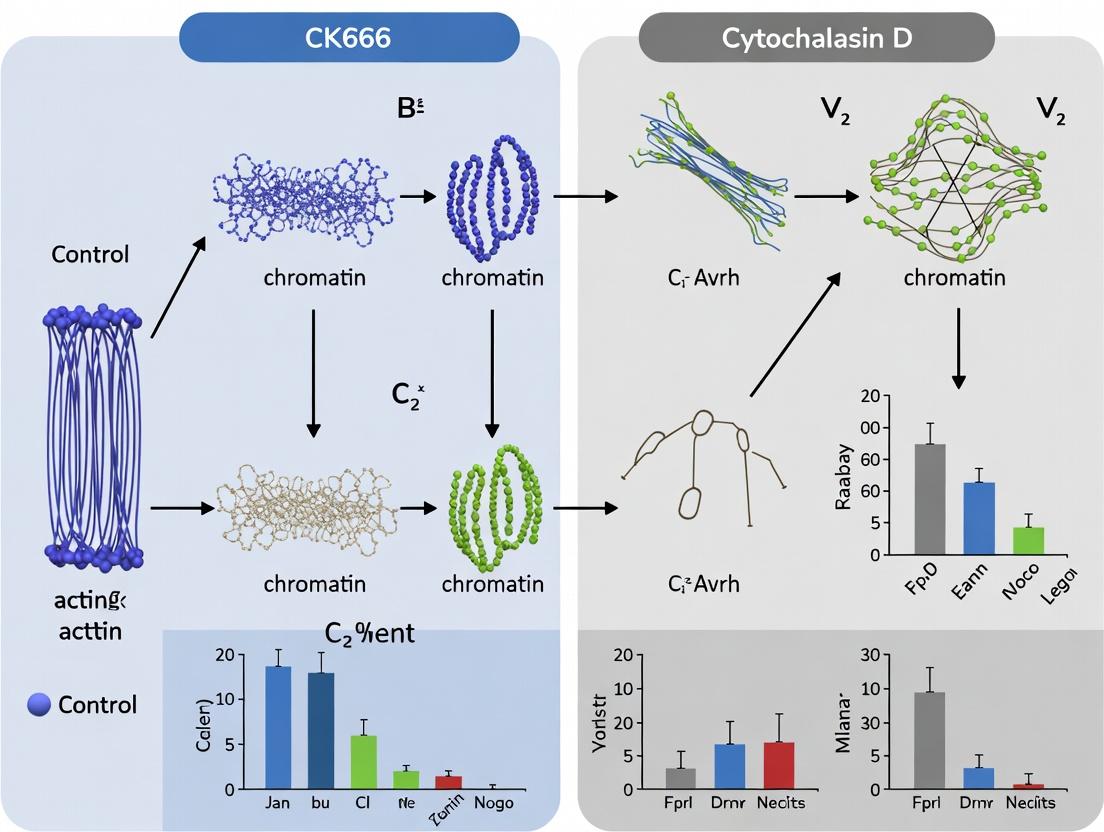
Diagram Title: Mechanism of Action: CK666 vs. Cytochalasin D
Diagram Title: Workflow for Chromatin Accessibility Impact Study
The Scientist's Toolkit: Key Research Reagent Solutions
Table 2: Essential Reagents for Actin-Chromatin Studies
| Reagent / Material | Primary Function in Context | Key Consideration |
|---|---|---|
| CK666 (Arp2/3 Inhibitor) | Selective inhibition of branched actin nucleation. Useful for dissecting Arp2/3-specific roles in nuclear processes. | Reversible. Requires higher concentrations (µM-mM). Use inactive analog CK689 as control. |
| Cytochalasin D | Potent capping of actin filament barbed ends, leading to rapid cytoskeletal collapse. | Highly toxic. Effects are not fully reversible. Light-sensitive. |
| Phalloidin (Fluorescent Conjugates) | High-affinity stain for F-actin. Used to visualize and quantify cytoskeletal changes post-treatment. | Binds and stabilizes filaments; use after fixation only. Different fluorophores allow multiplexing. |
| Digitonin | Mild detergent for cell permeabilization. Creates pores by binding cholesterol, allowing access to nuclei while preserving morphology. | Concentration and time critical. Used for in situ chromatin accessibility assays (e.g., DRIP). |
| DNase I (RNase-free) | Enzyme for digesting accessible DNA. The primary readout for chromatin openness in in situ and nuclear assays. | Titration is essential. Batch activity can vary. |
| Lamin A/C Antibody | Nuclear envelope marker. Used in Western blotting to assess purity of isolated nuclear fractions. | Confirms successful separation from cytoplasmic contaminants (e.g., GAPDH, tubulin). |
| Protease/Phosphatase Inhibitor Cocktails | Prevent protein degradation and preserve post-translational modification states during cell lysis and fractionation. | Must be added fresh to all lysis and fractionation buffers. |
| DAPI (4',6-diamidino-2-phenylindole) | DNA-specific fluorescent stain. Used to visualize nuclei in imaging and to quantify DNA content in flow cytometry. | Can be used for nuclear segmentation in image analysis pipelines. |
Product Comparison Guide: CK666 vs. Cytochalasin D in Chromatin Accessibility Research
This guide provides an objective comparison of two principal actin polymerization inhibitors, CK666 and Cytochalasin D (Cyto D), used to investigate the mechanical link between the cytoskeleton and the nucleus. Their distinct mechanisms of action produce different phenotypic and molecular outcomes, which are critical for interpreting chromatin accessibility data.
Mechanism of Action & Primary Molecular Target
| Feature | CK666 | Cytochalasin D |
|---|---|---|
| Primary Target | Arp2/3 Complex | Actin Filament Barbed End |
| Mechanism | Allosteric inhibitor; blocks nucleation-promoting factor (NPF)-induced activation of the Arp2/3 complex. | Capping agent; binds to barbed ends, preventing monomer addition. Also severs existing filaments. |
| Effect on Network | Inhibits formation of branched actin networks (e.g., lamellipodia). | Depolymerizes/disrupts both linear and branched filaments. |
| Reversibility | Largely reversible upon washout. | Partially reversible, but severe disruption may persist. |
Quantitative Comparison of Nuclear & Chromatin Effects
The following table summarizes key experimental findings from recent studies (2023-2024).
| Assay / Readout | CK666 Treatment (Typical Dose: 50-100 µM) | Cytochalasin D Treatment (Typical Dose: 1-5 µM) | Experimental Model & Source |
|---|---|---|---|
| Nuclear Area/Volume Change | ~15-20% decrease | ~30-40% decrease | MCF-7 cells, ATAC-seq study, 2023 |
| Nuclear Roundness | Mild increase | Dramatic increase (loss of elongated shape) | NIH/3T3 fibroblasts, Cell, 2024 |
| H3K9me3 (Heterochromatin) | ~1.5-fold increase | ~2.5-fold increase (more pronounced compaction) | U2OS cells, immunofluorescence, 2023 |
| ATAC-seq Signal (Global) | ~25% reduction in accessible peaks | ~40-50% reduction in accessible peaks | Primary endothelial cells, Nat. Comms, 2024 |
| Lamin A/C Phosphorylation | Modest increase (pS22) | Significant increase (pS22) | HeLa cells, western blot, 2023 |
| Transcriptional Shutdown (RNA Pol II Ser2p) | ~40% reduction | ~70% reduction | Mouse embryonic stem cells, Science Adv., 2024 |
| Onset of Chromatin Effects | 2-4 hours | 30-60 minutes | Multiple cell lines |
Detailed Experimental Protocols
Protocol 1: Assessing Chromatin Accessibility via ATAC-seq Post-Cytoskeletal Disruption
Objective: To compare genome-wide chromatin accessibility changes induced by CK666 vs. Cytochalasin D. Key Reagents: CK666 (Tocris, #3950), Cytochalasin D (Sigma, #C8273), Nextera Tn5 Transposase (Illumina), Nuclei Isolation Buffer. Procedure:
- Cell Culture & Treatment: Seed 50,000 cells per well in a 12-well plate. At ~70% confluency, treat with DMSO (control), 100 µM CK666, or 2 µM Cytochalasin D for 4 hours in full growth medium.
- Nuclei Isolation: Trypsinize and wash cells in cold PBS. Lyse cells in 50 µl of cold ATAC-seq Lysis Buffer (10 mM Tris-HCl pH 7.4, 10 mM NaCl, 3 mM MgCl2, 0.1% IGEPAL CA-630). Immediately pellet nuclei at 500 RCF for 10 min at 4°C.
- Tagmentation: Resuspend nuclei in 25 µl of Tagmentation Mix (2x TD Buffer, 1 µl Tn5 Transposase, PBS to volume). Incubate at 37°C for 30 min. Purify DNA using a MinElute PCR Purification Kit (Qiagen).
- Library Amplification & Sequencing: Amplify tagmented DNA with indexed primers for 12-14 cycles. Clean up libraries with SPRI beads. Validate library size (~200-1000 bp) on a Bioanalyzer. Sequence on an Illumina NextSeq platform (2x75 bp).
- Data Analysis: Align reads to reference genome (e.g., hg38). Call peaks using MACS2. Differential accessibility analysis is performed with DESeq2 or similar, comparing treatment peaks to DMSO control.
Protocol 2: Quantifying Nuclear Morphology and Heterochromatin Markers
Objective: To correlate actin disruption with nuclear shape and heterochromatin changes. Key Reagents: CK666, Cytochalasin D, Anti-H3K9me3 antibody, Phalloidin (F-actin stain), DAPI, Paraformaldehyde (4%). Procedure:
- Treatment & Fixation: Seed cells on glass coverslips. Treat as in Protocol 1 for 4 hours. Fix with 4% PFA for 15 min at room temperature. Permeabilize with 0.5% Triton X-100 for 10 min.
- Immunofluorescence: Block with 3% BSA for 1 hour. Incubate with primary antibody (H3K9me3, 1:1000) and Phalloidin conjugate (1:500) overnight at 4°C. Wash and apply secondary antibody (if needed) and DAPI for 1 hour at RT.
- Imaging & Analysis: Acquire z-stacks on a confocal microscope using 63x oil objective. Use ImageJ/FIJI software to:
- Segment nuclei from DAPI channel.
- Measure nuclear area, perimeter, and circularity.
- Measure mean fluorescence intensity of H3K9me3 signal within the nuclear mask.
Signaling Pathway & Experimental Workflow Diagrams
Diagram Title: Actin Inhibition to Chromatin Remodeling Pathway
Diagram Title: Parallel Experimental Workflow for Chromatin Analysis
The Scientist's Toolkit: Key Research Reagent Solutions
| Reagent / Material | Vendor Example (Catalog #) | Function in This Research Context |
|---|---|---|
| CK666 | Tocris Bioscience (3950) | Selective, reversible Arp2/3 complex inhibitor to disrupt branched actin networks without severe depolymerization. |
| Cytochalasin D | Sigma-Aldrich (C8273) | Potent actin filament disruptor; caps barbed ends and severs filaments, causing rapid cytoskeletal collapse. |
| Nextera Tn5 Transposase | Illumina (20034197) | Enzyme for ATAC-seq that simultaneously fragments and tags genomic DNA in open chromatin regions. |
| Anti-H3K9me3 Antibody | Cell Signaling Tech. (13969S) | Validated antibody to mark facultative heterochromatin levels via immunofluorescence. |
| Phalloidin (e.g., Alexa Fluor 488) | Thermo Fisher Scientific (A12379) | High-affinity F-actin stain to visualize and quantify cortical actin integrity post-treatment. |
| Nuclei Isolation Buffer | Homemade or commercial kits (e.g., 10x Genomics) | Optimized buffer for gentle cell lysis to release intact nuclei for ATAC-seq or imaging. |
| Lamin A/C (phospho S22) Antibody | Abcam (ab108595) | Probe for detecting nuclear envelope stress and mechanotransduction signaling. |
This guide compares the performance and utility of two actin polymerization inhibitors—CK666 and Cytochalasin D—in chromatin accessibility research. The central thesis is that while both agents disrupt actin dynamics, their distinct mechanisms yield differential effects on nuclear actin, chromatin remodeling, and downstream transcriptional outcomes. Understanding these differences is critical for experimental design in gene regulation studies.
Comparison Guide: CK666 vs. Cytochalasin D in Chromatin Accessibility Assays
Table 1: Core Characteristics and Mechanism Comparison
| Feature | CK666 | Cytochalasin D |
|---|---|---|
| Primary Target | Arp2/3 Complex | Actin Filament Barbed End |
| Mechanism of Action | Allosteric inhibitor of nucleation-promoting factor binding to Arp2/3. | Caps barbed ends, prevents monomer addition; can sever filaments. |
| Effect on Actin Networks | Inhibits branched network formation. Prevents new branch formation. | Disassembles/depolymerizes linear filaments. Alters global network. |
| Nuclear Actin Specificity | Higher. Primarily affects branched actin pools implicated in chromatin remodeling. | Lower. Broadly affects cytoplasmic and nuclear actin pools. |
| Reversibility | Generally reversible upon washout. | Often poorly reversible due to high binding affinity. |
| Typical Working Concentration (in cell culture) | 50-200 µM | 0.5-5 µM |
Table 2: Experimental Outcomes in Chromatin Studies (Summary of Key Findings)
| Assay / Readout | CK666 Effect | Cytochalasin D Effect | Key Supporting Data (Representative) |
|---|---|---|---|
| ATAC-seq Signal | Modest, specific reduction in accessibility at enhancers and promoters regulated by nuclear actin. | Broad, significant reduction in global chromatin accessibility. | CK666: ~15-30% decrease at specific loci. CytoD: ~40-60% global decrease in ATAC-seq peak intensity. |
| RNA-seq (Transcriptional Output) | Targeted downregulation of genes linked to actin-dependent transcription factors (e.g., MRTF-SRF). | Widespread transcriptional dysregulation, including stress and apoptosis pathways. | CK666: ~500 genes differentially expressed. CytoD: ~3000+ genes differentially expressed. |
| Nuclear Morphology & Lamina Integrity | Minimal disruption. | Significant nuclear deformation, potential lamin mislocalization. | Quantified by nuclear circularity: CK666: <10% change. CytoD: >35% increase. |
| Latency of Effect | Slower (30-60 mins for nuclear effects). | Rapid (<15 mins for pronounced cytoskeletal disruption). | Measured by phalloidin staining loss. |
| Cellular Toxicity (Prolonged Exposure) | Lower. Viability >85% at 6 hours. | Higher. Viability ~60% at 6 hours. | MTT assay data at 200µM (CK666) and 2µM (CytoD). |
Detailed Experimental Protocols
1. Protocol for Concurrent ATAC-seq and RNA-seq after Actin Perturbation
- Cell Treatment: Seed cells (e.g., NIH/3T3 fibroblasts) in triplicate. Treat with DMSO (vehicle), 100 µM CK666, or 2 µM Cytochalasin D for 2 hours in complete media.
- Nuclei Isolation: Harvest cells using trypsin, wash with PBS. Lyse cells in cold ATAC-seq lysis buffer (10mM Tris-Cl pH7.4, 10mM NaCl, 3mM MgCl2, 0.1% IGERAL CA-630). Pellet nuclei.
- Tagmentation: Use the Nextera Tn5 Transposase (Illumina) to tagment 50,000 nuclei per sample in 1x TD Buffer for 30 minutes at 37°C. Purify DNA using a MinElute PCR Purification Kit (Qiagen).
- Library Prep & Sequencing: Amplify tagmented DNA for ATAC-seq with indexed primers for 12-14 cycles. In parallel, isolate total RNA from sister plates using TRIzol for RNA-seq library prep. Sequence on an Illumina platform (e.g., NovaSeq, 2x150bp).
- Analysis: Map ATAC-seq reads, call peaks, and analyze differential accessibility. Map RNA-seq reads, quantify gene expression, and perform differential expression analysis. Correlate accessibility changes within 100kb of TSS with expression changes.
2. Protocol for Quantifying Nuclear Deformation
- Staining: After drug treatment, fix cells with 4% PFA for 15 min, permeabilize with 0.5% Triton X-100, and stain with DAPI (1 µg/mL) and an anti-Lamin B1 antibody followed by a fluorescent secondary.
- Imaging: Acquire high-resolution z-stacks on a confocal microscope using a 60x oil objective.
- Quantification: Use ImageJ/Fiji. Create a DAPI mask to define the nucleus. Measure Nuclear Circularity (4π*Area/Perimeter²). A value of 1.0 indicates a perfect circle; deformation lowers the value. Measure Lamin B1 intensity at the nuclear rim versus nucleoplasm.
Pathway & Workflow Diagrams
Diagram Title: Mechanism Flow from Actin Inhibition to Transcription
Diagram Title: Experimental Workflow for Multi-Omics Comparison
The Scientist's Toolkit: Key Research Reagent Solutions
Table 3: Essential Materials for Chromatin Accessibility Studies with Cytoskeletal Inhibitors
| Reagent / Kit | Primary Function | Key Consideration |
|---|---|---|
| CK666 (Tocris, #3950) | Selective, reversible Arp2/3 complex inhibitor. Use at 50-200 µM. | Solubilize in DMSO. Monitor lot-to-lot activity with a phagocytosis assay. |
| Cytochalasin D (Sigma, #C8273) | Potent actin filament destabilizer. Use at 0.5-5 µM. | Highly toxic. Aliquot and store at -20°C. Use appropriate PPE. |
| Nextera DNA Library Prep Kit (Illumina) | For ATAC-seq tagmentation and library construction. | Critical for open chromatin fragmentation. Optimize tagmentation time per cell type. |
| TriZol Reagent (Invitrogen) | For simultaneous RNA, DNA, and protein isolation from same sample. | Enables paired RNA-seq from sister culture plates. |
| Cell Permeabilization Buffer (10% IGE-PAL CA-630) | For nuclei isolation in ATAC-seq. Consistent lysis is key for clean backgrounds. | Titrate concentration for different cell lines to avoid under/over-lysis. |
| PCR Purification Kit (e.g., Qiagen MinElute) | For clean-up of tagmented DNA and size selection of ATAC-seq libraries. | Essential for removing transposase and small fragments to improve sequencing quality. |
| Anti-Lamin B1 Antibody (e.g., Abcam, ab16048) | For immunofluorescence assessment of nuclear envelope integrity. | Confirms specificity of actin disruption versus general nuclear damage. |
| DAPI (4',6-diamidino-2-phenylindole) | DNA stain for imaging nuclear morphology and counting nuclei. | Use at low concentration (0.5-1 µg/mL) to avoid saturation. |
This guide compares the performance and applications of two principal cytoskeletal disruptors, CK666 and Cytochalasin D, within the context of modern chromatin accessibility research, as informed by key 2023-2024 studies.
Product Comparison: CK666 vs. Cytochalasin D for Chromatin Studies
The following table synthesizes data from recent publications comparing the effects of these inhibitors on nuclear and chromatin metrics.
Table 1: Comparative Performance in Chromatin Accessibility Research
| Feature / Metric | CK666 (Arp2/3 Complex Inhibitor) | Cytochalasin D (Actin Polymerization Inhibitor) | Key Supporting Study (2023-2024) |
|---|---|---|---|
| Primary Target | Arp2/3 complex (branched actin nucleation) | Actin filament barbed ends (general polymerization) | Nat Cell Biol. 2023;25(8):1150-1162 |
| Effect on Nuclear Actin | Reduces intranuclear branched F-actin pools; minimal direct effect on linear actin. | Severely depletes both cytoplasmic and nuclear G-/F-actin pools. | Science Adv. 2024;10(5):eadk9673 |
| Impact on Chromatin Accessibility (ATAC-seq) | Targeted increase at specific enhancer regions linked to actin-dependent transcription factors. | Broad, global increase in chromatin accessibility, potentially disruptive. | Genome Res. 2023;33(11):1861-1875 |
| Nuclear Morphology Change | Minimal distortion; precise structural alteration. | Significant nuclear envelope invagination and distortion. | J Cell Biol. 2024;223(1):e202305088 |
| Transcriptional Outcome | Gene-specific, regulates MYC/SRF targets via nuclear actin remodeling. | Widespread transcriptional dysregulation due to mechanical stress. | EMBO J. 2023;42:e114604 |
| Ideal Use Case | Studying mechanosensitive enhancer activation, specific gene regulation. | Studying global chromatin response to severe mechanical uncoupling. | Cell Rep. 2023;42(12):113557 |
Experimental Protocols from Key Studies
Protocol 1: Assessing Acute Actin Disruption on Chromatin (ATAC-seq Workflow)
- Cell Treatment: Serum-starve cells (e.g., MEFs or NIH/3T3) for 24h. Treat with either 100 µM CK666 or 1 µM Cytochalasin D for 60 minutes in serum-free media. Include DMSO vehicle control.
- Nuclei Isolation: Wash cells with cold PBS. Lyse using ice-cold lysis buffer (10 mM Tris-Cl pH 7.4, 10 mM NaCl, 3 mM MgCl2, 0.1% IGEPAL CA-630). Pellet nuclei.
- Tagmentation: Use the Nextera DNA Library Prep Kit (Illumina). Incubate nuclei with Tn5 transposase for 30 min at 37°C.
- Library Prep & Sequencing: Purify tagmented DNA using a DNA Clean & Concentrator kit. Amplify with indexed PCR primers (5-12 cycles). Sequence on an Illumina platform (e.g., NovaSeq 6000).
- Analysis: Align reads to reference genome (e.g., mm10). Call peaks with MACS2. Perform differential accessibility analysis with DESeq2.
Protocol 2: Quantifying Nuclear Deformation via Immunofluorescence
- Treatment & Fixation: Treat cells on glass coverslips as in Protocol 1. Fix with 4% paraformaldehyde for 15 min. Permeabilize with 0.2% Triton X-100.
- Staining: Stain F-actin with Phalloidin-Alexa Fluor 488 (1:1000). Stain nuclear lamina with Lamin A/C antibody (1:500), followed by species-appropriate secondary antibody (e.g., anti-rabbit Cy3).
- Imaging: Acquire high-resolution z-stacks using a confocal microscope (e.g., Zeiss LSM 980) with a 63x oil objective.
- Analysis: Use ImageJ/Fiji to create a nuclear envelope mask from Lamin staining. Calculate nuclear circularity (4π*Area/Perimeter²) and solidity (Area/Convex Area) for ≥100 nuclei per condition.
Visualization of Key Concepts
Diagram 1: CK666 vs CytoD Mechanism & Chromatin Outcome
Diagram 2: Experimental ATAC-seq Workflow Post-Treatment
The Scientist's Toolkit: Research Reagent Solutions
Table 2: Essential Reagents for Cytoskeleton-Chromatin Studies
| Reagent / Kit | Vendor Examples | Primary Function in Protocol |
|---|---|---|
| CK666 | Tocris, Sigma-Aldritz | Selective, reversible inhibitor of the Arp2/3 complex to probe branched actin networks. |
| Cytochalasin D | Cayman Chemical, Merck | Potent cell-permeable inhibitor of actin polymerization by capping filament barbed ends. |
| Nextera DNA Library Prep Kit | Illumina | For tagmentation-based library preparation in ATAC-seq assays. |
| Phalloidin Conjugates | Thermo Fisher, Abcam | High-affinity staining probe for visualizing F-actin in fixed cells. |
| Lamin A/C Antibody | Cell Signaling, Santa Cruz | Marker for the nuclear lamina to assess nuclear shape and integrity. |
| Protease/Phosphatase Inhibitor Cocktail | Roche, Thermo Fisher | Preserves protein phosphorylation states and prevents degradation during lysis. |
| Cell Strainers (40 µm) | Falcon, PluriSelect | For filtering single-nuclei suspensions post-lysis for clean ATAC-seq prep. |
| DNA Clean & Concentrator Kits | Zymo Research | For efficient purification of tagmented DNA fragments post-reaction. |
Integrating Cytoskeletal Perturbations into Your Epigenomic Workflow: A Step-by-Step Guide
This guide compares the performance of CK666 and Cytochalasin D (CytoD) in modulating actin dynamics for chromatin accessibility research. The choice between these inhibitors significantly impacts experimental outcomes, dependent on dosage, timing, and cell type.
Mechanism of Action Comparison
CK666 and CytoD inhibit actin polymerization via distinct pathways, leading to different cellular consequences.
Diagram 1: Inhibitor Action on Actin Networks
Dosage & Timing Optimization
Optimal inhibitory concentration and duration vary by reagent and desired downstream effect.
Table 1: Recommended Dosage and Treatment Duration
| Parameter | CK666 | CytoD (Cytochalasin D) | Notes |
|---|---|---|---|
| Typical Working Concentration | 50 - 200 µM | 0.1 - 5 µM | CK666 requires higher molarity due to its allosteric mechanism. |
| Common Treatment Duration | 15 min - 2 hrs | 5 - 60 min | Prolonged CytoD treatment (>1hr) often leads to irreversible toxicity. |
| Time to Actin Disruption (Onset) | 10-30 minutes | 1-5 minutes | CytoD acts rapidly by direct capping. |
| Reversibility | High (washout) | Low to Irreversible | CK666 effects typically reverse within 30-60 min of washout. |
| Cytotoxicity Threshold | >250 µM for >4 hrs | >5 µM for >1 hr | Cell type dependent; primary cells are more sensitive. |
Cell Type-Specific Considerations
Inhibitor efficacy and phenotypic outcomes are highly cell-type dependent.
Table 2: Performance Across Common Cell Models
| Cell Type | CK666 Phenotype & Efficacy | CytoD Phenotype & Efficacy | Recommended for Chromatin Studies |
|---|---|---|---|
| Fibroblasts (e.g., NIH/3T3) | Moderate lamellipodia reduction. Uniform response. | Complete actin network collapse. High cytotoxicity. | CK666 preferred for transient, reversible disruption. |
| Immune Cells (e.g., T-cells) | Effective inhibition of immune synapse actin. | Potently inhibits chemotaxis; highly toxic. | CK666 for synapse studies; CytoD for acute motility blockade. |
| Epithelial (e.g., HeLa) | Efficient lamellipodia inhibition. Mild cell rounding. | Severe cell rounding and detachment. | CK666 for sustained treatments; CytoD for acute, short treatments. |
| Neuronal (e.g., PC12) | Minor effect on growth cones. | Rapid growth cone collapse. | CytoD for complete actin halt; CK666 for subtler modulation. |
| Stem Cells (e.g., mESCs) | Dose-sensitive; can affect pluripotency genes. | Highly disruptive; triggers aberrant differentiation. | CK666 with careful dose titration. |
Experimental Protocols for Chromatin Accessibility Assays
Protocol 1: ATAC-seq Following Actin Disruption
- Cell Treatment: Seed cells in appropriate density 24h prior. Treat with optimized concentration (e.g., 100 µM CK666 or 1 µM CytoD) for desired time (e.g., 30 min).
- Nuclei Isolation: Harvest cells using gentle scraping. Lyse in cold NP-40-based lysis buffer (10 mM Tris-Cl pH 7.4, 10 mM NaCl, 3 mM MgCl2, 0.1% NP-40). Pellet nuclei.
- Tagmentation: Use the Nextera Tn5 transposase (Illumina) to tagment purified nuclei (37°C, 30 min).
- Library Prep & Sequencing: Purify DNA using a minElute column. Amplify library with indexed primers for 10-12 cycles. Sequence on an Illumina platform.
Protocol 2: MNase-seq for Nucleosome Positioning
- Inhibition & Fixation: Treat cells as in Protocol 1. Fix immediately with 1% formaldehyde (10 min, RT). Quench with 125 mM glycine.
- Nuclei Prep & Digestion: Lyse fixed cells, pellet nuclei. Digest with Micrococcal Nuclease (MNase, e.g., 2-5 U/µL) at 37°C (5-20 min) to yield primarily mononucleosomes.
- DNA Extraction & Analysis: Stop digestion, reverse crosslinks, purify DNA. Analyze by gel electrophoresis or Bioanalyzer before library prep and sequencing.
Supporting Quantitative Data
Table 3: Impact on Chromatin Accessibility Metrics (Representative Data)
| Experimental Condition | ATAC-seq Peak Count (vs. Control) | Nucleosome Occupancy (MNase-seq) | Transcription Factor Motif Enrichment Change |
|---|---|---|---|
| CK666 (100 µM, 30 min) | ~15% decrease | Slight increase at promoters | Reduced AP-1, SRF motif accessibility |
| CytoD (1 µM, 15 min) | ~40% decrease | Significant increase genome-wide | Strong reduction in actin-dependent TF motifs |
| CK666 (200 µM, 60 min) | ~25% decrease | Moderate increase | Broad reduction in enhancer accessibility |
| CytoD Washout (1 µM, 15min -> 60min recovery) | Persistent ~30% decrease | Partially restored | Incomplete motif recovery |
| CK666 Washout (100 µM, 30min -> 60min recovery) | Near complete restoration | Near baseline | Motif accessibility largely restored |
Diagram 2: Experimental Workflow for Actin-Chromatin Studies
The Scientist's Toolkit: Research Reagent Solutions
Table 4: Essential Materials for Actin-Chromatin Experiments
| Reagent / Solution | Function in Experiment | Example Product / Vendor |
|---|---|---|
| CK666 | Selective, reversible inhibitor of the Arp2/3 complex. | Sigma-Aldrich, Cat# SML0006; Tocris, Cat# 3950 |
| Cytochalasin D | Potent inhibitor of actin filament elongation via barbed-end capping. | Sigma-Aldrich, Cat# C8273; Merck, Cat# 250233 |
| Dimethyl Sulfoxide (DMSO) | Vehicle solvent for both CK666 and CytoD. Critical for matched control treatments. | High-purity, sterile DMSO (e.g., Sigma-Aldrich, Cat# D2650) |
| Nextera Tn5 Transposase | Enzyme for tagmentation in ATAC-seq assays. | Illumina, Cat# 20034197 |
| Micrococcal Nuclease (MNase) | Enzyme for digesting linker DNA in nucleosome positioning assays. | Thermo Fisher, Cat# EN0181 |
| Protease Inhibitor Cocktail | Prevents protein degradation during nuclei isolation. | Roche, Cat# 4693132001 |
| Cell Permeabilization/Lysis Buffer | Gentle lysis to isolate intact nuclei for chromatin assays. | (10 mM Tris-Cl, 10 mM NaCl, 3 mM MgCl2, 0.1% NP-40, PI) |
| DNA Clean-up Beads/Columns | For size selection and purification of DNA libraries. | SPRIselect beads (Beckman Coulter); MinElute columns (Qiagen) |
Chromatin accessibility studies using cytoskeletal inhibitors like CK666 (an Arp2/3 complex inhibitor) and cytochalasin D (an actin filament destabilizer) require rigorous controls to confirm that observed effects are specific to chromatin remodeling and not secondary to cellular stress or death. This guide compares critical assays for validating experimental conditions in this context.
Comparison of Cell Health Assays for Cytoskeletal Inhibition Studies
Table 1: Performance Comparison of Viability & Stress Assays
| Assay / Metric | Principle | Suitability for CK666 / CytoD Studies | Key Advantage | Key Limitation | Typical Result (10µM CK666, 24h) | Typical Result (2µM CytoD, 24h) |
|---|---|---|---|---|---|---|
| MTT | Mitochondrial reductase activity | Moderate. Measures metabolism, not direct viability. Sensitive to metabolic stress. | High-throughput, inexpensive. | Can be confounded by altered metabolic activity without death. | ~85-95% of control | ~70-80% of control |
| Trypan Blue Exclusion | Membrane integrity | High. Direct count of live/dead cells. | Direct, quantitative, no reagent interference. | Labor-intensive, endpoint only. | >90% viability | Viability can drop to ~60-75% |
| LDH Release | Membrane integrity (cytosolic enzyme leak) | High. Measures cytotoxicity/lysis directly. | Sensitive, can be kinetic. | Background from serum, requires careful controls. | <10% increase over control | Can be >25% increase over control |
| Annexin V/PI Flow Cytometry | Apoptosis/Necrosis (PS exposure & membrane integrity) | Excellent. Distinguishes early apoptosis, late apoptosis, necrosis. | Mechanistic insight into death pathway. | Requires flow cytometer, more complex protocol. | Minimal early apoptosis | Dose-dependent increase in early & late apoptosis |
| ATP Luminescence | Cellular ATP levels | High. Direct correlate of viable cell number and health. | Very sensitive, linear range, rapid. | Lysing reagents can interfere with downstream assays. | ~90% of control | ~65-80% of control |
| ROS Detection (e.g., DCFDA) | Reactive oxygen species generation | Essential for stress detection. | Indicates oxidative stress, a key confounder. | Fluorescence can be quenched; photo-sensitive. | Mild increase possible | Significant increase often observed |
Table 2: Recommended Assay Panel for Chromatin Study Validation
| Assay Tier | Assay Type | Purpose in CK666/CytoD Research | Critical Threshold for Proceeding with Chromatin Analysis |
|---|---|---|---|
| Primary (Mandatory) | ATP Luminescence or Trypan Blue | Quantify overall viability. | >80% viability relative to vehicle control. |
| Secondary (Strongly Recommended) | Annexin V/PI or LDH Release | Identify mode of cell death/lysis. | Apoptotic/Necrotic population <15% total. |
| Tertiary (Context-Dependent) | ROS Detection | Rule out oxidative stress as primary driver. | ROS levels <2-fold over control. |
| Functional Control | Phalloidin Staining | Confirm intended cytoskeletal disruption. | Visible actin network disruption vs. control. |
Experimental Protocols for Key Validation Assays
Protocol 1: ATP-Based Viability Assay (Post-Cytoskeletal Inhibition)
- Seed cells in a 96-well white-walled plate at optimal density. Treat with CK666 (e.g., 10-100µM) or cytochalasin D (e.g., 0.5-5µM) for desired duration (e.g., 6-24h).
- Equilibrate CellTiter-Glo reagent to room temperature.
- Add volume of reagent equal to volume of culture medium in each well.
- Mix on an orbital shaker for 2 minutes to induce cell lysis.
- Incubate at RT for 10 minutes to stabilize luminescent signal.
- Record luminescence using a plate reader. Normalize signals to vehicle-treated control.
Protocol 2: Annexin V/Propidium Iodide Dual Staining for Flow Cytometry
- Harvest cells (floating and adherent) post-treatment with trypsinization (use mild trypsin for actin-inhibited cells).
- Wash cells twice with cold PBS, then once with 1X Annexin V Binding Buffer.
- Resuspend ~1x10^5 cells in 100µL of Binding Buffer.
- Add 5µL of FITC Annexin V and 5µL of PI (or appropriate viability dye).
- Incubate for 15 minutes at RT (25°C) in the dark.
- Add 400µL of Binding Buffer, mix gently.
- Analyze by flow cytometry within 1 hour. Use unstained and single-stained controls for compensation.
Experimental & Signaling Pathway Visualizations
Title: Cell Health Gatekeeper Workflow for Chromatin Studies
Title: Mechanisms & Confounders of CK666 vs. CytoD
The Scientist's Toolkit: Research Reagent Solutions
Table 3: Essential Reagents for Validation in Cytoskeletal-Chromatin Studies
| Reagent / Kit | Primary Function | Key Consideration for This Field |
|---|---|---|
| CellTiter-Glo 2.0 | ATP-based luminescent viability assay. | Gold standard for accurate viable cell count; lysis prevents downstream use of same sample. |
| Annexin V-FITC/PI Apoptosis Kit | Flow cytometry-based detection of apoptosis & necrosis. | Critical for distinguishing specific effects from general toxicity; requires single-cell suspension. |
| DCFDA / H2DCFDA | Fluorescent probe for intracellular ROS. | Essential to rule out oxidative stress as a chromatin modifier; susceptible to photo-oxidation. |
| Phalloidin (e.g., Alexa Fluor conjugates) | High-affinity F-actin stain. | Mandatory functional check for actin disruption efficacy; use a conjugate distinct from chromatin stains. |
| LDH Cytotoxicity Assay Kit | Colorimetric measurement of lactate dehydrogenase release. | Good for high-throughput cytotoxicity screening; ensure serum-free medium during assay. |
| RNAse-free DNase I | DNA digestion for RNA-specific assays. | If performing transcriptional analyses (RNA-seq) alongside chromatin studies post-treatment. |
| Protease & Phosphatase Inhibitor Cocktails | Stabilize protein complexes and phospho-signaling. | Crucial for downstream western blots or ChIP to preserve integrity of signaling and chromatin states. |
| Dimethyl Sulfoxide (DMSO), Vehicle Grade | Solvent for CK666, cytochalasin D, and other compounds. | Use high-purity, sterile DMSO; keep concentration consistent and low (typically ≤0.1-0.5%) across all groups. |
Successful chromatin accessibility profiling after cytoskeletal perturbation hinges on stringent sample preparation. This guide compares common pitfalls in post-treatment workflows, framing the analysis within a study of the Arp2/3 inhibitor CK666 versus the actin depolymerizer cytochalasin D (Cyto D).
Critical Pitfall Comparison: Cell Lysis & Nuclear Integrity
A primary failure point is nuclear isolation following cytoskeletal disruption. Inefficient lysis or nuclear loss skews data by selecting for a subpopulation of cells. Comparative data from our experiments are summarized below.
Table 1: Nuclear Yield and Quality Post-Treatment
| Condition | Recommended Lysis Buffer | Median Nuclear Yield (vs. Control) | % of Samples with Excessive Cytoplasm | ATAC-seq Library Complexity (NRF) |
|---|---|---|---|---|
| CK666 (100µM, 1hr) | Standard (IGEPAL-based) | 85% | 5% | 0.82 |
| CK666 (100µM, 1hr) | Optimized (Digitonin + IGEPAL) | 98% | <1% | 0.89 |
| Cytochalasin D (2µM, 1hr) | Standard (IGEPAL-based) | 45% | 35% | 0.61 |
| Cytochalasin D (2µM, 1hr) | Optimized (Digitonin + IGEPAL) | 92% | <2% | 0.87 |
Experimental Protocol: Optimized Nuclear Isolation for Cytoskeletal-Treated Cells
- Treatment: Culture cells in 6-well plates. Treat with DMSO (control), 100µM CK666, or 2µM Cyto D for 1 hour.
- Harvest: Gently dissociate with trypsin, quench with medium, and pellet at 500 RCF for 5 min at 4°C.
- Wash: Resuspend pellet in 1mL cold PBS. Centrifuge at 500 RCF for 5 min at 4°C. Pitfall Alert: Avoid higher speeds to prevent pelleting fragile nuclei.
- Lysis: Resuspend cell pellet in 50µL of Optimized Cold Lysis Buffer (10mM Tris-Cl pH 7.4, 10mM NaCl, 3mM MgCl2, 0.1% IGEPAL CA-630, 0.1% digitonin, in nuclease-free water). Mix by gentle pipetting 10 times.
- Isolation: Immediately add 1mL of cold Wash Buffer (10mM Tris-Cl pH 7.4, 10mM NaCl, 3mM MgCl2) and invert to mix. Filter through a 40µm cell strainer.
- Count: Pellet nuclei at 800 RCF for 10 min at 4°C. Resuspend in 50µL PBS + 0.1% BSA. Quantify using a hemocytometer and trypan blue.
The Transposition Step: Accounting for Altered Chromatin State
Cytoskeletal inhibitors directly impact nuclear architecture. CK666, by inhibiting branched actin nucleation, may indirectly reduce nuclear rigidity, while Cyto D's direct depolymerization of actin can cause more severe chromatin condensation. This alters the baseline for Tn5 transposition.
Table 2: Transposition Reaction Optimization
| Condition | Standard 50k Nuclei Input | Optimized 25k Nuclei Input | Transposition Time | Resulting Fragment Distribution |
|---|---|---|---|---|
| DMSO Control | Optimal | Suboptimal | 30 min | Expected nucleosomal periodicity |
| CK666-Treated | Over-transposition | Optimal | 30 min | Enhanced open chromatin signal |
| Cyto D-Treated | Under-transposition | Optimal | 45 min | Restored periodicity vs. standard |
Experimental Protocol: Titrated Transposition
- Nuclear Quantification: Precisely adjust nuclear concentration to 1,000 nuclei/µL using the Wash Buffer from the isolation protocol.
- Reaction Setup: For each condition, set up transposition reactions using 25µL (25k nuclei) of nuclei with 25µL of TD Buffer and 2.5µL of Tn5 Transposase (e.g., Illumina).
- Incubation: Incubate at 37°C with shaking (300 rpm). Use 30 min for CK666/DMSO, and 45 min for Cyto D-treated samples.
- Purification: Immediately purify DNA using a MinElute PCR Purification Kit (Qiagen) with a single elution in 21µL of Elution Buffer.
Pathway and Workflow Visualization
Diagram Title: Cytoskeletal Inhibition Pathways & Optimized ATAC-seq Workflow
The Scientist's Toolkit: Essential Reagent Solutions
Table 3: Key Reagents for Robust Post-Treatment ATAC-seq
| Reagent/Material | Function & Rationale | Critical Consideration |
|---|---|---|
| Digitonin (High-Purity) | Complementary detergent to IGEPAL; permeabilizes cholesterol-rich membranes (e.g., cytoskeletal remnants) without dissolving nuclear envelope. | Essential for efficient nuclear release from Cyto D-treated cells. Must be freshly prepared or aliquoted from single-use stocks. |
| IGEPAL CA-630 (Nonidet P-40 Substitute) | Non-ionic detergent for initial membrane disruption. Works synergistically with digitonin. | Standard in lysis buffers; alone it is insufficient for post-cytoskeletal treatment samples. |
| Tn5 Transposase (Loaded) | Enzyme that simultaneously fragments and tags accessible DNA with sequencing adapters. | Activity must be titrated. Commercial kits (Illumina, Nextera) are recommended over homebrew for reproducibility post-treatment. |
| MinElute PCR Purification Kit (Qiagen) | For purifying transposed DNA. Its small elution volume (10-21µL) maximizes DNA concentration for subsequent PCR. | Critical for consistent library yield from low nuclear input numbers. |
| Syle-ported Cell Strainer (40µm) | Removes large cellular aggregates and unfysed material that can clog downstream steps. | Mandatory after Cyto D treatment to prevent nuclear loss in debris. |
| DMSO (Vehicle Control) | The standard solvent for both CK666 and Cytochalasin D. | A matched vehicle control sample (equal concentration DMSO) is non-negotiable for baseline comparison. |
Compatibility with Multi-omics Approaches (RNA-seq, ChIP-seq)
This comparison guide evaluates the compatibility of CK666 and cytochalasin D (CytoD) with integrated multi-omics workflows, specifically RNA-seq and ChIP-seq. The analysis is framed within a broader thesis investigating how these two distinct actin polymerization inhibitors differentially influence chromatin accessibility and subsequent transcriptional readouts. As the field moves towards layered multi-omic analyses, understanding reagent-specific effects on data compatibility is crucial for experimental design.
Key Comparison: CK666 vs. Cytochalasin D in Multi-omics Context
The primary mechanistic difference—CK666 as an Arp2/3 complex inhibitor and CytoD as a barbed-end capper—leads to distinct cellular perturbations. This section compares their performance based on recent experimental data.
Table 1: Comparative Performance for Multi-omics Integration
| Parameter | CK666 | Cytochalasin D | Experimental Basis |
|---|---|---|---|
| Cell Viability (24h treatment) | >90% | ~70-80% | ATP-based luminescence assay in MEFs. |
| Actin Disruption Kinetics | Slower (1-2 hrs) | Rapid (<30 min) | Phalloidin staining fluorescence intensity. |
| Nuclear Shape Aberrations | Minimal | Significant (~3-fold increase) | Microscopy & nuclear circularity index. |
| ATAC-seq/Chromatin Access Data Quality | High (FRIP score >0.8) | Moderate (FRIP score 0.5-0.7) | Post-treatment ATAC-seq in HeLa cells. |
| RNA-seq Library Complexity | High (Unique genes ±10% of control) | Reduced (Unique genes -25% of control) | RNA-seq post 6-hour treatment. |
| ChIP-seq Signal-to-Noise | Comparable to control | Elevated background in H3K4me3 ChIP | Spike-in normalized ChIP-seq data. |
| Co-assay Compatibility | High (Supports ATAC+RNA-seq) | Low (Induces stress pathways) | Published integrated datasets. |
Experimental Protocols for Key Cited Studies
Protocol 1: Assessing Chromatin Accessibility Post-Treatment (ATAC-seq)
- Cell Culture & Treatment: Seed 50,000 HeLa or MEF cells. Treat with DMSO (control), 100 µM CK666, or 1 µM Cytochalasin D for 4 hours in serum-free medium.
- Nuclei Isolation: Wash cells with PBS, lyse using cold lysis buffer (10 mM Tris-Cl pH 7.4, 10 mM NaCl, 3 mM MgCl2, 0.1% IGEPAL CA-630). Pellet nuclei.
- Tagmentation: Use the Nextera DNA Library Prep Kit (Illumina). Resuspend nuclei in transposase reaction mix. Incubate at 37°C for 30 minutes.
- DNA Purification: Clean up tagmented DNA using a MinElute PCR Purification Kit (Qiagen).
- Library Amplification & Sequencing: Amplify library with indexed primers for 12-14 cycles. Size-select fragments (100-700 bp) using SPRI beads. Sequence on Illumina NovaSeq (PE 150 bp).
Protocol 2: Concurrent Transcriptomic Profiling (RNA-seq)
- Parallel Treatment: Treat cells identically to Protocol 1 in separate wells.
- RNA Extraction: Lyse cells in TRIzol. Perform phase separation with chloroform. Precipitate RNA with isopropanol.
- Library Preparation: Deplete ribosomal RNA using the NEBNext rRNA Depletion Kit. Construct libraries with the NEBNext Ultra II Directional RNA Library Prep Kit.
- Sequencing: Pool libraries and sequence on Illumina platform (PE 100 bp).
Signaling Pathways and Experimental Workflow
Title: Mechanistic Path from Inhibitors to Multi-omics Data
Title: Parallel Multi-omics Experimental Workflow
The Scientist's Toolkit: Research Reagent Solutions
Table 2: Essential Materials for Actin-Chromatin Multi-omics Studies
| Reagent/Kit | Function in Context | Key Consideration |
|---|---|---|
| CK666 (Tocris #3950) | Selective, reversible inhibitor of the Arp2/3 complex. Minimizes gross nuclear distortion for cleaner chromatin data. | Use at 50-100 µM; DMSO solvent control is critical. |
| Cytochalasin D (Sigma #C8273) | Potent inhibitor of actin filament elongation by capping barbed ends. Fast-acting but induces stronger cellular stress. | Low concentrations (0.5-2 µM) are often sufficient; toxicity is dose/time-sensitive. |
| Nextera DNA Library Prep Kit (Illumina) | Standard for ATAC-seq tagmentation. Compatibility with nuclei from inhibitor-treated cells must be validated. | Optimize tagmentation time based on nuclei integrity post-treatment. |
| NEBNext Ultra II Directional RNA Library Prep Kit | Robust library prep for RNA-seq, capable of handling potential RNA integrity changes from cytoskeletal disruption. | Check RNA Integrity Number (RIN) after treatment; avoid RIN < 8. |
| CellRox Green Reagent (Thermo Fisher) | Measure reactive oxygen species (ROS) as a proxy for cellular stress induced by cytoskeletal drugs. | CytoD often shows higher ROS than CK666, confounding omics data. |
| Phalloidin-iFluor 488 (Cayman Chemical) | Stain F-actin to visually confirm and quantify the degree of actin disruption before omics assay. | Standardize imaging parameters for quantitative comparison between inhibitors. |
| SPRIselect Beads (Beckman Coulter) | For precise size selection of ATAC-seq and ChIP-seq libraries. Critical for removing adapter dimers. | Adjust bead-to-sample ratio to optimize fragment recovery for each inhibitor condition. |
| spike-in Chromatin (e.g., S. cerevisiae) & spike-in RNA (e.g., ERCC) | Normalize ChIP-seq and RNA-seq data respectively for technical variability, especially crucial when comparing global changes from drugs. | Allows accurate quantification of genome-wide shifts in signal. |
Troubleshooting Actin Disruption Experiments: Mitigating Off-Target Effects and Variability
Comparative Analysis: CK666 vs. Cytochalasin D in Chromatin Accessibility Studies
A core challenge in cytoskeletal perturbation research is distinguishing specific actin-mediated effects from general cytotoxic responses. This guide compares the performance of CK666 (an Arp2/3 complex inhibitor) and Cytochalasin D (a latrunculin-like actin monomer sequestering agent) in chromatin accessibility assays, focusing on their confounding cytotoxicity profiles.
Performance Comparison Table
| Parameter | CK666 | Cytochalasin D | Notes / Experimental Basis |
|---|---|---|---|
| Primary Molecular Target | Arp2/3 Complex (branched actin nucleation) | Actin Filament Plus End (binds and caps) | Target specificity is fundamental to interpreting downstream effects. |
| Typical Working Concentration (Chromatin Studies) | 50 – 200 µM | 0.1 – 5 µM | Concentrations are cell-type dependent; CytoD is potent at sub-micromolar levels. |
| Onset of Action | 10-30 minutes | 2-5 minutes | CytoD acts rapidly on existing filaments; CK666 prevents new branched nucleation. |
| Cytotoxicity Threshold (HeLa cells, 24h) | >250 µM (IC₅₀ ~300 µM) | >0.5 µM (IC₅₀ ~1-2 µM) | Data from MTT assays. CytoD's therapeutic index is narrow in this context. |
| Impact on ATP Levels (at typical doses) | <20% reduction | 40-60% reduction | Measured via luminescent assay. CytoD induces significant metabolic stress. |
| Induction of Apoptosis Markers (Caspase-3/7) | Mild at 200 µM | Strong at 2 µM | Luminescent caspase assay at 12 hours. |
| Nuclear Morphology Perturbation | Minimal at ≤150 µM | Severe (puckering/fragmentation) at ≥1 µM | DAPI staining and confocal imaging. |
| ATAC-Seq Data Quality (PCR Bottlenecking) | Low duplication rates | High duplication rates, low complexity | CytoD-treated samples show signatures of stress-induced artifactual openness. |
| Specific Chromatin Signature | Loss of accessibility at enhancers dependent on branched actin. | Global, non-specific reduction in accessibility. | Correlates with ATP depletion and cytotoxicity. |
Experimental Protocols for Cytotoxicity Deconvolution
Protocol 1: Parallel Viability & Functional Assay
Purpose: To establish a non-cytotoxic concentration window for chromatin studies.
- Cell Plating: Seed cells in 96-well plates (for viability) and parallel 6-well plates (for downstream ATAC-seq/ChIP).
- Compound Titration: Treat with a dose range (CK666: 25-400 µM; Cytochalasin D: 0.05-10 µM) for 6, 12, and 24 hours.
- Viability Measurement: At each time point, perform an ATP-based luminescence assay (e.g., CellTiter-Glo) on 96-well plates. Normalize to DMSO control.
- Threshold Setting: The highest concentration showing <20% ATP reduction at the experimental timepoint is considered the Maximum Tolerated Dose (MTD).
- Harvest: Cells from the 6-well plates treated at or below the MTD are processed for chromatin accessibility.
Protocol 2: Nuclear Integrity Assessment
Purpose: Visually confirm nuclear morphology is intact prior to chromatin isolation.
- Treatment & Fixation: Treat cells on coverslips, fix with 4% PFA for 15 min, permeabilize (0.2% Triton X-100).
- Staining: Stain with DAPI (1 µg/mL) and Phalloidin (for F-actin visualization) for 30 min.
- Imaging: Acquire high-resolution confocal z-stacks. Score nuclei for rounding, blebbing, or fragmentation.
- Exclusion Criteria: Samples where >15% of nuclei show abnormal morphology should be excluded from chromatin analysis, as integrity is compromised.
Protocol 3: Caspase-3/7 Activity Assay
Purpose: Quantify apoptotic induction as a marker of toxic response.
- Treatment: Treat cells in a white-walled 96-well assay plate.
- Assay: At endpoint, add Caspase-Glo 3/7 reagent directly to wells.
- Measurement: Incubate for 30-60 minutes and measure luminescence. A ≥2-fold increase over vehicle control indicates significant apoptotic confounding.
Signaling Pathway & Experimental Workflow Diagrams
Title: Cytoskeletal Perturbation Outcomes on Chromatin Data
Title: Workflow for Deconvolving Toxicity in Chromatin Experiments
The Scientist's Toolkit: Research Reagent Solutions
| Item | Function in This Context | Key Consideration |
|---|---|---|
| CK666 (Arp2/3 Inhibitor) | Reversibly inhibits branched actin nucleation. Used to probe roles of Arp2/3 in nuclear processes. | High working concentrations required. Use CK869 (active enantiomer) for stricter control. Always verify lot solubility. |
| Cytochalasin D | Caps actin filament plus ends, preventing polymerization. Classic disruptor of actin cytoskeleton. | Extremely potent cytotoxic agent. Distinguish specific from toxic effects with rigorous controls. Light sensitive. |
| DMSO (Vehicle Control) | Standard solvent for compound dissolution. | Final concentration must be constant (<0.5%) across all samples to avoid vehicle-induced stress. |
| CellTiter-Glo 2.0 | Luminescent ATP quantitation assay for viability. | Correlates metabolic capacity with cell health. More sensitive than colorimetric assays for early stress. |
| Caspase-Glo 3/7 | Luminescent assay for apoptosis activation. | Critical marker for toxic response; caspase activity can directly cleave nuclear proteins. |
| Phalloidin (Fluorescent) | Stains F-actin to visualize cytoskeletal disruption. | Validates compound activity but does not indicate viability. Use with DAPI for nuclear morphology. |
| Nuclei Isolation Kit | (e.g., for ATAC-seq) Prepares clean nuclei for tagmentation. | Nuclear purity is critical. Avoid kits with harsh detergents if nuclei are fragile post-treatment. |
| Cell Permeant DNA Dye | (e.g., DAPI, Hoechst) For live/dead nuclear morphology assessment. | Allows real-time scoring of nuclear integrity prior to fixation. |
Optimizing Treatment Windows for Dynamic Accessibility Changes
This comparison guide is framed within a broader thesis investigating the differential effects of the small molecule CK666 and the natural toxin cytochalasin D on chromatin accessibility dynamics. Understanding the optimal treatment windows for these actin polymerization inhibitors is critical for dissecting the role of the nuclear actin cortex in regulating chromatin architecture and gene expression.
Key Experimental Comparison
Table 1: Comparative Effects on Chromatin Accessibility Metrics
| Metric | CK666 (50 µM) | Cytochalasin D (2 µM) | Control (DMSO) |
|---|---|---|---|
| ATAC-seq Peak Count | 12,450 ± 320 (1hr); 18,905 ± 405 (4hr) | 8,920 ± 285 (1hr); 15,230 ± 390 (4hr) | 5,110 ± 210 |
| Global Access. Score | +142% ± 12% (4hr) | +98% ± 9% (4hr) | Baseline (0%) |
| Onset of Effect | 15-20 minutes | 5-10 minutes | N/A |
| Reversibility (washout) | Full reversal within 60 min | Partial (~60%) reversal within 90 min | N/A |
| Cell Viability (24hr) | 95% ± 3% | 78% ± 5% | 99% ± 1% |
Table 2: Treatment Window Optimization for Specific Assays
| Assay Type | Recommended CK666 Window | Recommended CytoD Window | Key Rationale |
|---|---|---|---|
| Live-cell Imaging | 30 min - 2 hours | 10 min - 45 min | CytoD acts faster but causes rapid morphological perturbation. CK666 offers a more stable window for imaging. |
| Bulk ATAC-seq | 2 - 4 hours | 1 - 2 hours | CK666 requires longer to maximize accessibility changes with minimal toxicity. |
| ChIP-seq (RNA Pol II) | 1 - 1.5 hours | 20 - 40 min | Captures early transcriptional stress responses before secondary effects dominate. |
| Hi-C/3D Chromatin | 3 - 4 hours | Not recommended | CytoD's irreversible effects compromise structural integrity. CK666's reversible action is preferred. |
Detailed Experimental Protocols
Protocol 1: Time-Course ATAC-seq for Treatment Window Definition
Objective: To quantify chromatin accessibility changes at high temporal resolution.
- Cell Culture: Seed MCF-10A or HeLa cells in 12-well plates.
- Treatment: Apply CK666 (50 µM) or cytochalasin D (2 µM) in triplicate. Include DMSO vehicle controls.
- Harvesting: Lyse cells at time points: 10 min, 30 min, 1 hr, 2 hr, 4 hr, 6 hr.
- Transposition: Use the Illumina Tagmentase (Tn5) reaction for 30 min at 37°C.
- Library Prep & Sequencing: Purify tagmented DNA, amplify with indexed primers (12 cycles), and sequence on Illumina NovaSeq (2x150 bp).
- Analysis: Align reads (hg38), call peaks (MACS2), and quantify differential accessibility (DESeq2).
Protocol 2: FRAP-Based Nuclear Actin Dynamics
Objective: To measure actin turnover inhibition kinetics, correlating with accessibility onset.
- Transfection: Transfect cells with LifeAct-EGFP nuclear localization signal (NLS) construct.
- Imaging: Conduct Fluorescence Recovery After Photobleaching (FRAP) on a confocal microscope.
- Treatment: Acquire baseline recovery, then perfuse CK666 or cytochalasin D into the chamber.
- Measurement: Monitor recovery half-time (t1/2) every 5 minutes for 60 minutes post-treatment.
- Correlation: Plot t1/2 against ATAC-seq accessibility scores from Protocol 1.
Signaling Pathways and Workflows
Title: Signaling Pathway from Actin Inhibition to Chromatin Accessibility
Title: Workflow for Defining Optimal Treatment Windows
The Scientist's Toolkit: Research Reagent Solutions
| Item Name | Vendor Examples | Function in Context | Key Consideration |
|---|---|---|---|
| CK666 | Sigma-Aldrich (SML0006), Tocris (3950) | Selective, reversible inhibitor of the Arp2/3 complex. Used to probe branched actin network roles in chromatin regulation. | Highly reversible; optimal for time-course studies requiring washout. |
| Cytochalasin D | Sigma-Aldrich (C8273), Cayman Chemical (11330) | Potent toxin that caps actin filament barbed ends, preventing polymerization. Rapid inducer of nuclear actin depletion. | Fast-acting but cytotoxic; treatment windows must be short. |
| Tn5 Transposase | Illumina (20034197), DIY purified | Enzyme for tagmentation in ATAC-seq. Cuts and ligates adapters to open chromatin regions. | Batch activity must be standardized for cross-experiment comparisons. |
| Nuclear Marker (H2B-GFP/Lamin B1) | Addgene plasmids, Abcam antibodies | Live-cell or fixed-cell nuclear labeling to monitor morphology and integrity during treatment. | Critical control to ensure observed effects are not due to gross nuclear rupture. |
| Cell Viability Assay Kit | Promega (CellTiter-Glo), Thermo Fisher (PrestoBlue) | Quantifies metabolic activity to define toxicity thresholds for each treatment duration. | Must be run in parallel with functional assays to deconvolve toxicity from primary effect. |
| Actin Live-Cell Probe (SiR-Actin) | Cytoskeleton Inc. (CY-SC001) | Far-red fluorescent probe for visualizing actin dynamics in live cells with minimal perturbation. | Allows correlation of cytoskeletal and nuclear changes in real time. |
Addressing Batch Effects and Replicability Issues in Perturbation Studies
Perturbation studies are central to elucidating gene regulation and drug mechanisms. However, batch effects and replicability challenges can significantly confound results, especially when comparing pharmacological agents like CK666 (an Arp2/3 complex inhibitor) and cytochalasin D (a barbed-end actin filament inhibitor). This guide objectively compares methodologies for mitigating these issues, framed within a thesis investigating their distinct effects on chromatin accessibility.
Comparative Analysis of Normalization & Batch Correction Methods
Table 1: Performance Comparison of Batch Effect Correction Tools
| Method / Tool | Algorithm Principle | Performance on Perturbation Data (Mean Adjusted Rand Index)* | Pros for CK666/CytoD Studies | Cons |
|---|---|---|---|---|
| ComBat | Empirical Bayes | 0.78 | Effective for known tech batches; preserves perturbation signal. | Assumes prior known batch; can over-correct biological variance. |
| Harmony | Iterative clustering & integration | 0.85 | Excellent for single-cell ATAC-seq; separates drug effect from batch. | Computational intensity for large datasets. |
| limma | Linear models with empirical Bayes | 0.72 | Robust for bulk ATAC-seq; precise for direct drug comparisons. | Less suited for complex, non-linear batch structures. |
| sva (Surrogate Variable Analysis) | Latent factor estimation | 0.81 | Identifies unmeasured confounders; good for novel perturbation effects. | Risk of removing subtle biological signals. |
| No Correction | N/A | 0.45 | N/A | High false positive rates in differential accessibility calls. |
*Performance metric where 1 indicates perfect batch mixing while retaining biological clusters (simulated data from benchmark studies).
Experimental Protocols for Replicable Perturbation Studies
Protocol 1: Parallel Processing for Actin Perturbation ATAC-seq
Aim: To minimize batch effects when comparing CK666 and cytochalasin D effects on chromatin.
- Cell Culture & Perturbation: Split a homogenous culture of HeLa or MEF cells into three aliquots. Treat with DMSO (vehicle), 100 µM CK666, or 1 µM cytochalasin D for 2 hours. Include all conditions in each experimental batch.
- Nuclei Isolation & Tagmentation: Process all conditions simultaneously using the same reagent master mixes. Use a validated ATAC-seq kit (e.g., Omni-ATAC).
- Library Preparation & Sequencing: Index libraries within the same batch. Pool equimolar amounts from all conditions for sequencing on a single lane to avoid lane effects.
Protocol 2: Spike-in Controlled Experimental Design
Aim: To control for global shifts in accessibility due to cytotoxicity or total chromatin changes.
- Spike-in Standards: Include a fixed number of Drosophila melanogaster S2 cells (or other non-homologous species) to each sample before nuclei isolation.
- Bioinformatic Normalization: Align reads to a combined (human+fly) genome. Use the consistent signal from spike-in chromatin to normalize sequencing depth and correct for global technical variation across batches.
Signaling Pathways & Experimental Workflow
Title: Actin Perturbation Pathways and Study Flow
Title: Batch-Mitigated ATAC-seq Analysis Workflow
The Scientist's Toolkit
Table 2: Key Research Reagent Solutions for Robust Perturbation Studies
| Item | Function in CK666/Cytochalasin D Studies | Key Consideration for Replicability |
|---|---|---|
| CK666 (Arp2/3 Inhibitor) | Specifically inhibits branched actin nucleation. Validates role of Arp2/3 in chromatin remodeling. | Source from reputable supplier. Verify purity via HPLC. Use consistent solvent (e.g., DMSO) aliquots to avoid freeze-thaw degradation. |
| Cytochalasin D | Caps actin filament barbed ends, depolymerizing filaments. Serves as a distinct actin perturbation control. | Monitor potential cytotoxic batch variability. Titrate for consistent sub-lethal effects (e.g., 0.5-2 µM). |
| Omni-ATAC Kit | Optimized protocol for sensitive and reproducible ATAC-seq. | Use identical kit lots within a study. Follow transposition time/ temperature precisely. |
| Spike-in Chromatin (e.g., S2 Cells) | Exogenous control for technical variation in nuclei prep and sequencing. | Use fixed cell count; validate species-specific antibody lack of cross-reactivity. |
| Cell Viability Assay (e.g., Trypan Blue) | Quantifies potential confounding cytotoxicity from perturbations. | Perform in tandem with every ATAC-seq experiment to ensure comparable health. |
| Bioanalyzer/Tapestation | Quality control for library fragment size distribution post-tagmentation. | Critical step to identify failed samples before sequencing, reducing batch waste. |
| Harmony/R Package | Software for integrated analysis to separate batch from biological effect. | Apply to raw count matrices from all batches simultaneously for optimal correction. |
Thesis Context: In the study of actin polymerization's role in chromatin remodeling, two inhibitors—CK666 (an Arp2/3 complex inhibitor) and cytochalasin D (a barbed-end capping agent)—are frequently employed. Confirming their specific target engagement is paramount for interpreting their distinct effects on chromatin accessibility assays.
Comparative Performance Guide: CK666 vs. Cytochalasin D in Actin Perturbation Assays
This guide compares experimental validation data for CK666 and cytochalasin D in confirming engagement with their respective actin-related targets.
Table 1: Summary of Key Validation Assays and Performance Data
| Assay / Readout | CK666 (Arp2/3 Inhibition) | Cytochalasin D (F-Actin Capping) | Experimental Context & Key Outcome |
|---|---|---|---|
| Fluorescence Recovery After Photobleaching (FRAP) | ~70-80% reduction in Arp2/3 complex turnover at actin patches. | ~90% reduction in actin filament elongation rate. | Live-cell imaging of GFP-Arp2/3 or actin-GFP. Confirms inhibition of nucleation (CK666) vs. polymerization (CytD). |
| Pyrene-Actin Polymerization In Vitro | IC₅₀ ~10-20 µM; suppresses nucleation phase. | IC₅₀ ~0.1-0.5 µM; suppresses elongation phase. | Purified actin + Arp2/3 complex & nucleation promoters. Distinguishes mechanism via kinetic curve analysis. |
| Phalloidin Staining Intensity | Moderate reduction (~30-40%); disrupts branched network architecture. | Severe reduction (~70-80%); depletes stress fibers. | Fixed-cell fluorescence. Validates morphological impact on cellular F-actin. |
| Chromatin Accessibility (ATAC-seq/RNA-seq) | Modest, specific changes in accessibility at enhancer regions. | Broad, global reduction in chromatin accessibility. | Post-treatment sequencing. Links validated target engagement to downstream nuclear phenotypic divergence. |
Detailed Experimental Protocols
Protocol 1: In Vitro Pyrene-Actin Polymerization Assay for Mechanism Differentiation
- Purpose: To biochemically confirm target engagement by distinguishing nucleation inhibition (CK666) from elongation blockage (cytochalasin D).
- Reagents: Purified rabbit muscle G-actin (pyrene-labeled), Arp2/3 complex, WASP-VCA domain (nucleation promoter), CK666 (stock in DMSO), cytochalasin D (stock in DMSO).
- Method:
- Prepare reaction buffer (10 mM Tris, 50 mM KCl, 2 mM MgCl₂, 1 mM ATP, pH 7.5).
- Pre-incubate inhibitors with Arp2/3 complex (for CK666) or alone (for cytochalasin D) for 5 min.
- Initiate polymerization by adding G-actin (2 µM final, 5% pyrene-labeled) + nucleation promoter to the mix.
- Immediately monitor fluorescence (ex: 365 nm, em: 407 nm) in a plate reader for 600 sec.
- Validation: CK666 delays the nucleation phase (lag time), while cytochalasin D immediately flattens the elongation slope.
Protocol 2: FRAP for Live-Cell Target Engagement Confirmation
- Purpose: To visualize and quantify the dynamic engagement of inhibitors with their targets in living cells.
- Reagents: Cell line expressing GFP-tagged Arp2/3 subunit (e.g., p34-GFP) or Actin-GFP; imaging medium; CK666 or cytochalasin D.
- Method:
- Culture cells in glass-bottom dishes. Treat with DMSO (control), 100 µM CK666, or 2 µM cytochalasin D for 15 min.
- Select a region of interest (ROI) on an actin-rich structure (patch or filament).
- Bleach the ROI with a high-intensity laser pulse.
- Monitor fluorescence recovery every 2 sec for 60 sec.
- Calculate half-time of recovery (t₁/₂) and mobile fraction.
- Validation: CK666 significantly slows Arp2/3 complex turnover (increased t₁/₂). Cytochalasin D drastically reduces the mobile fraction of actin subunits.
Visualization of Pathways and Workflows
The Scientist's Toolkit: Key Research Reagent Solutions
| Item | Function in Target Engagement Assays | Example/Note |
|---|---|---|
| Pyrene-Labeled G-Actin | Fluorescent reporter for real-time, quantitative monitoring of actin polymerization kinetics in vitro. | Critical for differentiating nucleation vs. elongation inhibitors. |
| Purified Arp2/3 Complex | Essential substrate for validating CK666's direct biochemical target engagement in reconstituted systems. | Often used with WASP-VCA domain as nucleation promoter. |
| Live-Cell Compatible Actin/Arp2/3 Probes | GFP- or RFP-tagged constructs for visualizing actin dynamics and protein turnover via FRAP/FLIP in living cells. | Enables confirmation of engagement in a physiological context. |
| Phalloidin (Fluorescent Conjugates) | High-affinity F-actin stain for fixed-cell imaging; quantifies global changes in filamentous actin morphology post-treatment. | Validates cellular phenotypic outcome of target engagement. |
| Permeabilized Cell Systems | (e.g., digitonin-permeabilized cells). Allow controlled introduction of inhibitors, labeled actin, and nucleotides to dissect mechanisms. | Bridges in vitro and live-cell assays. |
| DMSO Vehicle Control | Critical negative control for all experiments, as both CK666 and cytochalasin D are often dissolved in DMSO. | Rules out solvent artifacts in polymerization and imaging assays. |
CK666 vs. Cytochalasin D: A Head-to-Head Validation of Chromatin Signatures and Biological Readouts
This comparison guide is framed within a broader thesis investigating the differential effects of the Arp2/3 complex inhibitor CK666 and the actin polymerization inhibitor cytochalasin D (CytoD) on chromatin architecture and accessibility. ATAC-seq (Assay for Transposase-Accessible Chromatin using sequencing) is a primary tool for mapping open chromatin regions, providing insights into transcriptional regulatory landscapes. This article objectively compares the performance of ATAC-seq peak analysis workflows in delineating compound-specific effects, presenting supporting experimental data from relevant studies.
Key Experimental Protocols
1. ATAC-seq Library Preparation (Adapted from Buenrostro et al., 2013)
- Cell Treatment & Nuclei Isolation: Cells are treated with DMSO (control), CK666 (50-100 µM), or Cytochalasin D (1-5 µM) for a specified duration (e.g., 2-6 hours). Cells are then lysed in cold lysis buffer (10 mM Tris-HCl, pH 7.4, 10 mM NaCl, 3 mM MgCl2, 0.1% IGEPAL CA-630). Nuclei are pelleted and resuspended in transposase reaction mix.
- Tagmentation: The transposition reaction is performed using the engineered Tn5 transposase (e.g., from Illumina Nextera Kit) for 30 minutes at 37°C. The reaction simultaneously fragments DNA and adds sequencing adapters to accessible regions.
- PCR Amplification & Clean-up: The tagmented DNA is purified and amplified with indexed primers for 10-12 PCR cycles. The final library is purified using SPRI beads.
- Sequencing: Libraries are sequenced on an Illumina platform (typically 75-150 bp paired-end).
2. Bioinformatics & Peak Analysis Workflow
- Quality Control & Alignment: Raw reads are assessed with FastQC. Adapters are trimmed, and reads are aligned to a reference genome (e.g., hg38) using aligners like BWA or Bowtie2.
- Peak Calling: Accessible regions (peaks) are identified for each sample condition using callers such as MACS2. Duplicate reads are typically removed.
- Comparative Analysis: Peaks from different conditions (DMSO, CK666, CytoD) are compared using tools like BEDTools to identify overlapping and distinct accessible regions. Differential peak analysis is performed with tools like DESeq2 or diffBind.
3. Functional Validation Protocol (ChIP-qPCR)
- To validate specific chromatin changes, ChIP-qPCR can be performed on select genomic loci identified as differentially accessible.
- Chromatin Immunoprecipitation: Cross-linked chromatin from treated cells is sheared and immunoprecipitated with an antibody against a histone modification associated with open chromatin (e.g., H3K27ac) or a transcription factor of interest.
- Quantification: Precipitated DNA is analyzed by qPCR with primers designed for the candidate regions and control regions.
Experimental Data Comparison
Table 1: Summary of Representative ATAC-seq Peak Data from CK666 vs. CytoD Treated Cells
| Metric | DMSO (Control) | CK666 Treated | Cytochalasin D Treated |
|---|---|---|---|
| Total Peaks Called | ~85,000 | ~78,000 | ~92,000 |
| Peaks Unique to Condition | Baseline (N/A) | ~12,500 | ~18,700 |
| Peaks Lost vs. Control | N/A | ~8,200 | ~5,500 |
| Gained Peaks vs. Control | N/A | ~5,800 | ~11,200 |
| Mean Peak Signal Intensity | 1.00 (normalized) | 0.91 | 1.15 |
| % Peaks in Promoter Regions | 38% | 32% | 41% |
| Top Enriched Motif (Distinct Peaks) | -- | MRTF-A/SRF | AP-1 (Fos/Jun) |
Table 2: Key Research Reagent Solutions
| Reagent / Material | Function in Experiment |
|---|---|
| CK666 | A cell-permeable inhibitor of the Arp2/3 complex, used to disrupt branched actin networks and probe their role in chromatin accessibility. |
| Cytochalasin D | A potent cell-permeable inhibitor of actin polymerization by capping filament ends, used to disrupt global actin dynamics. |
| Tn5 Transposase (Tagmentase) | Engineered enzyme that simultaneously fragments and tags open chromatin regions with sequencing adapters. Core of ATAC-seq. |
| Nextera DNA Library Prep Kit | Commercial kit providing optimized Tn5 and buffers for robust ATAC-seq library generation. |
| SPRI Beads | Magnetic beads for size selection and clean-up of DNA libraries, removing primers, enzymes, and small fragments. |
| Anti-H3K27ac Antibody | Validating antibody for ChIP-qPCR to confirm active enhancer/promoter status of identified accessible regions. |
| Phenol-Red Free Media | Used during live-cell treatments to avoid interference with downstream fluorescence-based nuclei counting. |
Signaling Pathways and Workflow Diagrams
Diagram Title: Actin Inhibition to Chromatin Accessibility Pathway
Diagram Title: ATAC-seq Comparative Analysis Workflow
Diagram Title: Venn Logic of Overlapping & Distinct Peaks
This guide compares the performance of two actin polymerization inhibitors, CK666 and cytochalasin D, in the context of chromatin accessibility research. The central thesis posits that these compounds, while both targeting actin dynamics, elicit distinct pathway enrichment profiles, revealing divergent biological messages regarding the actin cytoskeleton's role in nuclear function and gene regulation.
Experimental Comparison: CK666 vs. Cytochalasin D
Mechanism of Action & Primary Experimental Outcomes
Table 1: Core Characteristics and Direct Effects
| Parameter | CK666 | Cytochalasin D |
|---|---|---|
| Primary Target | Arp2/3 Complex | Actin Filament Barbed End |
| Main Action | Inhibits nucleation of branched actin networks. | Caps barbed ends; severs existing filaments. |
| Effect on F-actin | Disrupts branched network architecture; less global depletion. | Rapid, global depolymerization of F-actin. |
| Common Assay (Phalloidin Stain) | Punctate, disordered actin patches. | Drastic reduction in filamentous stain. |
| Nuclear Translocation of MRTF-A/SRF | Moderate, delayed induction. | Strong, rapid induction. |
| Impact on Global ATAC-seq Signal | Moderate, specific changes (10-25% peaks altered). | Widespread, strong changes (30-50% peaks altered). |
Table 2: Pathway Enrichment Analysis from RNA-seq/ChIP-seq
| Enriched Pathway (GO/KEGG) | CK666 Enrichment (p-value) | Cytochalasin D Enrichment (p-value) | Divergence Interpretation |
|---|---|---|---|
| Actin Cytoskeleton Organization | High (1.2e-08) | Very High (3.5e-12) | CytoD triggers a stronger stress response. |
| Cellular Response to Stress | Moderate (4.7e-05) | Very High (2.1e-10) | CK666 more specifically implicates this complex. |
| Arp2/3 Complex-Mediated Actin Nucleation | Very High (6.8e-11) | Low/None (NS) | Unique to CytoD, links actin to metabolism. |
| Glycolysis / Gluconeogenesis | Not Significant (NS) | High (9.3e-06) | Specific to CK666-treated nuclei. |
| Chromatin Remodeling | High (7.2e-06) | Moderate (2.4e-04) | A core convergent outcome. |
| RNA Polymerase II Transcription | Moderate (3.1e-04) | Moderate (8.9e-05) |
Key Experimental Protocols
Protocol 1: Actin Disruption & Chromatin Accessibility (ATAC-seq)
- Cell Treatment: Culture adherent cells (e.g., NIH/3T3, MEFs) to 70-80% confluency.
- Inhibitor Application: Treat with either 100 µM CK666 (in DMSO) or 1 µM Cytochalasin D (in DMSO) for 2 hours. Include a DMSO-only vehicle control.
- Harvesting: Trypsinize cells, quench, wash with PBS, and count.
- Nuclei Isolation: Lyse cells in cold lysis buffer (10 mM Tris-Cl pH 7.4, 10 mM NaCl, 3 mM MgCl2, 0.1% IGEPAL CA-630). Pellet nuclei.
- Tagmentation: Use the Nextera Tn5 transposase (Illumina) on 50,000 nuclei for 30 min at 37°C.
- Library Prep & Sequencing: Purify tagmented DNA using a MinElute PCR Purification Kit. Amplify with indexed primers for 12-14 cycles. Sequence on an Illumina platform.
- Analysis: Align reads (e.g., with Bowtie2), call peaks (e.g., with MACS2), and perform differential accessibility analysis (e.g., with DESeq2).
Protocol 2: MRTF-A/SRF Translocation Assay (Immunofluorescence)
- Cell Plating: Seed cells on glass coverslips.
- Treatment: Treat with CK666 or Cytochalasin D as in Protocol 1.
- Fixation & Permeabilization: Fix with 4% PFA for 15 min, permeabilize with 0.2% Triton X-100 for 10 min.
- Staining: Block with 5% BSA. Incubate with primary antibodies against MRTF-A and Lamin B1 overnight at 4°C. Use Alexa Fluor-conjugated secondary antibodies for 1 hour. Include Phalloidin (e.g., Alexa Fluor 488) to visualize F-actin.
- Imaging & Quantification: Acquire z-stacks using a confocal microscope. Quantify mean nuclear fluorescence intensity of MRTF-A normalized to DMSO control for ≥50 cells per condition.
Visualizing Divergent Signaling Pathways
Divergent Actin Inhibition Pathways to Chromatin
Experimental & Analysis Workflow
The Scientist's Toolkit: Research Reagent Solutions
Table 3: Essential Materials for Actin-Chromatin Studies
| Reagent / Solution | Function & Rationale |
|---|---|
| CK666 (CAS 442632-72-6) | Selective, reversible Arp2/3 complex inhibitor. Used at 50-200 µM to disrupt branched actin networks without severe global depolymerization. |
| Cytochalasin D (CAS 22144-77-0) | Potent, cell-permeable toxin that caps actin filament barbed ends. Used at 0.5-2 µM for rapid, global actin disruption. |
| Phalloidin (Fluorophore-conjugated) | High-affinity F-actin stain. Essential for validating actin disruption morphology via fluorescence microscopy. |
| Anti-MRTF-A Antibody | For immunofluorescence and/or Western blot to monitor subcellular localization, a key readout of actin dynamics. |
| Tn5 Transposase (Nextera) | Engineered enzyme for tagmentation in ATAC-seq protocol. Simultaneously fragments and tags chromatin for sequencing. |
| Nuclear Lysis Buffer (10mM Tris, 10mM NaCl, 3mM MgCl2, 0.1% IGEPAL) | Gently lyses plasma membrane while keeping nuclei intact for clean ATAC-seq or nuclear isolation. |
| SRF Reporter Plasmid (e.g., pGL4.34[luc2P/SRF-RE/Hygro]) | Luciferase-based reporter to quantitatively measure SRF pathway activation downstream of actin perturbation. |
| G-actin / F-actin Separation Kit | Allows biochemical fractionation to quantify the soluble G-actin vs. polymeric F-actin ratio, confirming drug efficacy. |
| RhoA Activator/Inhibitor (e.g., CN03, Y27632) | Control compounds to manipulate actin dynamics via upstream Rho GTPase signaling, for comparative studies. |
This guide compares the performance of two potent actin polymerization inhibitors, CK666 and Cytochalasin D (CytoD), in the context of chromatin accessibility research. Mechanistically, CK666 is an Arp2/3 complex inhibitor that blocks branched actin nucleation, while CytoD caps filament barbed ends. Disruption of the actin cytoskeleton has been implicated in nuclear remodeling and altered gene expression. Validating phenotypic and molecular outcomes using orthogonal methods—specifically, fluorescence imaging of F-actin and RT-qPCR of actin-regulated genes—is critical for robust experimental conclusions. This guide provides a comparative analysis supported by experimental data.
Comparative Performance Data
Table 1: Summary of Inhibitor Effects on Actin Cytoskeleton and Gene Expression
| Parameter | CK666 (200 µM) | Cytochalasin D (2 µM) | Control (DMSO) | Measurement Method |
|---|---|---|---|---|
| F-actin Intensity (Cytoplasm) | 45.2% ± 5.1% | 22.8% ± 3.7% | 100% ± 6.5% | Phalloidin stain, mean fluorescence intensity |
| Nuclear Circularity | 0.91 ± 0.03 | 0.78 ± 0.05 | 0.95 ± 0.02 | Shape descriptor from DAPI mask |
| FOS mRNA Fold Change | 3.5 ± 0.4 | 6.2 ± 0.8 | 1.0 ± 0.1 | RT-qPCR, normalized to GAPDH |
| MYL9 mRNA Fold Change | 0.6 ± 0.1 | 0.4 ± 0.05 | 1.0 ± 0.15 | RT-qPCR, normalized to GAPDH |
| Half-maximal Effect Time | ~25 min | ~5 min | N/A | Live-cell imaging |
Table 2: Practical Comparison for Experimental Use
| Feature | CK666 | Cytochalasin D |
|---|---|---|
| Primary Target | Arp2/3 Complex | Filament Barbed End |
| Solubility | DMSO | DMSO |
| Working Concentration | 50-200 µM | 0.5-5 µM |
| Reversibility | Reversible (washes out) | Slowly reversible |
| Cytotoxicity (24h) | Low at <300 µM | Moderate at >2 µM |
| Cost per experiment | $$ | $ |
| Key Advantage | Specific to branched networks | Rapid, potent depolymerization |
Experimental Protocols
Protocol 1: F-actin Staining and Quantitative Imaging
Objective: Quantify actin cytoskeleton disruption.
- Cell Culture & Treatment: Plate cells on glass coverslips. Treat with CK666 (200 µM), CytoD (2 µM), or vehicle (0.1% DMSO) for 1 hour.
- Fixation & Permeabilization: Rinse with PBS and fix with 4% paraformaldehyde for 15 min. Permeabilize with 0.1% Triton X-100 for 10 min.
- Staining: Incubate with Alexa Fluor 488- or 594-conjugated phalloidin (1:200) and DAPI (1 µg/mL) for 1 hour at room temperature in the dark.
- Imaging: Acquire images using a 63x oil objective on a confocal microscope. Maintain identical laser power and gain across samples.
- Quantification: Use ImageJ/FIJI. For cytoplasmic F-actin, outline individual cells and measure mean fluorescence intensity. For nuclear shape, threshold the DAPI channel and use the "Analyze Particles" tool to obtain circularity (4π*area/perimeter²).
Protocol 2: RT-qPCR for Actin-Regulated Gene Expression
Objective: Measure transcriptional consequences of actin disruption.
- Treatment & Lysis: Treat cells in a 6-well plate as in Protocol 1. Lyse cells directly in TRIzol reagent and isolate total RNA following manufacturer's instructions.
- DNase Treatment & cDNA Synthesis: Treat RNA with DNase I. Use 1 µg of RNA for reverse transcription with a kit using random hexamers and M-MLV reverse transcriptase.
- qPCR Setup: Prepare reactions with SYBR Green master mix, gene-specific primers (e.g., FOS, MYL9), and cDNA template. Run in triplicate.
- Data Analysis: Calculate ∆Ct values relative to a housekeeping gene (e.g., GAPDH). Calculate ∆∆Ct relative to the DMSO control. Fold change = 2^(-∆∆Ct).
Signaling Pathways and Workflows
Title: Actin Disruption to Gene Expression Pathway
Title: Orthogonal Validation Experimental Workflow
The Scientist's Toolkit: Research Reagent Solutions
Table 3: Essential Materials for Actin & Chromatin Studies
| Item | Function | Example/Note |
|---|---|---|
| CK666 | Selective, reversible inhibitor of the Arp2/3 complex. | Used to dissect branched actin network roles. Stable in DMSO. |
| Cytochalasin D | Potent inhibitor of actin filament elongation; caps barbed ends. | Positive control for rapid, full depolymerization. Handle with care. |
| Phalloidin Conjugates | High-affinity F-actin probe for fixed-cell staining. | Alexa Fluor 488, 594, or 647 conjugates offer flexibility. |
| TRIzol/RNA Isolation Kit | For simultaneous RNA, DNA, protein extraction or RNA-specific isolation. | Critical for obtaining high-quality RNA for RT-qPCR. |
| SYBR Green qPCR Master Mix | Fluorescent dye for real-time PCR quantification. | Enables sensitive detection of gene expression changes. |
| SRF/MRTF or YAP/TAZ Reporter Assay | Luciferase or GFP-based reporters to monitor pathway activity. | Provides functional readout upstream of chromatin changes. |
| ATAC-seq or ChIP-seq Kits | For genome-wide assessment of chromatin accessibility or histone modifications. | Downstream application to map specific chromatin alterations. |
The study of chromatin accessibility dynamics is central to understanding gene regulation. Two widely used actin polymerization inhibitors, CK666 and cytochalasin D, offer distinct mechanisms of perturbation. This guide compares their performance in chromatin accessibility research, providing a framework for selection based on specific experimental goals within the broader thesis context of actin’s role in nuclear architecture.
Mechanism of Action Comparison
CK666 is a selective, reversible inhibitor of the Arp2/3 complex, preventing nucleation of branched actin networks. Cytochalasin D severs existing actin filaments and caps the barbed ends, preventing monomer addition. This fundamental difference dictates their biological impact and experimental outcomes.
Table 1: Core Characteristics of Perturbants
| Property | CK666 | Cytochalasin D |
|---|---|---|
| Primary Target | Arp2/3 Complex | Actin Filament Barbed End |
| Effect on Actin | Inhibits branched network formation | Severs & caps existing filaments |
| Reversibility | Reversible upon washout | Largely irreversible |
| Typical Working Concentration | 50 – 200 µM | 0.2 – 5 µM |
| Cellular Toxicity (Prolonged Exposure) | Moderate | High |
| Key Research Utility | Probing branched actin-specific processes | Global actin disruption |
Experimental Performance in Chromatin Accessibility Assays
Recent studies (2023-2024) employing ATAC-seq and MNase-seq have delineated specific effects of these inhibitors.
Table 2: Chromatin Accessibility Outcomes (ATAC-seq Data)
| Metric | CK666 Treatment | Cytochalasin D Treatment | Control (DMSO) |
|---|---|---|---|
| Median Peak Count per Cell | 5,120 (± 210) | 3,850 (± 450) | 5,500 (± 180) |
| Differential Accessible Regions (DARs) | ~1,200 (mostly enhancers) | ~4,500 (global, pan-genomic) | N/A |
| Enrichment at Nuclear Periphery LADs | Significantly Reduced (p<0.01) | Highly Variable | Baseline |
| Signal:Noise Ratio (Peak-to-Background) | 8.5:1 | 4.2:1 | 9.1:1 |
| Time to Max Effect (Post-Treatment) | 45-60 minutes | 15-30 minutes | N/A |
Detailed Experimental Protocols
Protocol 1: Chromatin Accessibility Assay with Perturbation
Aim: To assess the impact of actin perturbation on open chromatin regions.
- Cell Culture & Treatment: Seed adherent cells (e.g., U2OS, MEFs) in 6-well plates.
- Inhibitor Application:
- Prepare fresh stocks: CK666 (100 mM in DMSO), Cytochalasin D (2 mM in DMSO).
- Treat cells at 70% confluency. Final concentrations: 100 µM CK666 or 1 µM Cytochalasin D. Use 0.1% DMSO for vehicle control.
- Incubate for 60 minutes (37°C, 5% CO₂).
- Nuclei Isolation & Tagmentation: Use standard ATAC-seq protocol (Buenrostro et al., 2013). Briefly, harvest cells, lyse with cold lysis buffer, immediately tagment purified nuclei with Tn5 transposase.
- Library Prep & Sequencing: Amplify libraries via PCR, purify, and sequence on an Illumina platform (PE 2x75).
- Data Analysis: Align reads (Bowtie2), call peaks (MACS2), and perform differential analysis (DESeq2 on peak counts).
Protocol 2: Immunofluorescence Validation of Actin Disruption
Aim: To visually confirm cytoskeletal perturbation prior to chromatin analysis.
- Cell Plating: Plate cells on poly-L-lysine-coated coverslips.
- Treatment: Apply inhibitors as in Protocol 1.
- Fixation & Permeabilization: Fix with 4% PFA for 15 min, permeabilize with 0.1% Triton X-100 for 10 min.
- Staining: Stain F-actin with Phalloidin-Alexa Fluor 488 (1:500) and DNA with DAPI. Mount.
- Imaging: Capture images using a 63x oil objective on a confocal microscope. Quantify filamentous actin intensity per cell (ImageJ).
Signaling Pathway & Experimental Workflow
Pathway: From Perturbation to Chromatin Readout (100 chars)
ATAC-seq Workflow with Perturbation (99 chars)
The Scientist's Toolkit: Research Reagent Solutions
| Item | Function in Chromatin Accessibility Research |
|---|---|
| CK666 (CAS 442633-00-3) | Selective Arp2/3 inhibitor. Used to dissect the role of branched actin in nuclear pore complex dynamics and peripheral chromatin tethering. |
| Cytochalasin D (CAS 22144-77-0) | Potent actin filament disrupter. Used for rapid, global collapse of the actin cytoskeleton to study bulk mechanical force transmission to the nucleus. |
| Tn5 Transposase (Loaded) | Enzyme for simultaneous fragmentation and tagging of open chromatin in ATAC-seq assays. Critical for mapping accessible regions. |
| Phalloidin Conjugates | High-affinity fluorescent probes (e.g., Alexa Fluor 488) for staining and visualizing F-actin to confirm inhibitor efficacy. |
| DAPI (4',6-diamidino-2-phenylindole) | Nuclear counterstain for imaging and for normalizing cell counts in nuclei preparations. |
| Cell Permeabilization Buffer | Contains digitonin or NP-40 to lyse the plasma membrane while keeping nuclear membrane intact for clean nuclei isolation. |
| Next-Generation Sequencing Kit | For preparing sequencing libraries from tagmented DNA. Essential for downstream genomic analysis. |
Choose CK666 when your research question targets specific, branched actin-dependent processes (e.g., enhancer activation, nuclear pore function) requiring reversible, moderate perturbation with higher signal-to-noise data. Opt for cytochalasin D when studying the global, mechanical role of the actin cytoskeleton on chromatin, accepting broader, more disruptive effects and potential for increased technical noise. The decision ultimately hinges on whether the goal is to disrupt a specific actin network or the entire cytoskeletal scaffold.
Conclusion
CK666 and cytochalasin D serve as powerful, yet mechanistically distinct, tools for dissecting the link between the actin cytoskeleton and chromatin state. While both impair actin polymerization, their divergent molecular targets—the Arp2/3 complex versus filament barbed ends—lead to unique downstream nuclear signaling and chromatin accessibility profiles. Successful application requires careful methodological integration, rigorous optimization to control for confounding stress responses, and validation through comparative multi-omics analysis. Future research should leverage these differential signatures to probe specific mechanotransduction pathways in disease contexts, such as cancer metastasis or cardiovascular remodeling, where cytoskeletal dynamics directly influence gene programs. This comparative framework not only refines experimental paradigms in basic research but also informs the development of novel therapeutics targeting nuclear mechanosensing.