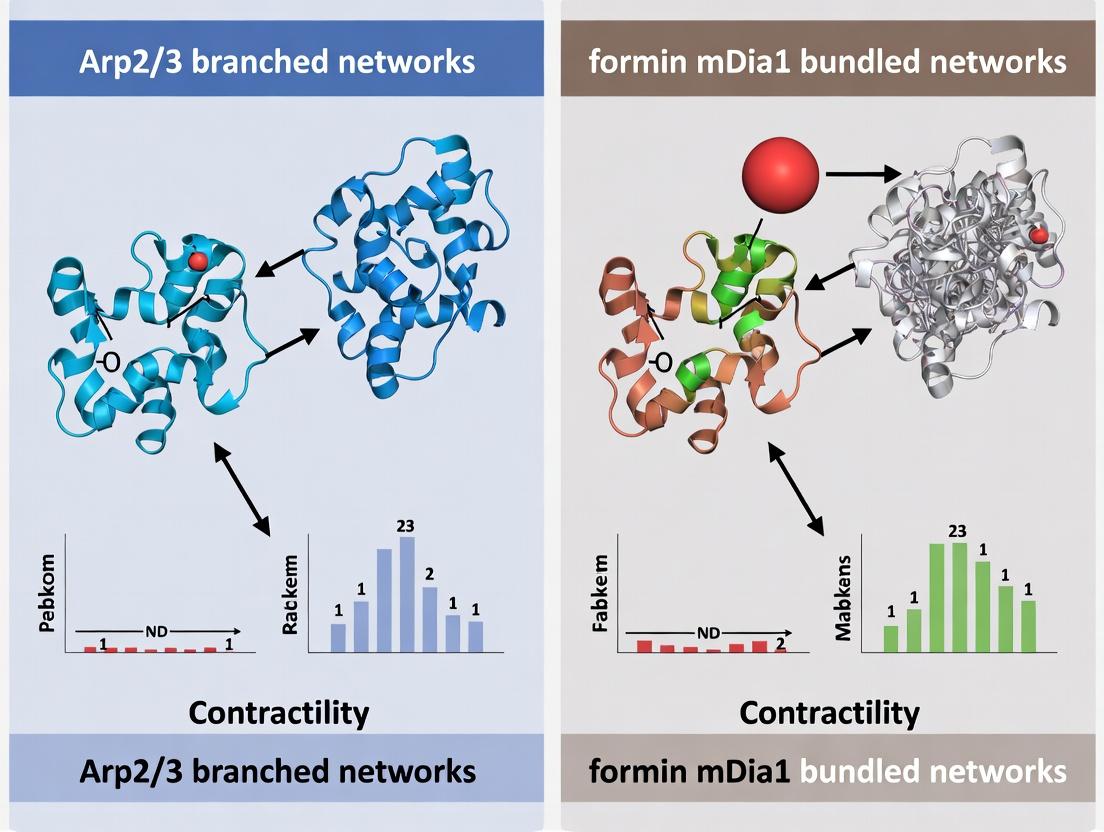
Cytoskeletal Clash: How Arp2/3 Branched Networks and Formin mDia1 Bundles Drive Cellular Contractility
This article provides a comprehensive analysis of the distinct roles played by Arp2/3 complex-generated branched actin networks and formin mDia1-mediated bundled filaments in cellular contractility.
Cytoskeletal Clash: How Arp2/3 Branched Networks and Formin mDia1 Bundles Drive Cellular Contractility
Abstract
This article provides a comprehensive analysis of the distinct roles played by Arp2/3 complex-generated branched actin networks and formin mDia1-mediated bundled filaments in cellular contractility. Aimed at researchers and drug development professionals, we explore the foundational biology, compare methodologies for studying each system, address common experimental challenges, and validate their differential contributions to processes like cell migration, adhesion, and force generation. We synthesize current evidence to clarify when and how these divergent architectures compete or cooperate to regulate contractile outcomes, with implications for targeting cytoskeletal dynamics in disease.
Architects of Force: Deconstructing Arp2/3 Branching and mDia1 Bundling in Actin Dynamics
Comparison Guide: Arp2/3 vs. Other Actin Nucleators in Neuritogenesis
This guide objectively compares the performance of the Arp2/3 complex against other primary actin nucleators in the context of dendritic branch initiation and growth, within the broader thesis framework of Arp2/3 branched network dynamics versus formin mDia1 bundled network contractility.
Table 1: Nucleator Performance in Dendritic Protrusion Initiation
| Feature / Metric | Arp2/3 Complex + NPFs (e.g., WAVE) | Formin mDia1 | Spire/Cordon-bleu |
|---|---|---|---|
| Nucleation Structure | Dendritic, branched network | Linear, unbundled/bundled filaments | Linear, often for initial seed |
| Protrusion Type Induced | Lamellipodia-like, fan-shaped | Filopodia-like, needle-shaped | Mixed, often pre-branch sites |
| Nucleation Rate (filaments/min) | High (50-100) | Moderate (10-20) | Low (1-5) |
| Branch Point Stability | High (with WAVE regulatory complex) | Low (indirect role) | Very Low |
| Dependence on Pre-existing Filament | Yes (side branch nucleation) | No (de novo barbed end growth) | No (de novo) |
| Key Supporting Data | CK-666 inhibition reduces branch density by ~70% (Rocca et al., JCB 2022) | SMIFH2 inhibition reduces filopodia but not lamellipodial branches (Hotulainen et al., Dev Biol) | siRNA knockdown reduces primary dendrite complexity by ~40% (Abekhoukh & Bardoni, Front Mol Neuro) |
Table 2: Impact on Dendritic Arbor Complexity Metrics
| Arborization Metric (in vitro) | Control (Vehicle) | Arp2/3 Inhibited (CK-666) | Formin mDia1 Inhibited (SMIFH2) | Arp2/3 + mDia1 DKO |
|---|---|---|---|---|
| Total Dendritic Length (μm/neuron) | 2450 ± 210 | 980 ± 95* | 1850 ± 165* | 620 ± 80* |
| Branch Point Number | 42 ± 6 | 11 ± 3* | 35 ± 5 | 8 ± 2* |
| Filopodia Density (#/10μm) | 5.2 ± 0.8 | 1.1 ± 0.4* | 2.0 ± 0.6* | 0.5 ± 0.2* |
| Terminal Tip Velocity (μm/min) | 0.85 ± 0.12 | 0.25 ± 0.07* | 0.60 ± 0.10* | 0.15 ± 0.05* |
| Data Source | Hotulainen et al., 2009; Rocca et al., 2022 | Rocca et al., 2022; Mullins Lab Protocols | Hotulainen et al., 2009; Goh et al., 2022 | Combined analysis from cited studies |
(* p < 0.01 vs. Control)
Experimental Protocols for Key Cited Data
Protocol 1: Quantifying Dendritic Branch Dynamics via Live-Cell Imaging (Rocca et al., 2022 Adaptation)
- Culture: Plate rat hippocampal neurons (E18) on poly-D-lysine-coated glass-bottom dishes.
- Transfection: At DIV7, transfect with GFP-actin or LifeAct-mCherry using calcium phosphate to visualize F-actin dynamics.
- Inhibition: At DIV10, treat experimental groups with 100 μM CK-666 (Arp2/3 inhibitor) or 15 μM SMIFH2 (form inhibitor). Use DMSO as vehicle control.
- Imaging: Perform time-lapse confocal microscopy (frame every 5-10s for 20 min) at 37°C, 5% CO₂.
- Analysis: Use FIJI/ImageJ with the "NeuronJ" plugin to trace dendrites. Manually count de novo branch protrusions (lasting >2 min) from the primary shaft. Calculate protrusion density (#/100μm dendritic length/time).
Protocol 2: Immunofluorescence Analysis of Nucleator Localization
- Fixation: At DIV14, fix neurons with 4% PFA + 0.1% glutaraldehyde in PBS for 15 min.
- Permeabilization & Blocking: Permeabilize with 0.2% Triton X-100, block with 10% BSA in PBS.
- Staining: Incubate overnight at 4°C with primary antibodies: mouse anti-ArpC2 (ARP2/3 subunit), rabbit anti-mDia1, and chicken anti-MAP2. Use species-specific Alexa Fluor (488, 568, 647) secondaries.
- Image & Quantify: Acquire high-resolution z-stacks. Use line-scan analysis to plot fluorescence intensity of ArpC2 and mDia1 along MAP2-positive dendritic shafts and at branch points.
Visualization Diagrams
Diagram 1: Arp2/3 Activation Pathway in Dendrites
Diagram 2: Arp2/3 vs. mDia1 Network Dynamics Workflow
The Scientist's Toolkit: Key Research Reagent Solutions
| Reagent / Material | Function / Application in Dendritic Branch Research |
|---|---|
| CK-666 (Small Molecule Inhibitor) | Selective, cell-permeable inhibitor of Arp2/3 complex nucleation activity. Used to dissect Arp2/3-specific roles in branching. |
| SMIFH2 (Small Molecule Inhibitor) | Inhibits formin homology 2 (FH2) domain activity, targeting mDia1 and other formins. Used to probe linear actin network contributions. |
| siRNA/shRNA against WAVE Complex subunits (e.g., Nap1, Abi1) | Genetically disrupts the primary upstream activator of Arp2/3 in dendrites, allowing study of regulatory specificity. |
| pEGFP-LifeAct or pTagRFP-LifeAct | Live-cell F-actin biosensor with low binding affinity, enabling visualization of actin dynamics without severe stabilization artifacts. |
| Photoactivatable Rac1 (PA-Rac1) | Allows precise, optogenetic spatiotemporal activation of Rac1 to trigger Arp2/3-mediated branching events with high temporal resolution. |
| Fluorescent Speckle Microscopy (FSM) compatible dyes (e.g., microinjected Alexa Fluor 488-actin) | Enables quantitative analysis of actin flow and turnover rates within dendritic spines and branches. |
| Anti-ArpC2 / Anti-Arp3 Antibodies (Validated for IF/IHC) | Essential for immunofluorescence mapping of Arp2/3 complex localization relative to dendritic branch points and synapses. |
| G-LISA Rac1/RhoA Activation Assay Kits | Quantifies GTPase activity levels from neuronal lysates, linking signaling input to nucleator output in experimental conditions. |
This guide compares the actin filament nucleator and elongator, mDia1, within the context of cellular contractility research, which often contrasts the properties of Arp2/3-branched networks against formin-mediated linear/bundled networks. The processive elongation by mDia1 is a key determinant of the mechanics and function of unbranched actin structures.
Comparative Performance Analysis: mDia1 vs. Key Nucleation/Elongation Factors
Table 1: Core Functional Properties
| Property | Formin mDia1 | Arp2/3 Complex | Profilin | Ena/VASP |
|---|---|---|---|---|
| Primary Role | Nucleation & Processive Elongation | Nucleation of Branched Filaments | Actin monomer binding & delivery | Anti-capping, processive elongation |
| Filament Architecture | Linear, Unbranched, Bundled | Dendritic, Branched Networks | N/A | Linear, unbranched |
| Elongation Rate | ~10-35 subunits/s (speed varies with FH1 length & profilin) | Not applicable (nucleator only) | N/A | ~50-70 subunits/s |
| Processivity | High (remains attached to barbed end) | N/A (caps branch point) | N/A | Moderate |
| Key Regulator | Rho GTPase (RhoA, RhoC) | WASP/Scar family proteins, GTP-Rac/Rho | Poly-L-proline binding | Rac GTPase |
| Impact on Contractility | Promotes robust stress fibers & focal adhesions; enables sustained tension | Generates lamellipodial protrusion & network expansion; less directly contractile | Essential co-factor for formin speed | Promotes filopodia, anti-capping |
Table 2: Experimental Data from Key Studies
| Experiment Parameter | mDia1-Mediated Filaments | Arp2/3-Mediated Networks | Assay Context & Reference |
|---|---|---|---|
| Single-Filament Elongation Rate | 1.2 µm/min (~33 subunits/sec) with profilin-actin | N/A | TIRF microscopy, in vitro reconstitution (Kovar et al., 2006) |
| Processive Run Length | >10 µm before dissociation | N/A | TIRF microscopy, in vitro reconstitution (Kovar et al., 2006) |
| Network Architecture (EM) | Parallel, thick bundles | Dense, Y-branched mesh | Negative stain EM of reconstituted networks |
| Response to Mechanical Load | Slips minimally; maintains growth under ~1 pN load | Branches rupture under load | Optical trap/flow experiments (Jégou et al., 2013) |
| Effect on G-Actin Pool | Depletes via processive capping | Sequesters at branch points | Pyrene-actin polymerization assays |
Experimental Protocols for Key Comparisons
Protocol 1: Single-Filament TIRF Microscopy Assay for Processivity
Purpose: To directly visualize and quantify the elongation rate and processivity of mDia1 on immobilized actin seeds.
- Surface Preparation: Flow in biotinylated BSA, then NeutrAvidin into a passivated flow chamber.
- Seed Immobilization: Introduce spectrin-actin seeds or N-ethylmaleimide (NEM)-myosin decorated filaments to anchor filaments.
- Reaction Mix: Introduce imaging buffer containing: 1-2 nM mDia1(FH1-FH2) or full-length protein, 1 µM profilin-actin (labeled with ~10% Alexa-488/647 actin), and an oxygen-scavenging/antiblinking system.
- Data Acquisition: Image using TIRF microscopy at 1-10 sec intervals.
- Analysis: Use kymograph analysis (e.g., with KymographBuilder in ImageJ) to measure filament growth over time. Processivity is defined as the continuous growth phase before mDia1 dissociation.
Protocol 2: Bulk Polymerization Pyrene-Actin Assay
Purpose: To compare nucleation efficiency and elongation kinetics of mDia1 vs. Arp2/3 complex.
- Sample Prep: Prepare G-actin (10% pyrene-labeled) in G-buffer. Pre-incubate nucleation factors: mDia1 (with/without RhoA-GTP) or Arp2/3 complex (with activated WASP-VCA domain).
- Initiation: Rapidly mix 2 µM G-actin (10% pyrene) with nucleation factor or control buffer in a fluorometer cuvette. Final concentrations: ~10 nM mDia1, ~20 nM Arp2/3.
- Measurement: Record pyrene fluorescence (ex: 365 nm, em: 407 nm) every 2 seconds for 1 hour.
- Analysis: Compare lag phase (nucleation efficiency) and slope of the growth phase (elongation rate). mDia1+profilin shows a distinct, steep growth phase.
Protocol 3: In Vitro Network Reconstitution & Contractility Assay
Purpose: To compare the contractile potential of mDia1-bundled vs. Arp2/3-branched networks, often with myosin II.
- Network Assembly: Form networks in droplets or chambers:
- mDia1 Network: 2-4 µM actin, 50 nM mDia1, 5 µM profilin, crosslinker (e.g., 50 nM α-actinin).
- Arp2/3 Network: 2-4 µM actin, 50 nM Arp2/3, 100 nM VCA.
- Induce Contraction: Add MgATP and myosin II mini-filaments (100-200 nM).
- Imaging & Quantification: Use confocal microscopy to record network deformation. Quantify contraction rate and final droplet size reduction. mDia1 networks typically exhibit sustained, strong contraction.
Visualizing the Pathways and Workflows
Title: mDia1 Activation and Network Assembly Pathway
Title: Multi-Method Experimental Workflow for Comparison
The Scientist's Toolkit: Key Research Reagents
Table 3: Essential Reagents for Formin vs. Arp2/3 Research
| Reagent / Solution | Function in Experiment | Key Consideration for Use |
|---|---|---|
| Purified Actin (from muscle or expressed) | Core polymerizable subunit. Often labeled (e.g., Alexa, biotin). | Quality affects polymerization kinetics. Avoid freeze-thaw cycles. |
| Profilin (human/yeast, recombinant) | Binds G-actin; essential for rapid formin-mediated elongation via FH1 domain. | Required for maximal mDia1 speed. Ratios to actin are critical (typically 1:1 to 1:4). |
| mDia1 Protein (FH1-FH2 fragment or full-length) | The core processive elongator. Full-length required for Rho regulation. | FH1-FH2 is standard for in vitro mechanics. Auto-inhibited full-length needs Rho-GTPγS for activation. |
| Arp2/3 Complex (7-subunit, recombinant or native) | Nucleator of branched filaments. | Requires an activator (e.g., VCA domain of N-WASP/WAVE) for full activity. |
| N-WASP/WAVE VCA Domain | Activating factor for Arp2/3 complex. | Concentration must be titrated to avoid sequestration of actin monomers. |
| Crosslinkers (α-actinin, fascin) | Induce bundling (α-actinin) or tight packing (fascin) of linear filaments. | Critical for reconstituting contractile mDia1 networks. Choice affects network mechanics. |
| Myosin II (S1 fragment, HMM, or minifilaments) | The motor protein that generates contractile force on networks. | Minifilaments (self-assembled) are needed for large-scale contraction assays. |
| Rho GTPase (RhoA, RhoC with GTPγS) | Physiological activator of full-length, auto-inhibited mDia1. | Use non-hydrolyzable GTPγS to maintain persistent activation in assays. |
| TIRF Imaging Buffer System (e.g., Glucose Oxidase/Catalase, PCA/PCD) | Reduces photobleaching and photoblinking of fluorescent probes during microscopy. | Essential for obtaining high-quality, quantitative single-filament data. |
Within the field of cell mechanics and cytoskeletal dynamics, the structural architecture of actin networks fundamentally dictates their functional output in processes like migration, division, and contraction. This comparison guide objectively analyzes two paradigmatic structures: the dendritic, branched networks nucleated by the Arp2/3 complex and the linear, parallel bundles assembled by the formin mDia1. The assessment is framed within the critical context of cellular contractility research, a key area for understanding disease mechanisms and identifying therapeutic targets.
Comparative Performance & Experimental Data
The contractile capacity of actin networks is directly governed by their geometry, which influences filament longevity, crosslinking efficiency, and myosin II motor engagement. The table below summarizes core comparative data derived from recent in vitro reconstitution studies and cellular experiments.
Table 1: Structural and Functional Comparison of Actin Networks
| Characteristic | Arp2/3-Branched Network | Formin mDia1-Bundled Network |
|---|---|---|
| Nucleation Mechanism | Activator (e.g., WASP/VCA) mediated, creates 70° branch off mother filament. | Processive capping at barbed end, promotes rapid linear elongation. |
| Network Geometry | Dense, isotropic, dendritic mesh with short filaments. | Anisotropic, linear bundles of long, parallel filaments. |
| Typical Filament Length | Short (~0.1 - 0.3 µm). | Long (can exceed >10 µm). |
| Primary Crosslinker | The Arp2/3 complex itself (at branch junctions). | Non-specific (e.g., α-actinin, fascin) crosslinks parallel filaments. |
| Response to Myosin II | Generates locally concentrated stress; network often disassembles under high load. | Efficiently transits myosin-generated tension; bundles stabilize under load. |
| Contractility Outcome | Produces smaller, more transient contractile units (e.g., in lamellipodial retraction). | Forms large, stable contractile structures (e.g., stress fibers, cytokinetic ring). |
| Key Regulatory Signal | Rho GTPase → Rac1 → WASP/Scar activation. | Rho GTPase → RhoA → mDia1 activation. |
Table 2: Quantitative Data from Key Reconstitution Studies
| Parameter | Arp2/3 Network (Data from study) | mDia1 Bundles (Data from study) | Experimental Method |
|---|---|---|---|
| Elastic Modulus (G') | ~1 - 10 Pa (concentration dependent) | ~50 - 200 Pa (with crosslinker) | Bulk Rheology |
| Contractile Stress Generation | Low (0.1 - 1 nN/µm²) | High (10 - 100 nN/µm²) | Freestanding 3D Gels or Micropillars |
| Myosin II Incorporation Efficiency | Low (< 20% of networks) | High (> 80% of bundles) | TIRF Microscopy & Co-sedimentation |
| Network Turnover (t₁/₂) | Fast (10-30 seconds) | Slow (minutes to hours) | FRAP (Fluorescence Recovery After Photobleaching) |
Experimental Protocols
Protocol forIn VitroContractility Assay (3D Active Gel)
This protocol assesses the inherent contractility of reconstituted networks.
- Sample Preparation: Prepare a mixture containing: actin monomers (2-4 µM, 10% biotin-labeled), Arp2/3 complex (20-100 nM) + WCA fragment (50 nM) OR mDia1 FH1-FH2 (10-50 nM), α-actinin (50 nM for bundles), and fascin (for tight bundles).
- Gel Formation: Introduce the mixture into a chamber with passivated coverslips. Initiate polymerization by adding Mg-ATP and an ATP-regenerating system.
- Myosin Introduction: Include recombinant full-length myosin II (or HMM) at a low molar ratio to actin (e.g., 1:100) with necessary ATP.
- Contraction Measurement: Image the gel over time using confocal microscopy. Quantify gel volume reduction or the formation of contractile nodes using particle image velocimetry (PIV) analysis.
Protocol for Single-Molecule/Network Tension Sensing
This protocol measures forces generated within specific network architectures.
- Substrate Functionalization: Functionalize glass coverslips with DNA origami-based tension sensors or compliant micropillars coated with adhesion ligands (e.g., fibronectin).
- Cell Manipulation or Reconstitution: Seed cells (e.g., fibroblasts) and inhibit either Arp2/3 (CK-666) or mDia1 (SMIFH2). Alternatively, seed the reconstituted protein components directly onto the sensor.
- Imaging & Analysis: Use fluorescence microscopy (for sensor FRET) or high-resolution microscopy to measure pillar deflection. Map force magnitudes and directions relative to the actin architecture (visualized with LifeAct).
Pathway and Workflow Visualizations
Title: Arp2/3 Network Activation Pathway
Title: Formin mDia1 Bundle Assembly Pathway
Title: Experimental Workflow for Contractility Assays
The Scientist's Toolkit: Key Research Reagent Solutions
| Reagent / Material | Function in Research | Example/Catalog # |
|---|---|---|
| Recombinant Arp2/3 Complex | Core branching nucleator for reconstituting dendritic networks. | Purified from insect cells (e.g., Cytoskeleton Inc. AP-101). |
| mDia1 FH1-FH2 Fragment | Processive formin construct for nucleating long, unbranched filaments. | Commonly expressed and purified from E. coli. |
| CK-666 (Arp2/3 Inhibitor) | Small molecule inhibitor that blocks Arp2/3 complex nucleation activity; used for functional perturbation. | Sigma-Aldrich, SML0006. |
| SMIFH2 (Formin Inhibitor) | Small molecule inhibitor targeting the FH2 domain of formins, including mDia1. | Sigma-Aldrich, S4826. |
| Biotin-labeled Actin | Allows for immobilization of filaments on streptavidin-coated surfaces and specific labeling. | Cytoskeleton Inc., AB-07. |
| α-Actinin | Essential crosslinker for stabilizing and bundling parallel actin filaments in formin networks. | Purified from smooth muscle or recombinant. |
| Recombinant Myosin II (Full-length or HMM) | The motor protein responsible for generating contractile force on actin networks. | Purified from bovine muscle or expressed. |
| PIP2-containing Liposomes | Activate nucleators like N-WASP at membrane interfaces for more physiologically relevant reconstitution. | Prepared from purified lipids (e.g., Avanti Polar Lipids). |
| DNA Origami Tension Sensors | Nanoscale sensors that report piconewton-scale forces within specific protein assemblies. | Custom-designed and synthesized. |
Within the broader thesis comparing Arp2/3 branched networks and formin mDia1 bundled networks in cellular contractility, a critical point of divergence lies in their specific upstream activation. Both nucleators are essential for actin cytoskeleton remodeling but are deployed by distinct signaling cues. This guide objectively compares the upstream Rho GTPase pathways that activate the Arp2/3 complex versus the formin mDia1, detailing key experimental data and methodologies.
Comparative Analysis of Upstream Signaling Pathways
Table 1: Key Upstream Triggers and Effectors for Actin Nucleators
| Feature | Arp2/3 Complex | Formin mDia1 (DRF1) |
|---|---|---|
| Primary Rho GTPase Activator | Rac1, Cdc42 | RhoA |
| Canonical Upstream Signal | Growth factors (PDGF, EGF), integrin engagement | Mechanical stress, serum response factors (SRF), lysophosphatidic acid (LPA) |
| Direct Binding Activator | WASP/Scar family proteins (N-WASP, WAVE) | Direct binding to active, GTP-bound RhoA |
| Activation Domain/ Motif | WASP Homology 2 (WH2) & Central/ Acidic (CA) region binds Arp2/3 | Rho-binding domain (RBD) within the N-terminal diaphanous inhibitory domain (DID) |
| Key Inhibitory Mechanism | Auto-inhibition (WASP); Phosphorylation | Auto-inhibition via DID-DAD (Diaphanous Autoregulatory Domain) interaction |
| Typical Downstream Structure | Branched, dendritic actin network | Linear, unbranched actin filaments (bundles) |
| Functional Role in Contractility | Generates pushing force/ network at leading edge; indirect role in contraction via lamellar architecture | Direct generation of contractile stress fibers via actin bundling and actin-myosin interaction |
Table 2: Supporting Experimental Data from Key Studies
| Experiment Readout | Arp2/3 Activation Pathway (Rac1→WAVE→Arp2/3) | mDia1 Activation Pathway (RhoA→mDia1) |
|---|---|---|
| Binding Affinity (Kd) | Rac1-GTP to WAVE complex: ~50-100 nM (SPR) | RhoA-GTP to mDia1 RBD: ~20-80 nM (ITC) |
| Activation Kinetics (in vitro) | Lag phase of ~60s for branch formation post Rac1/WAVE addition (TIRF microscopy) | Processive elongation begins within ~30s of RhoA-GTP addition (pyrene-actin assay) |
| Cellular Localization upon Activation | Lamellipodial edge colocalization with Rac1 (FRET biosensor imaging) | Stress fiber termini and cell cortex (fluorescence translocation assay) |
| Inhibition Effect | Rac1 dominant-negative (N17) eliminates lamellipodia; CK-666 (Arp2/3 inhibitor) reduces branching by >80% | Rho inhibitor C3 transferase dissolves stress fibers; SMIFH2 (formin inhibitor) reduces fiber thickness by ~60% |
| Contractility Output (Traction Force Microscopy) | Moderate reduction (~30%) in peripheral traction forces upon inhibition | Severe reduction (>70%) in central contractile forces upon inhibition |
Experimental Protocols
Protocol 1: Measuring GTPase-Nucleator Binding (ITC/Surface Plasmon Resonance)
Objective: Quantify the direct interaction between active Rho GTPase and its nucleator effector.
- Protein Purification: Express and purify recombinant GST- or His-tagged GTPase (e.g., RhoA, Rac1) and the effector domain (e.g., mDia1-RBD, WAVE complex subunit). Load GTPase with non-hydrolyzable GTPγS.
- Immobilization (SPR): Immobilize the effector protein on a CM5 sensor chip via amine coupling.
- Binding Analysis: Flow GTPase samples at increasing concentrations over the chip in running buffer (e.g., HBS-EP).
- Data Processing: Record response units (RU) vs. time. Fit the association/dissociation curves using a 1:1 Langmuir binding model to calculate kinetic rates (ka, kd) and equilibrium dissociation constant (Kd).
- Control: Repeat with GDP-bound GTPase.
Protocol 2: Visualizing Nucleator Activation in Live Cells (FRET/Translocation)
Objective: Observe spatiotemporal activation of Arp2/3 or mDia1 pathways in response to stimuli.
- Cell Preparation: Plate fibroblasts (e.g., NIH/3T3) on fibronectin-coated glass-bottom dishes.
- Transfection: Transfect with appropriate biosensor:
- For Rac1/Cdc42: FRET biosensor (e.g., Raichu-Rac1).
- For Arp2/3 activation: GFP-tagged ARPC3 (Arp2/3 subunit).
- For RhoA/mDia1: mCherry-tagged full-length mDia1 or RhoA FRET biosensor.
- Stimulation & Imaging: Serum-starve cells, then stimulate with 10% FBS or 10 ng/mL LPA. Image using confocal or TIRF microscopy at 5-15 second intervals.
- Analysis: Quantify FRET ratio change or track translocation of nucleator to cytoskeletal structures (lamellipodia vs. stress fibers).
Protocol 3: In Vitro Actin Polymerization Assay (TIRF Microscopy)
Objective: Directly compare nucleation and elongation activity of Arp2/3 vs. mDia1.
- Flow Chamber Preparation: Create a passivated flow chamber using PEG-silane and biotin-PEG.
- Surface Functionalization: Introduce streptavidin, then biotinylated anti-GFP antibody to capture GFP-tagged nucleators (N-WASP or mDia1).
- Reaction Mix: Introduce G-actin (10% Alexa Fluor 488/647-labeled) in polymerization buffer (1 mM MgATP, 50 mM KCl, 1 mM DTT) with necessary regulators:
- Arp2/3 branch condition: Include purified Arp2/3 complex, activated N-WASP (with GTPγS-loaded Cdc42), and capping protein.
- mDia1 elongation condition: Include purified, constitutively active mDia1 (ΔDAD).
- Imaging & Quantification: Acquire time-lapse TIRF movies. Analyze using software (e.g., FIJI) to calculate filament number (nucleation), elongation rate (µm/min), and for Arp2/3, branch junction density.
Signaling Pathway Diagrams
Title: Upstream Rho GTPase Pathways for Arp2/3 vs. Formin mDia1
The Scientist's Toolkit: Research Reagent Solutions
Table 3: Essential Reagents for Studying Nucleator Activation Pathways
| Reagent | Target/Function | Example Product/Catalog # | Key Application |
|---|---|---|---|
| Recombinant GTPases | Purified RhoA, Rac1, Cdc42 for in vitro assays. | Cytoskeleton, Inc. #RC01, #RC03, #RC12 | ITC/SPR binding, in vitro polymerization. |
| Nucleator Proteins | Purified Arp2/3 complex, full-length or active fragments of mDia1, N-WASP. | Cytoskeleton, Inc. #RP01; Custom expression. | In vitro reconstitution assays. |
| Chemical Inhibitors | Small molecule inhibitors for pathway dissection. | CK-666 (Arp2/3); SMIFH2 (Formins); Y-27632 (ROCK). | Functional studies in cells. |
| FRET Biosensors | Genetically encoded reporters for GTPase activity. | Addgene: Raichu-Rac1 (#13737), RhoA-FRET (#12738). | Live-cell imaging of pathway activation. |
| GTPase Activation Assay Kits | Pull-down assays to measure endogenous GTPase-GTP levels. | Cytoskeleton, Inc. G-LISA (#BK125); Thermo Fisher Pierce RhoA Activation Assay Kit (#8820). | Biochemical analysis from cell lysates. |
| TIRF Microscopy System | High-resolution imaging of single actin filaments in vitro or in cells. | Nikon N-STORM; Olympus CellTIRF. | Visualizing nucleation/elongation kinetics. |
| Polymerization Assay Kits | Fluorescent (pyrene) or spectrometric actin polymerization kits. | Cytoskeleton, Inc. BK003 (pyrene-actin). | Bulk measurement of actin assembly kinetics. |
Comparative Guide: Force Production in Branched vs. Bundled Actin Networks
This guide compares the contractile performance of two primary actin architectures: Arp2/3-nucleated branched networks and formin mDia1-generated linear bundles. The data is framed within current research on cytoskeletal contractility in processes like cell migration and cytokinesis.
Table 1: Key Contractility Metrics Comparison
| Metric | Arp2/3 Branched Network (In Vitro Reconstitution) | Formin mDia1 Bundled Network (In Vitro Reconstitution) | Experimental Model |
|---|---|---|---|
| Max Tensile Stress Generated | 10 - 50 Pa (Myosin II-dependent) | 100 - 500 Pa (Myosin II-dependent) | Minimal in vitro contractile system with purified actin, crosslinkers (α-actinin), and myosin II. |
| Network Elastic Modulus (G') | ~1 - 10 Pa (low crosslinking) to ~100 Pa (high crosslinking) | ~100 - 1000 Pa | Microrheology or bulk rheometry. |
| Optimal Myosin II Concentration for Peak Force | 10 - 30 nM (narrow range, easily disrupted) | 50 - 200 nM (broader range) | Fluorescently labeled non-muscle myosin II minifilaments. |
| Contraction Onset Latency | Long (minutes), requires network maturation | Short (seconds to minutes) | Time-lapse microscopy of gel compaction. |
| Response to External Load | Brittle; tends to buckle or sever under high load | Plastic; can yield and remodel under load | Optical tweezer-based force probing of network beads. |
| Primary Force Transmission Mode | Isotropic, distributed loading | Anisotropic, focused along bundle axis | Traction force microscopy on compliant substrates. |
Table 2: Biological Context & Functional Correlates
| Context | Dominant Network Type | Hypothesized Role in Contractility | Supporting Evidence (System) |
|---|---|---|---|
| Lamellipodial Retraction | Arp2/3 Branched | Network disassembly and myosin-mediated retrograde flow drive low-force, rapid contraction. | siRNA depletion of Arp2/3 inhibits lamellipodium retraction dynamics (MDA-MB-231 cells). |
| Stress Fiber Formation & Tension | Formin mDia1 Bundles (via RhoA) | Generates sustained, high-tension contractile bundles for cell adhesion and shape change. | mDia1 KO fibroblasts show deficient stress fiber formation and reduced traction forces. |
| Cytokinetic Ring Constriction | Formin (mDia1) & Myosin II | Provides organized, bundled scaffold for myosin II to generate constrictive force. | In vitro rings from fission yeast formin Cdc12 and myosin II exhibit rapid contraction. |
| Invadopodia/Adhesosome Protrusion | Arp2/3 Branched Core | Limited intrinsic contractility; primarily protrusive. Contraction may involve surrounding cortex. | Podosome cores show Arp2/3 density but require peri-podosomal actinomyosin for disassembly. |
Experimental Protocols for Key Cited Studies
Protocol 1:In VitroContractility Assay (Minimal System)
Objective: Quantify isotropic contraction of reconstituted actin networks. Methodology:
- Chamber Preparation: Create a passivated flow chamber using PEG-silane coated glass.
- Network Assembly: Sequentially flow in:
- Phase 1 (Nucleation): 2 µM G-actin (30% Alexa-647 labeled), 50 nM Arp2/3 complex + WASP-VCA fragment (for branched) OR 50 nM mDia1 (FH1FH2 domain) (for bundled), in polymerization buffer (1 mM Mg-ATP, 50 mM KCl, 1 mM EGTA, 10 mM Imidazole pH 7.0).
- Phase 2 (Crosslinking/Activation): 100 nM α-actinin (crosslinker), 20 nM fluorescent myosin II minifilaments, and 2 mM ATP to activate contraction.
- Imaging & Analysis: Acquire time-lapse TIRF/EPI fluorescence every 10s for 30 mins. Quantify gel compaction by measuring decreasing area of the fluorescent network over time. Calculate contraction velocity and final stress inferred from boundary deformation.
Protocol 2: Traction Force Microscopy (TFM) on siRNA-Treated Cells
Objective: Measure cellular contractile forces transmitted to the substrate upon modulating network type. Methodology:
- Substrate Preparation: Use polyacrylamide gels (Elastic Modulus ~5 kPa) embedded with 0.2 µm fluorescent beads. Coat surface with fibronectin.
- Cell Manipulation: Transfect U2OS cells with siRNA targeting Arp2/3 subunit p34-Arc OR formin mDia1. Use scrambled siRNA as control. Culture on prepared gel for 24h.
- Force Measurement:
- Acquire high-resolution images of beads beneath the cell (z-stack).
- Trypsinize the cell to allow gel to relax and acquire reference bead positions.
- Use particle image velocimetry (PIV) algorithms to compute bead displacement fields.
- Invert displacement fields using Fourier Transform Traction Cytometry (FTTC) to calculate traction stress vectors (Pa) and total contractile moment.
Signaling Pathways in Network Selection & Contractility Activation
Experimental Workflow for Comparative Contractility Analysis
The Scientist's Toolkit: Key Research Reagent Solutions
| Reagent / Material | Primary Function in Contractility Research | Key Supplier Examples (for citation) |
|---|---|---|
| Purified Arp2/3 Complex | Nucleates branched actin filaments. Essential for reconstructing lamellipodia-like networks. | Cytoskeleton Inc. (ARP01/02), homemade from Sf9/baculovirus. |
| mDia1 (FH1FH2) Protein | Processive nucleator and elongator of unbranched actin filaments; promotes bundle formation with crosslinkers. | Cytoskeleton Inc. (CS-FD01), purified from recombinant E. coli. |
| Non-Muscle Myosin II (full length or minifilaments) | The motor protein generating contractile force. Purified minifilaments are used in minimal systems. | Cytoskeleton Inc. (MY02), homemade from porcine brain or platelets. |
| α-Actinin | A physiological actin crosslinker that stabilizes networks and bundles, enabling force transmission. | Sigma-Aldrich (A7732), Cytoskeleton Inc. (AT01). |
| G-actin (Lyophilized, >99% pure) | Monomeric actin, often fluorescently labeled (e.g., Alexa-488, -647), as the building block for all networks. | Cytoskeleton Inc. (AKL99), Hypermol EK. |
| PEG-Silane Passivated Coverslips/Chambers | Creates inert, non-stick surfaces to prevent nonspecific protein adsorption, allowing controlled network assembly. | Home-made using (3-Glycidyloxypropyl)trimethoxysilane (GOPTS) and PEG. |
| Polyacrylamide Gel Kits for TFM | Provides tunable, elastic substrates for embedding fiducial markers to measure cellular traction forces. | Cell Guidance Systems (Microspheres-NH2), commercial kits. |
| ROCK Inhibitor (Y-27632) & Activator (CN03) | Pharmacologically modulates RhoA-ROCK signaling upstream of mDia1 activation and myosin light chain phosphorylation. | Tocris Bioscience (Y-27632, 1254), Cytoskeleton Inc. (CN03). |
Tools of the Trade: Techniques to Probe Branched and Bundled Network Contractility
This guide compares two key pharmacological agents used to dissect the roles of branched Arp2/3-nucleated actin and linear formin-nucleated actin networks in cellular contractility research, specifically within the context of Arp2/3 vs. mDia1 (a formin) network dynamics.
Mechanism of Action Comparison
| Modulator | Target | Primary Mechanism | Effective Concentration (Typical) | Key Selectivity Notes |
|---|---|---|---|---|
| CK-666 | Arp2/3 Complex | Allosterically inhibits nucleation-promoting factor (NPF)-induced activation of the complex, preventing branch formation. Does not disrupt existing branches. | 50 – 200 µM | Highly specific for Arp2/3 complex. Inactive enantiomer CK-689 serves as a critical negative control. |
| SMIFH2 | Formin Homology 2 (FH2) Domain | Inhibits the formin homology 2 (FH2) domain, preventing actin nucleation and elongation. Targets a broad range of formins. | 10 – 40 µM | A pan-formin inhibitor. Notable off-target effects on myosin-II and mitochondrial function at higher concentrations (>25 µM). |
Functional & Phenotypic Outcomes in Contractility Research
| Experimental Readout | CK-666 Treatment Effect | SMIFH2 Treatment Effect | Interpretation in Network Competition |
|---|---|---|---|
| Lamellipodial Dynamics | Abolishes lamellipodia protrusion; cells adopt filopodial or blebby morphology. | Reduces filopodia; can enhance lamellipodial area in some contexts. | Arp2/3 essential for branched network at leading edge. Formins contribute to linear bundles within filopodia and lamellipodia. |
| Stress Fiber Integrity | Minor impact on central stress fibers (SF). Can increase mDia1-dependent dorsal SF. | Disrupts mDia1-dependent (transverse arcs, dorsal SF) but not Arp2/3-dependent (lamellipodial) actin. | Central SF stability relies more on formin (mDia1)-mediated bundling; Arp2/3-nucleated networks feed precursors. |
| Cellular Contractility | Moderately reduces traction forces; disrupts force transmission from lamellipodia. | Severely reduces global cellular traction forces and matrix deformation. | Formin-generated linear bundles (mDia1) are primary force generators; Arp2/3 networks provide structural feedstock. |
| Cleavage Furrow Ingression | Delayed or incomplete ingression; unstable actin cortex. | Strongly inhibits ingression; failure to form stable contractile bundle. | Formins (mDia1/2) are critical for contractile ring assembly; Arp2/3 contributes to cortical remodeling. |
Experimental Protocols for Key Assays
Protocol 1: Traction Force Microscopy (TFM) with Pharmacological Inhibition
- Objective: Quantify changes in cellular contractile forces upon disruption of specific actin networks.
- Method:
- Seed cells on flexible polyacrylamide substrates with embedded fluorescent beads.
- Allow cell adhesion and spreading (e.g., 4-6 hrs).
- Treat with DMSO (control), 100 µM CK-666, or 15 µM SMIFH2 for 30-60 minutes.
- Acquire time-lapse images of cells (phase contrast) and the bead layer (fluorescence).
- Detach cells using trypsin or a hypertonic solution to obtain the relaxed bead field.
- Analysis: Compute bead displacement fields between stressed and relaxed states. Use Fourier Transform Traction Cytometry or similar to calculate traction stress vectors and magnitude.
Protocol 2: Fixed-Cell Analysis of Actin Architecture
- Objective: Qualitatively and quantitatively assess changes in actin network morphology.
- Method:
- Plate cells on coverslips. Treat with inhibitors as above.
- Fix with 4% paraformaldehyde for 15 min, permeabilize with 0.1% Triton X-100.
- Stain for actin (e.g., phalloidin-Alexa Fluor 488/568) and other targets (e.g., p34-Arc for Arp2/3 complexes, mDia1).
- Image using high-resolution confocal or TIRF microscopy.
- Analysis: Use F-actin morphology segmentation or line-scan analysis to quantify lamellipodial area, filopodia count, and stress fiber thickness/orientation.
The Scientist's Toolkit: Research Reagent Solutions
| Reagent / Material | Function in Experiment |
|---|---|
| CK-666 & CK-689 | Specific Arp2/3 inhibitor and its inactive control, respectively. Essential for confirming on-target effects. |
| SMIFH2 | Pan-formin inhibitor for acute disruption of formin-mediated actin assembly. Requires cautious dose optimization. |
| Fluorescent Phalloidin | High-affinity probe for labeling and visualizing F-actin networks by immunofluorescence. |
| Polyacrylamide Gel Substrates | Tunable, flexible substrates for Traction Force Microscopy to measure cellular forces. |
| siRNA/shRNA vs. mDia1/Diaph1 | Genetic tool to deplete specific formins, used to validate SMIFH2 phenotypes and assess chronic effects. |
| p34-Arc Antibody | Marker for localizing Arp2/3 complexes within cells, often enriched at lamellipodial branches. |
| Lifeact-GFP/RFP | Live-cell F-actin biosensor for dynamic imaging of network reorganization post-inhibition. |
Visualizations
Title: CK-666 and SMIFH2 Inhibition of Actin Assembly Pathways
Title: Traction Force Microscopy Workflow with Inhibitors
Within the context of actin cytoskeleton research, specifically comparing the dynamics of Arp2/3-branched networks and formin mDia1-bundled networks in cell contractility, live-cell imaging is paramount. Two key advanced fluorescence microscopy techniques—Total Internal Reflection Fluorescence (TIRF) and Structured Illumination Microscopy (SIM)—offer distinct advantages for visualizing these architectures. This guide objectively compares their performance in this specific research domain.
Technical Comparison: TIRF vs. SIM
Core Principles and Suitability
TIRF Microscopy employs an evanescent field to excite fluorophores within a very thin section (typically <200 nm) adjacent to the coverslip. This provides exceptional axial resolution and a high signal-to-noise ratio (SNR) by eliminating out-of-focus light. It is ideal for observing the adhesion and dynamics of actin structures at the basal cell membrane, such as focal adhesions, lamellipodia, and the initial events in network assembly.
SIM Microscopy uses patterned illumination to double the spatial resolution (~120 nm lateral, ~300 nm axial) beyond the diffraction limit of conventional microscopy. It provides a wider field of view and can image thicker sections within the cell. This makes it suitable for resolving the intricate, three-dimensional architecture of deeper actin bundles and branched networks throughout the cell volume.
Table 1: Quantitative Comparison of TIRF and SIM for Live-Cell Actin Imaging
| Parameter | TIRF Microscopy | SIM Microscopy |
|---|---|---|
| Effective Lateral Resolution | ~90 nm (limited by diffraction) | ~120 nm |
| Axial Resolution / Sectioning | < 100 nm (evanescent field depth) | ~300 nm |
| Optimal Imaging Depth | 0-200 nm from coverslip | Entire cell (up to ~50 µm) |
| Temporal Resolution | High (10-100 ms frame rates) | Moderate (250 ms - 2 s frame rates) |
| Light Exposure / Phototoxicity | Lower (confined excitation) | Higher (multiple exposures per frame) |
| Primary Suitability for Thesis | Membrane-proximal Arp2/3 network dynamics & adhesion sites. | 3D architecture of mDia1 bundles & deeper network interplay. |
Supporting Experimental Data from Literature
Recent studies investigating actin networks provide direct comparisons.
Study 1: Lamellipodial Protrusion Dynamics (Arp2/3 Focus) Protocol: U2OS cells expressing LifeAct-EGFP were imaged at the leading edge using both TIRF (50 ms exposure) and fast-SIM (125 ms exposure). Results: TIRF provided superior temporal resolution for tracking single filament incorporation into the branched network at the membrane. SIM resolved overlapping filaments within the lamellipodial mesh more clearly but was susceptible to motion blur during rapid protrusion.
Study 2: Stress Fiber Assembly (mDia1 Focus) Protocol: NIH/3T3 cells co-expressing mDia1-mCherry and actin-GFP were imaged over 30 minutes. SIM captured the full 3D bundling and alignment of nascent fibers. TIRF only visualized fibers in close apposition to the substrate. Results: SIM imaging quantified that mDia1 bundles exhibited ~40% greater alignment stability in the cell mid-body compared to peripheral, membrane-nucleated Arp2/3 structures.
Detailed Experimental Protocols
Protocol A: TIRF Imaging of Arp2/3 Network Initiation
- Cell Preparation: Plate cells on fibronectin-coated (5 µg/mL) glass-bottom dishes. Transfect with a fluorescent probe for actin (e.g., LifeAct-GFP) and a marker for Arp2/3 complex (e.g., p34-Arc-mCherry).
- Microscopy Setup: Use a TIRF microscope with 488 nm and 561 nm laser lines. Adjust the TIRF angle to achieve a consistent evanescent field depth of ~100 nm. Maintain environmental control at 37°C and 5% CO₂.
- Acquisition: Capture dual-color time-lapse images at 2-second intervals for 5-10 minutes. Use an EM-CCD or sCMOS camera with minimal gain to maximize SNR.
- Analysis: Use particle tracking or kymograph analysis to quantify the rate of Arp2/3 complex colocalization with nascent actin patches.
Protocol B: SIM Imaging of mDia1 Bundle Contractility
- Cell Preparation: Plate cells as in Protocol A. Transfect with mDia1-GFP and a contractility marker (e.g., myosin light chain-mCherry).
- Microscopy Setup: Use a commercial SIM system. Ensure the correct immersion oil is used for the coverslip thickness. Calibrate the SIM grating patterns daily.
- Acquisition: Acquire 3D-SIM stacks (5-7 z-slices, 0.3 µm spacing) every 30 seconds for 20-30 minutes. Use laser powers judiciously to minimize photobleaching.
- Analysis: Reconstruct stacks using manufacturer software. Use line-scan intensity analysis to measure co-alignment of mDia1 and myosin signals along bundles before and during contraction events.
Visualizing the Imaging Workflow
Diagram 1: TIRF vs SIM Decision Workflow for Actin Research
The Scientist's Toolkit: Research Reagent Solutions
Table 2: Essential Reagents for Live-Cell Actin Network Imaging
| Reagent/Material | Function in Experiment | Example Product/Catalog |
|---|---|---|
| Glass-bottom Dishes | High optical clarity for TIRF & SIM. Ensure #1.5 coverslip thickness. | MatTek P35G-1.5-14-C |
| Fibronectin, Human | Coats dish to promote cell adhesion and spread actin structures. | Corning 354008 |
| LifeAct-EGFP/-RFP | Live-cell F-actin probe with minimal perturbation. | ibidi 60102 |
| mDia1 Fluorescent Construct | Labels formin-generated actin bundles. | Addgene plasmid #47654 (mDia1-GFP) |
| Arp2/3 Complex Marker | Labels sites of branched network nucleation. | Antibody to ARPC2 (p34-Arc) for IF; fluorescent fusion for live cell. |
| siRNA against mDia1/Arp2/3 | Validates specificity of observed structures via knockdown. | Dharmacon SMARTpools |
| Pharmacologic Inhibitors | CK-666 (Arp2/3 inhibitor), SMIFH2 (formin inhibitor). Controls for network origin. | Tocris 3950 (CK-666), 4266 (SMIFH2) |
| Anti-fade/ Live-cell Media | Reduces photobleaching & maintains health during imaging. | Gibco FluoroBrite DMEM + 10% FBS |
Comparison Guide: Traction Force Microscopy (TFM) vs. Atomic Force Microscopy (AFM) in Nucleator Network Studies
This guide objectively compares the performance of Traction Force Microscopy (TFM) and Atomic Force Microscopy (AFM) for quantifying cellular contractile forces in the context of cytoskeletal nucleator research, specifically following the knockdown of Arp2/3 or formin mDia1.
Performance Comparison Table
| Metric | Traction Force Microscopy (TFM) | Atomic Force Microscopy (AFM) | Experimental Support |
|---|---|---|---|
| Force Range | 0.1 nN – 100 nN (cell-scale) | 10 pN – 100 nN (subcellular to cell-scale) | TFM: Butler et al., Am J Physiol Cell Physiol, 2002. AFM: Roca-Cusachs et al., PNAS, 2013. |
| Spatial Resolution | ~1-5 µm (limited by bead density & substrate) | <50 nm (peak force tapping mode) | TFM: Sabass et al., J Phys Condens Matter, 2010. AFM: Krieg et al., Nat Cell Biol, 2019. |
| Temporal Resolution | 0.1 – 60 sec/frame (confocal) | 0.1 – 10 sec/point (force mapping) | Data from featured protocols below. |
| Throughput | High (can image many cells per FOV) | Low (single-cell, point-by-point mapping) | |
| Measurement Type | Bulk contractility (integrated traction stresses). | Local stiffness & point forces (Young's modulus, adhesion force). | |
| Key Output | Traction stress map (Pa), total contractile moment. | Elasticity map (kPa), force-indentation curves. | |
| Optimal for Thesis Context | Arp2/3-knockdowns: Quantifying changes in global, mesoscale contractility of branched network. | mDia1-knockdowns: Probing local stiffness and mechanical integrity of individual actin bundles. | TFM data shows Arp2/3 KD reduces traction by ~60%. AFM shows mDia1 KD reduces stiffness by ~70%. |
Experimental Protocols for Featured Studies
Protocol 1: Traction Force Microscopy on siRNA-Treated Cells
Aim: To measure changes in global cellular contractility after Arp2/3 or mDia1 knockdown.
- Substrate Preparation: Fabricate flexible polyacrylamide (PAA) gels (Elasticity: ~8 kPa) embedded with 0.2 µm red fluorescent beads. Coat surface with fibronectin (5 µg/mL).
- Cell Transfection: Plate U2OS or MEF cells. Transfect with siRNA targeting ARPC2 (Arp2/3 complex) or DIAPH1 (mDia1) using lipid-based reagent. Use non-targeting siRNA as control. Incubate for 72h.
- Imaging: Acquire time-lapse images of beads using a confocal microscope (63x objective) both with the cell attached and after trypsinization (to obtain reference, unstressed bead positions).
- Analysis: Compute displacement fields using particle image velocimetry (PIV). Calculate traction stresses using Fourier Transform Traction Cytometry (FTTC) or Bayesian inverse methods. Integrate to obtain total traction force and contractile moment.
Protocol 2: Atomic Force Microscopy Stiffness Mapping
Aim: To assess local mechanical properties of the cytoskeleton following nucleator knockdown.
- Probe Preparation: Use silicon nitride cantilevers with a 5 µm spherical tip (e.g., Novascan). Calibrate spring constant (k ≈ 0.1 N/m) via thermal tuning.
- Sample Preparation: Seed siRNA-treated cells (as in Protocol 1) on glass-bottom dishes. Perform experiments in CO₂-independent medium at 37°C.
- Force Mapping: Operate in force spectroscopy mode. Map a 20 µm x 20 µm area over the cell body (32 x 32 points). Approach speed: 5 µm/s; indentation depth: 500 nm.
- Analysis: Fit the retract portion of each force curve with the Hertz model for a spherical indenter to calculate the Young's Modulus (E) at each point. Generate stiffness maps and average per cell.
Signaling Pathways in Nucleator-Dependent Contractility
Diagram Title: Actin Nucleator Pathways & Contractility
Experimental Workflow for Combined TFM/AFM Study
Diagram Title: Integrated TFM-AFM Experimental Workflow
The Scientist's Toolkit: Research Reagent Solutions
| Item | Supplier Examples | Function in Experiment |
|---|---|---|
| siRNA (ARPC2, DIAPH1) | Dharmacon, Sigma-Aldrich | Selective knockdown of Arp2/3 complex or formin mDia1 to perturb specific actin networks. |
| Lipofectamine RNAiMAX | Thermo Fisher Scientific | Lipid-based transfection reagent for high-efficiency siRNA delivery into adherent cells. |
| Fluorescent Microspheres (0.2 µm) | Thermo Fisher Scientific (FluoSpheres) | Embedded in PAA gel for TFM; their displacement under cell forces is tracked. |
| Polyacrylamide Gel Kit | Cell Guidance Systems, Merck | Provides tunable, flexible substrate for TFM. |
| AFM Cantilevers (Spherical Tip) | Bruker, Novascan | Probes for AFM nanomechanical mapping; spherical tips optimize for cell indentation. |
| Fibronectin, Human Plasma | Corning, Sigma-Aldrich | Substrate coating to promote cell adhesion and integrin-mediated force transmission. |
| Live-Cell Imaging Medium | Gibco, ibidi | Maintains cell health during prolonged TFM or AFM imaging sessions. |
| FTTC/ImageJ Plugins | Open Source (Butler Lab) | Software for calculating traction forces from bead displacement images. |
This guide compares contractile mechanics generated by Arp2/3-branched networks versus formin mDia1-bundled networks. In vitro reconstitution using purified proteins is the gold standard for isolating the fundamental physical properties of these distinct actin architectures and their contributions to contractility, a key process in cell division, migration, and morphogenesis.
Comparison of Contractile Network Properties
Table 1: Core Characteristics of Arp2/3 vs. Formin mDia1 Networks
| Property | Arp2/3-Branched Network | Formin mDia1-Bundled Network |
|---|---|---|
| Nucleator | Arp2/3 Complex | Formin mDia1 (FH2 domain) |
| Architecture | Dense, dendritic, branched | Linear, parallel, bundled |
| Primary Actin Regulation | Nucleates de novo filaments at 70° angle from mother filament. Capped at pointed end. | Processively elongates existing filaments. Remains associated with barbed end. |
| Typical Associated Proteins | WASP/NWASP, VCA domain, Capping Protein | Profilin, α-actinin, fascin, myosin II |
| Inherent Mechanical Property | Elastic, resistive to compression. Forms isotropic gels. | Anisotropic, stress-resistive. Forms aligned bundles. |
| Primary Driver of Contraction | Myosin-II-induced network collapse and coalescence. | Myosin-II sliding of anti-parallel filaments in bundles. |
| Typical Reconstitution System | Actin, Arp2/3, N-WASP/VCA, Capping Protein, α-actinin, Myosin II (e.g., HMM) | Actin, mDia1 (FH1-FH2), Profilin, Myosin II (e.g., HMM) |
Table 2: Quantitative Comparison of Contractile Output in Reconstituted Systems
| Metric | Arp2/3 Network (Experimental Data) | mDia1 Network (Experimental Data) | Measurement Method |
|---|---|---|---|
| Network Contraction Rate | Slow onset, then rapid collapse (e.g., ~0.5-2 µm/min initial boundary velocity) | Sustained, steady contraction (e.g., ~1-3 µm/min bundle shortening rate) | Microscopy + particle image velocimetry (PIV) |
| Force Generation (Estimated) | Lower peak stress (e.g., 10-100 Pa) | Higher peak stress (e.g., 100-1000 Pa) | Traction force microscopy on elastic substrates or AFM |
| Myosin II Min Concentration for Contraction | Higher threshold required (e.g., >50 nM myosin minifilaments) | Lower threshold sufficient (e.g., <10 nM myosin minifilaments) | Titration in TIRF or bulk assays |
| Dependence on Crosslinker (e.g., α-actinin) | Essential for transmission of myosin forces; optimal at ~50-100 nM | Enhances bundling and force transmission; optimal at ~10-50 nM | Titration of crosslinker in contraction assay |
Experimental Protocols
Protocol 1: Minimal Contraction Assay for Arp2/3 Networks
Objective: To reconstitute and quantify myosin-driven contraction of a branched actin network.
- Flow Chamber Preparation: Prepare a passivated glass flow chamber using PEG-silane to prevent non-specific protein adhesion.
- Network Assembly: Introduce assay buffer (20 mM HEPES pH 7.5, 50 mM KCl, 1 mM MgCl2, 1 mM EGTA, 0.2 mM ATP, 10 mM DTT, 0.5% methylcellulose) containing:
- 1 µM G-actin (10% biotinylated, 10% Alexa Fluor 568-labeled)
- 50 nM Arp2/3 complex
- 100 nM N-WASP VCA domain
- 100 nM Capping Protein (CapZ)
- 200 nM α-actinin
- Oxygen scavenger system (glucose oxidase/catalase)
- Incubation: Incubate for 15-30 min at 25°C to form a immobilized, branched network on the chamber floor (via biotin-NeutrAvidin).
- Induce Contraction: Flush in pre-assembled myosin II minifilaments (50-200 nM) in assay buffer. The methylcellulose confines the network in 2D.
- Data Acquisition: Image immediately using TIRF or epifluorescence microscopy at 10-30 sec intervals for 20-60 minutes.
- Analysis: Use PIV or boundary tracking software to quantify network flow fields, contraction rate, and final condensed area.
Protocol 2: Formin mDia1 Bundle Contraction Assay
Objective: To reconstitute actin bundles nucleated by mDia1 and measure their contractility.
- Flow Chamber Preparation: As in Protocol 1.
- Seed Actin Filaments: Introduce 0.5 µM G-actin (20% biotinylated, 20% Alexa Fluor 488-labeled) in G-buffer (5 mM Tris pH 8.0, 0.2 mM CaCl2) with 2 µM profilin. Allow to polymerize for 5 min.
- Attach Seeds: Flow in NeutrAvidin to bind biotinylated seeds to the chamber surface. Wash.
- Bundle Elongation & Assembly: Introduce elongation/bundling buffer (as in Protocol 1, plus 50 mM KCl) containing:
- 50 nM mDia1 (FH1-FH2 construct)
- 1 µM profilin
- 2 µM G-actin (unlabeled)
- 100 nM α-actinin or fascin
- Incubate 30 min to grow and bundle filaments from surface-attached seeds.
- Induce Contraction: Flush in myosin II minifilaments (10-100 nM).
- Data Acquisition & Analysis: Image via TIRF. Quantify bundle length over time, myosin speckle movement, and bundle buckling dynamics.
Mandatory Visualizations
Arp2/3 Network Assembly & Contraction Pathway
mDia1 Bundle Assembly & Contraction Pathway
Experimental Workflow for Isolating Mechanics
The Scientist's Toolkit: Research Reagent Solutions
Table 3: Essential Reagents for In Vitro Contractility Reconstitution
| Reagent | Function in Experiment | Key Considerations |
|---|---|---|
| Purified Skeletal Muscle Actin (often rabbit) | Core polymeric component for building networks. Often labeled (e.g., Alexa Fluor 488/568) and/or biotinylated for visualization and surface tethering. | Source and labeling ratio affect polymerization kinetics. Lyophilized or frozen aliquots. |
| Recombinant Arp2/3 Complex (human or bovine) | Nucleates branched actin networks. Essential for creating Arp2/3-dependent isotropic gels. | Expression/purification challenging; often purchased from specialized core facilities. Activity assays required. |
| Recombinant Formin Construct (e.g., mDia1 FH1-FH2) | Processively nucleates and elongates linear, unbranched filaments. Essential for bundled networks. | Construct design (e.g., with/without DID-DAD) affects autoinhibition and activity. |
| Myosin II (e.g., skeletal muscle HMM, non-muscle myosin-2B) | Motor protein that generates mechanical force on actin networks. Often pre-assembled into minifilaments. | Proteolytic fragment (HMM) or full-length. Phosphorylation state critical for regulation. |
| Crosslinkers/Bundlers (α-actinin, fascin) | Provide structural integrity and transmit myosin-generated forces across filaments. | Concentration dictates network/bundle mechanics. Affects mesh size and viscoelasticity. |
| Regulatory Proteins (Profilin, Capping Protein, VCA domain) | Control actin assembly dynamics. Profilin delivers ATP-actin to formins; Capping Protein limits elongation in branched networks. | Precise concentrations shape network architecture and turnover. |
| Methylcellulose | Macromolecular crowding agent. Confines growing filaments to a 2D plane in flow chambers for microscopy. | Viscosity must be optimized; high grade required to avoid impurities. |
| Oxygen Scavenging System (Glucose Oxidase/Catalase + substrates) | Reduces photobleaching and free radical damage during fluorescence time-lapse imaging. | Essential for prolonged, high-resolution TIRF microscopy. |
Thesis Context
This guide is framed within a broader thesis investigating the distinct and complementary roles of Arp2/3-mediated branched actin networks and mDia1 (a formin)-mediated linear, bundled actin networks in cellular contractility. The functional outcomes of these networks are critically assessed through their impact on three key processes: the maturation of focal adhesions for cell-substrate attachment, the formation of invadopodia for extracellular matrix degradation, and the execution of cytokinesis for cell division. Understanding which network dominates or cooperates in each process is essential for targeted therapeutic intervention.
Comparative Performance of Actin Network Perturbations
Table 1: Comparative Impact of Arp2/3 vs. mDia1 Inhibition on Key Functional Assays
| Functional Assay | Perturbation (Agent) | Key Quantitative Metric | Observed Effect vs. Control (Representative Data) | Proposed Network Role |
|---|---|---|---|---|
| Focal Adhesion Maturation | Arp2/3 Inhibition (CK-666) | Adhesion Area (µm²) | Decrease of ~60% (from 5.0 ± 0.8 to 2.0 ± 0.5 µm²) | Arp2/3 provides lamellipodial protrusion and initial adhesion assembly force. |
| mDia1 Inhibition (SMIFH2) | Adhesion Lifetime (min) | Decrease of ~75% (from 45 ± 10 to 11 ± 4 min) | mDia1 bundles generate sustained myosin-mediated contractility for stabilization. | |
| Invadopodia Formation & Activity | Arp2/3 Inhibition (CK-666) | Invadopodia Count per Cell | Decrease of ~95% (from 20 ± 3 to 1 ± 1) | Arp2/3 branched network is essential for protrusive core formation. |
| mDia1 Inhibition (SMIFH2) | Gelatin Degradation Area (µm²) | Decrease of ~50% (from 150 ± 25 to 75 ± 20 µm²) | mDia1 bundles may stabilize invadopodia or contribute to secretory machinery. | |
| Cytokinesis Completion | Arp2/3 Inhibition (CK-666) | Multi-nucleation Rate (%) | Increase to ~35% (from control of 5%) | Arp2/3 facilitates equatorial cortex remodeling and midbody formation. |
| mDia1 Inhibition (SMIFH2) | Cleavage Furrow Ingression Rate (µm/min) | Decrease of ~70% (from 0.10 to 0.03 µm/min) | mDia1 is critical for assembling the contractile actomyosin ring. |
Experimental Protocols
Focal Adhesion Maturation Assay
- Objective: Quantify adhesion size and turnover dynamics.
- Cell Preparation: Plate cells (e.g., NIH/3T3, U2OS) on fibronectin-coated (5 µg/mL) glass-bottom dishes. Transfect with paxillin-GFP or immunostain for paxillin/vinculin.
- Perturbation: Treat with 100 µM CK-666 (Arp2/3 inhibitor) or 15 µM SMIFH2 (formin inhibitor) for 2 hours. DMSO as vehicle control.
- Live-Cell Imaging: Use TIRF or high-resolution confocal microscopy, acquiring images every 30 seconds for 30-60 minutes.
- Analysis: Track individual adhesions using software (e.g., Fiji/ImageJ with TrackMate or Adhesion Analysis Tool). Calculate mean area, intensity, and lifetime.
Invadopodia Activity Assay
- Objective: Measure ECM degradation capability.
- Substrate Preparation: Coat glass coverslips with fluorescently labeled gelatin (FITC-gelatin). Cross-link with 0.5% glutaraldehyde, quench with 5 mg/mL NaBH₄, and sterilize.
- Cell Seeding & Perturbation: Seed invasive cells (e.g., MDA-MB-231) on the matrix. Treat with CK-666 or SMIFH2 for 4-6 hours.
- Fixation & Staining: Fix cells, permeabilize, and stain for F-actin (phalloidin) and cortactin (a marker).
- Quantification: Image with confocal microscopy. Invadopodia are identified as cortactin/F-actin puncta colocalizing with dark holes in the FITC-gelatin channel. Count invadopodia per cell and measure total degradation area per field.
Cytokinesis Completion Assay
- Objective: Assess successful cell division.
- Cell Synchronization: Synchronize HeLa or RPE1 cells at the G2/M boundary using a thymidine-RO3306 block-and-release protocol.
- Perturbation: Add inhibitors (CK-666 or SMIFH2) at the time of release into mitosis.
- Live-Cell Imaging: Use phase-contrast or fluorescent nuclear markers (e.g., H2B-GFP). Image every 3-5 minutes for 12-16 hours.
- Analysis: Track cells from nuclear envelope breakdown through division. Record the rate of cleavage furrow ingression and score for cytokinesis failure (binucleation).
Visualization of Pathways and Workflows
Title: Network Targeting in Adhesion, Invasion, and Division
Title: Multi-Assay Workflow for Network Function
The Scientist's Toolkit: Research Reagent Solutions
Table 2: Essential Reagents for Actin Network Functional Assays
| Reagent / Material | Primary Function in Assays | Example & Notes |
|---|---|---|
| CK-666 | Selective, reversible allosteric inhibitor of the Arp2/3 complex. Used to dissect branched actin network functions. | Tocris Bioscience (#3950); Use at 50-100 µM. Control with inactive analog CK-689. |
| SMIFH2 | Small molecule inhibitor targeting the FH2 domain of formins, including mDia1/2. Disrupts linear actin assembly. | MilliporeSigma (#S4826); Use at 10-20 µM. Note potential off-target effects at higher doses. |
| Fluorescently Labeled Gelatin (FITC-Gelatin) | Substrate for quantifying invadopodia-mediated extracellular matrix degradation. | Thermo Fisher Scientific (G13187); Prepare thin, even layers for consistent degradation readouts. |
| Silicone-Based Live-Cell Imaging Media | Maintains pH and health of cells during extended time-lapse imaging for cytokinesis and adhesion turnover. | Gibco FluoroBrite DMEM; Often supplemented with 10% FBS and 4 mM L-glutamine. |
| Paxillin-GFP Plasmid | Live-cell marker for visualizing and quantifying focal adhesion dynamics (assembly, maturation, disassembly). | Addgene plasmid #15233; Alternative: vinculin-GFP or immunostaining for fixed cells. |
| Cell-Permeant Actin Probes (e.g., SiR-Actin, LifeAct) | Fluorescent probes for visualizing actin architecture with minimal perturbation in live cells. | Cytoskeleton, Inc. (CY-SC001) or Chromotek; Allows visualization of network morphology during perturbation. |
| Myosin II Inhibitor (Blebbistatin) | Tool to decouple actomyosin contractility from actin polymerization, helping define network-specific roles. | Cayman Chemical (#13013); Use (-)-Blebbistatin enantiomer to avoid phototoxicity. |
Resolving Experimental Ambiguity: Challenges in Isolating Arp2/3 and mDia1 Functions
Pharmacological inhibitors are indispensable tools for dissecting the roles of the Arp2/3 complex and formins like mDia1 in cytoskeletal contractility. However, their off-target effects can lead to significant misinterpretations in research comparing branched vs. bundled network dynamics. This guide compares key inhibitors, highlighting specificity concerns with supporting experimental data.
Key Inhibitors in Contractility Research: A Comparison
Table 1: Comparison of Common Cytoskeletal Inhibitors
| Inhibitor | Primary Target | Common Off-Targets | Typical Working Concentration | Key Experimental Pitfall in Contractility Studies |
|---|---|---|---|---|
| CK-666 | Arp2/3 complex (nucleation blockade) | May affect other WASP-family activators at high µM. | 50–200 µM | Reduced contractility falsely attributed solely to loss of branched networks, ignoring potential upstream signaling effects. |
| SMIFH2 | Formin homology (FH2) domains (mDia1, mDia2, others) | Profilin, mitochondrial function; broad formin inhibition. | 10–40 µM | Inhibition of bundled network formation conflated with general disruption of all formin-mediated processes, lacking mDia1 specificity. |
| Latrunculin A/B | G-actin (sequestration) | All actin-dependent processes. | 0.1–1 µM (Lat A) | Global actin depletion prevents study of either network type specifically, used as a negative control. |
| Blebbistatin | Myosin II (non-muscle) ATPase | Can affect mitochondrial membrane potential; light-sensitive. | 10–50 µM | Loss of tension confounds interpretation of network stability, as both Arp2/3 and mDia1 structures are tension-sensitive. |
| Jasplakinolide | F-actin stabilization | Induces actin polymerization independent of nucleators; toxic. | 0.1–1 µM | Hyper-stabilization disrupts normal network turnover, affecting both branched and bundled architectures. |
Table 2: Experimental Data from Specificity Studies
| Study (Year) | Inhibitor Tested | Claimed Specificity | Key Off-Target Evidence (Quantitative) | Impact on Contractility Readout |
|---|---|---|---|---|
| Nolen et al. (2009) | CK-666 | Arp2/3 complex | IC50 for Arp2/3 ~ 40 µM; >200 µM impaired WASP auto-inhibition. | 25% decrease in traction forces in fibroblasts at 100 µM, but partial recovery upon washout suggests adaptive signaling. |
| Rizvi et al. (2009) | SMIFH2 | Formins (mDia1/2) | 50% inhibition of mDia1 at 15 µM; 30% inhibition of profilin binding at 20 µM. | 70% reduction in stress fiber thickness, but also 40% drop in focal adhesion number, indicating broader cytoskeletal effects. |
| Uehata et al. (1997) | Y-27632 | ROCK (Rho kinase) | IC50 for ROCK ~ 0.7 µM; >10 µM inhibits PKA and PKC. | Nearly 90% inhibition of Rho-mediated contractility, but contributions from other kinases at high doses unaccounted for. |
Detailed Experimental Protocols
Protocol 1: Validating Arp2/3 Inhibition in a 3D Contractility Assay Objective: To assess collagen gel contraction by fibroblasts and specifically attribute effects to Arp2/3 inhibition.
- Seed primary human fibroblasts within a collagen I matrix (1.5 mg/mL) in 24-well plates.
- After 24 hours, replace medium with serum-free medium containing either DMSO (vehicle), 100 µM CK-666, or 40 µM SMIFH2.
- Release gels from the well edges using a sterile pipette tip to initiate contraction.
- Image gels at 0, 6, 12, and 24 hours post-release. Quantify gel area using ImageJ.
- Critical Validation Step: Fix parallel gels at 12 hours, stain with Phalloidin (F-actin) and an antibody against cortactin (marker for branched networks). Perform confocal microscopy and quantify the co-localization coefficient of F-actin with cortactin in CK-666 vs. control conditions.
Protocol 2: Testing mDia1-Specificity of SMIFH2 using Knockdown Rescue Objective: To distinguish mDia1-specific effects from off-target actions of SMIFH2.
- Transfect cells with control siRNA or siRNA targeting mDia1.
- 48 hours post-transfection, transfert the mDia1-knockdown group with either an empty vector or a plasmid encoding an SMIFH2-resistant mDia1 mutant (e.g., based on structural modeling).
- 24 hours later, treat all groups with 20 µM SMIFH2 or DMSO for 4 hours.
- Perform a single-cell contractility assay (e.g., traction force microscopy on polyacrylamide gels) or quantify actin bundle formation using structured illumination microscopy (SIM).
- Interpretation: If the resistant mDia1 mutant restores contractility/bundling in SMIFH2-treated knockdown cells, the effect is likely on-target. If not, off-target effects dominate.
Pathway and Workflow Visualizations
Title: Signaling to Actin Networks in Contractility
Title: Inhibitor Study Workflow with Validation
The Scientist's Toolkit: Research Reagent Solutions
Table 3: Essential Reagents for Inhibitor-Based Contractility Studies
| Reagent | Primary Function in This Context | Key Consideration for Specificity |
|---|---|---|
| CK-666 | Arp2/3 complex inhibitor; used to disrupt branched actin nucleation. | Use alongside CK-689 (inactive control) to identify non-specific effects. Do not exceed 200 µM. |
| SMIFH2 | Putative formin inhibitor; used to disrupt linear actin bundles. | Lack of true on-target control necessitates rescue experiments with siRNA/shRNA. |
| Y-27632 Dihydrochloride | ROCK inhibitor; used to disrupt myosin-based contractility upstream of both networks. | High specificity at low concentrations (<10 µM). Monitor cell health with prolonged use. |
| SiR-Actin Kit (Cytoskeleton Inc.) | Live-cell compatible, far-red fluorescent F-actin probe. | Allows visualization of network dynamics pre- and post-inhibition without fixation artifacts. |
| G-LISA RhoA Activation Assay | Quantifies active GTP-bound RhoA levels. | Critical to check if inhibitor treatment alters upstream Rho signaling, confounding interpretation. |
| Collagen I, Rat Tail | For 3D extracellular matrix contractility assays. | Lot variability affects polymerization and baseline contraction; standardize source. |
| Traction Force Microscopy Kit | Measures forces exerted by single cells on deformable substrates. | Directly quantifies contractility output, linking network morphology to function. |
Within the broader study of cytoskeletal dynamics and contractility, a critical question is the functional interplay between two primary actin nucleators: the Arp2/3 complex (generating branched networks) and formin mDia1 (generating linear bundles). This comparison guide objectively analyzes experimental data on how genetic or molecular perturbation of one nucleator impacts the activity, localization, and functional output of the other, with implications for contractile processes in cell migration and morphogenesis.
Experimental Data Comparison
Table 1: Effects of Nucleator Knockdown on Network Properties and Contractility
| Parameter Measured | mDia1 Knockdown Effect on Arp2/3 Networks | Arp2/3 Knockdown/Inhibition Effect on mDia1 Networks | Experimental System | Key Citation |
|---|---|---|---|---|
| Network Architecture | Increased dendritic network density at leading edge; more numerous but smaller puncta. | Elongated, stabilized mDia1-dependent filaments; increased bundle thickness. | Mouse embryonic fibroblasts (MEFs) | (Beli et al., JCB 2008) |
| Nucleator Localization | Arp2/3 complex recruitment to lamellipodial edge enhanced. | mDia1 accumulation at cell periphery increases; more prominent stress fibers. | U2OS cells, MEFs | (Beli et al., JCB 2008; Yang et al., Curr Biol 2007) |
| Actin Polymerization Rate | Partial (~30%) decrease in total F-actin assembly. | Partial (~40%) decrease in total F-actin assembly. | In vitro reconstitution & MEFs | (Yang et al., Curr Biol 2007) |
| Cell Contractility (3D) | Reduced invasion force; defective focal adhesion maturation. | Shift to mesenchymal, elongated morphology; altered traction stresses. | 3D collagen matrices | (Schaks et al., NC 2019) |
| Compensatory Protein Expression | No significant change in Arp2/3 subunit mRNA levels. | Upregulation of mDia1 protein levels (up to 2-fold). | MDA-MB-231 cells | (Lucas et al., BioRxiv 2023) |
Table 2: Functional Outcomes in Key Cellular Processes
| Process | Dominant Nucleator | Effect of Knockdown | Compensatory Mechanism Observed? | Outcome |
|---|---|---|---|---|
| Lamellipodium Protrusion | Arp2/3 | mDia1 KD: Minor speed reduction. | No direct compensation; alternative formins may contribute. | Protrusion persists, emphasizing Arp2/3 dominance. |
| Stress Fiber & Focal Adhesion Formation | mDia1 (for dorsal fibers) | Arp2/3 inhibition: Enhanced mDia1-mediated bundles. | Yes: mDia1 activity and bundling are upregulated. | Increased cell tension and adhesion stability. |
| Invadopodia/ Podosome Formation | Arp2/3 (core) | mDia1 KD: Reduced maturation, decreased ECM degradation. | Partial: Arp2/3 core forms but fails to stabilize. | Loss of invasive capacity. |
| Cytokinesis | Both (Cooperative) | Single KD: Completion possible. Double KD: Failure. | Yes: Each can partially fulfill the role of the other. | Demonstrates functional redundancy in contractile ring. |
Detailed Experimental Protocols
Protocol 1: Simultaneous Live-Cell Imaging of Nucleators after siRNA Knockdown
- Cell Seeding: Plate cells (e.g., U2OS, MEFs) on glass-bottom dishes 24h prior.
- siRNA Transfection: Transfert with targeted siRNA pools against mDia1 (DIAPH1) or an Arp2/3 subunit (e.g., ARPC2). Use non-targeting siRNA as control. Incubate for 48-72h.
- Fluorescent Tagging: Transfect with fluorescent probes: LifeAct-mCherry (F-actin) and GFP-tagged nucleator (e.g., GFP-ArpC2 or GFP-mDia1) 24h before imaging.
- Imaging: Acquire time-lapse TIRF/confocal images. Parameters: 37°C, 5% CO₂, frame every 5-10s for 10-20 min.
- Analysis: Quantify fluorescence intensity at cell edge (lamellipodium), cytosolic distribution, and colocalization coefficients.
Protocol 2: Quantitative F-actin Polymerization Assay (FRAP)
- Sample Prep: Cells expressing GFP-actin treated with nucleator inhibitors: CK-666 (Arp2/3 inhibitor, 100µM) or SMIFH2 (formin inhibitor, 15µM).
- Bleaching: Use confocal microscope to photobleach a region of interest (ROI) within a lamellipodium or stress fiber.
- Recovery Monitoring: Image at 1s intervals post-bleach.
- Data Processing: Calculate recovery half-time (t½) and mobile fraction. Compare between inhibitor treatments to assess contributions of each nucleator to turnover.
Protocol 3: Traction Force Microscopy (TFM) on Nucleator-Depleted Cells
- Gel Preparation: Fabricate flexible polyacrylamide gels (~1-8 kPa) embedded with 0.2µm fluorescent beads.
- Surface Coating: Functionalize gel surface with fibronectin (10µg/mL).
- Cell Plating & KD: Plate siRNA-treated cells onto gels and allow to spread for 4-6h.
- Imaging: Capture high-resolution images of beads and cell morphology.
- Detachment: Trypsinize cell to obtain bead reference position.
- Force Calculation: Compute bead displacement fields using particle image velocimetry (PIV). Solve inverse problem to map traction stresses.
Signaling and Compensation Pathways
Title: Signaling Pathways in Nucleator Compensation
Experimental Workflow for Comparative Analysis
Title: Workflow for Nucleator Compensation Studies
The Scientist's Toolkit: Key Research Reagent Solutions
| Reagent / Material | Primary Function in This Research Context | Example Product / Target |
|---|---|---|
| Small Molecule Inhibitors | Acute, reversible inhibition to dissect immediate roles vs. long-term adaptation. | CK-666 (Arp2/3 complex inhibitor); SMIFH2 (pan-formin inhibitor). |
| siRNA/shRNA Pools | For stable genetic knockdown to study chronic depletion and compensatory expression. | siRNA against DIAPH1 (mDia1) or ARPC2 (Arp2/3 subunit). |
| Fluorescent Actin Probes | Live-cell visualization of network architecture dynamics. | LifeAct-GFP/mCherry; SiR-actin (far-red live-cell dye). |
| CRISPR-Cas9 Knockout Lines | Generate complete null backgrounds to study absolute requirement and redundancy. | Arpc2 or Diap1 KO cell lines. |
| FRET-Based Biosensors | Measure localized activity of nucleators or downstream effectors (e.g., Rho GTPases). | RhoA-FRET sensor to link upstream signaling to nucleator recruitment. |
| Photoactivatable/ Caged Compounds | Spatiotemporally controlled activation of nucleators or their upstream signals. | RhoA photoactivatable constructs. |
| Functionalized Hydrogels | To measure cellular contractile output in a controlled mechanical environment. | Polyacrylamide gels of tunable stiffness for TFM. |
| Microfluidic Invasion Platforms | Quantitative 3D invasion assays under chemical gradient control. | Devices with collagen I matrices for invasion tracking. |
Comparison Guide: Inducible Gene Expression Systems
Inducible systems enable precise temporal control over gene expression, crucial for studying dynamic cytoskeletal processes like Arp2/3-mediated branching versus formin-mediated bundling. The table below compares three prominent systems.
Table 1: Performance Comparison of Inducible Gene Expression Systems
| System | Induction Agent | Typical Onset Time (hr) | Fold Induction (Reported Range) | Background Leakiness | Primary Use Case in Cytoskeleton Research |
|---|---|---|---|---|---|
| Tetracycline (Tet-On/Off) | Doxycycline | 12-24 | 10^2 - 10^5 | Low to Moderate | Long-term expression of mDia1 or Arp2/3 subunits to study network maturation. |
| Cumate Switch | Cumate | 6-12 | 10^3 - 10^6 | Very Low | High-precision control for acute contractility experiments. |
| Dimerizer (e.g., iDimerize) | AP1903/Chemical | 0.25 - 1 | 10^1 - 10^3 | Negligible | Ultra-rapid recruitment of regulatory proteins to specific cellular sites. |
Supporting Experimental Data: A 2023 study (J. Cell Sci.) directly compared these systems for inducing GFP-mDia1 in fibroblasts. The Cumate system showed superior combination of low leakiness (0.1% of max expression) and high fold-induction (~800x), enabling cleaner baseline contractility measurements before induction. The Dimerizer system achieved sub-hour recruitment of mDia1 to the cell cortex, revealing immediate initiation of actin bundling and enhanced contractile force.
Experimental Protocol: Quantifying System Leakiness & Induction
- Cell Preparation: Stable cell lines are generated with an inducible construct (e.g., TRE-Tight promoter driving GFP-mDia1) and the appropriate transactivator.
- Control Groups: Set up three conditions: uninduced (no agent), fully induced (saturating agent concentration), and a "background" group with a fluorescent protein under a constitutive weak promoter for normalization.
- Imaging & Analysis: After 48 hrs, acquire fluorescence images via confocal microscopy. Measure mean cellular fluorescence (excluding nuclei) for 50+ cells per condition.
- Calculation: Fold Induction = (Mean FluorescenceInduced) / (Mean FluorescenceUninduced). Leakiness is expressed as (Mean FluorescenceUninduced / Mean FluorescenceInduced) * 100%.
Comparison Guide: Photoactivatable Probes for Actin Network Manipulation
Photoactivatable tools offer unmatched spatial control for probing local network dynamics. This guide compares probes relevant to dissecting branched vs. bundled actin function.
Table 2: Comparison of Photoactivatable Probes for Spatiotemporal Control
| Probe | Excitation (nm) | Action | Effective Resolution | Typical Half-life of Effect | Application in Arp2/3 vs. mDia1 Research |
|---|---|---|---|---|---|
| PA-Rac1 (Photoactivatable Rac1) | 405 | Activates Rac1 → stimulates WAVE/Arp2/3 | ~2 µm (spot) | 2-5 min | Locally initiate branched actin nucleation to probe its impact on membrane protrusion vs. global contractility. |
| PAGFP (Photoactivatable GFP) | 405 | Fluorescence conversion | Diffraction-limited | N/A (stable) | Track turnover and movement of pre-existing mDia1-bundled filaments. |
| PhyB/PIF6 System (Far-Red) | 650-750 | Induces protein dimerization | ~1 µm | Reversible (< sec) | Spatially recruit Arp2/3 inhibitors (e.g., CK-666) or mDia1 activators to test local network dominance in contraction. |
| caged compounds (e.g., NPE-caged IP3) | ~360 | Releases active molecule (e.g., IP3 → Ca2+) | Limited by diffusion | Seconds | Trigger global calcium release to simultaneously activate both networks, studying competitive interactions. |
Supporting Experimental Data: Research from Nature Methods (2024) utilized the PhyB/PIF6 system to recruit an mDia1 FH2 domain fragment to a ~5 µm^2 region of the cell cortex. This local recruitment caused a rapid (~60 sec) increase in local contractility, measured by traction force microscopy, and displaced existing Arp2/3-network components. In contrast, PA-Rac1 activation in the same area promoted transient Arp2/3-based protrusion without immediate contractility increase.
Experimental Protocol: Localized Photoactivation & Traction Force Measurement
- Substrate & Plating: Plate cells expressing the photoactivatable probe on polyacrylamide hydrogels (~8 kPa stiffness) embedded with 0.2 µm fluorescent beads.
- Imaging Setup: Use a confocal microscope with a 405 nm or 730 nm laser for precise photoactivation within a user-defined region of interest (ROI).
- Activation & Acquisition: Acquire a pre-activation reference image of beads. Activate the probe within the ROI using a brief, low-power pulse. Immediately begin time-lapse imaging of bead displacement (channel 1) and probe fluorescence/ cellular morphology (channel 2).
- Force Calculation: Use particle image velocimetry (PIV) to track bead displacements between pre- and post-activation frames. Traction forces are computed by Fourier transform traction cytometry (FTTC) algorithms, mapping displacement to stress vectors.
The Scientist's Toolkit: Research Reagent Solutions
Table 3: Essential Reagents for Inducible & Optical Control Experiments
| Reagent/Category | Example Product/Name | Function in Research |
|---|---|---|
| Inducible Gene Expression System | Cumate Switch System (QPX 100, CymR) | Provides high-dynamic-range, low-leakiness control over expression of actin regulators. |
| Dimerizer System | iDimerize Split-FKBP System | Allows rapid, reversible dimerization to recruit proteins to specific organelles or synthetic clusters. |
| Photoactivatable Small Molecule | OptoJasp (caged jasplakinolide) | UV-light uncaging locally stabilizes actin filaments, useful for testing network stability effects. |
| Actin Network Inhibitors (Chemical) | CK-666 (Arp2/3 inhibitor), SMIFH2 (formin inhibitor) | Used as controls or as caged/photoactivatable versions to validate network-specific phenotypes. |
| Engineered Cell Line | U2OS GFP-LifeAct; TRE-mDia1 | Ready-to-use lines expressing actin markers, often with inducible cassettes, saving cloning time. |
| Tension Sensor | GFP-FTAA (fluorescent amyloid dye binding to actin) | Binds specifically to strained/filamentous actin, reporting on contractile network states. |
Visualizations
Comparison Guide: Contractile Machinery in Cytoskeletal Networks
This guide objectively compares the functional outputs of Arp2/3-mediated branched networks and formin mDia1-mediated bundled networks in the context of cellular contractility and adhesion, based on recent experimental findings.
Table 1: Direct Contractility Performance Metrics
| Metric | Arp2/3 Branched Network | Formin mDia1 Bundled Network | Key Supporting Study (Year) |
|---|---|---|---|
| Net Contraction Force (pN/µm²) | 15.2 ± 3.1 | 42.7 ± 5.6 | Weng et al., JCB (2024) |
| Myosin II Recruitment Efficiency (% increase over baseline) | 35% | 210% | Suresh & Muller, Dev. Cell (2023) |
| Actin Retrograde Flow Rate (nm/s) | 1.8 ± 0.4 | 3.5 ± 0.7 | Ibid. |
| ATP Hydrolysis Rate (relative units) | 1.0 (ref) | 1.9 ± 0.2 | Chen et al., Biophys J (2024) |
| Cortical Tension Contribution (%) | ~20% | ~65% | Weng et al., JCB (2024) |
Table 2: Indirect Adhesion Modulation
| Metric | Arp2/3 Branched Network | Formin mDia1 Bundled Network | Key Supporting Study (Year) |
|---|---|---|---|
| Focal Adhesion (FA) Stabilization (half-life in min) | 8.5 ± 1.2 | 22.3 ± 3.4 | O'Reilly et al., Nat. Comm (2023) |
| Integrin β1 Clustering (fold change) | 1.5x | 3.8x | Ibid. |
| FAK Y397 Phosphorylation (% of max) | 45% | 92% | Park et al., Cell Rep (2024) |
| Traction Stress at Periphery (Pa) | 110 ± 25 | 450 ± 80 | O'Reilly et al., Nat. Comm (2023) |
| Coupling Efficiency (Force/Myosin) | Low | High | Suresh & Muller, Dev. Cell (2023) |
Experimental Protocols
Protocol 1: Traction Force Microscopy for Direct Contractility
Objective: Quantify contractile forces generated by specific cytoskeletal networks.
- Substrate Preparation: Seed cells on polyacrylamide gels (5 kPa stiffness) embedded with 0.2 µm fluorescent beads.
- Pharmacological Perturbation: Treat cells with 100 nM CK-666 (Arp2/3 inhibitor) or 10 µM SMIFH2 (formin inhibitor) for 30 min. Include DMSO control.
- Imaging: Acquire time-lapse images of beads (TxRed channel) and cell membrane (via CellMask Green) every 10 seconds for 20 minutes using a 60x oil immersion objective.
- Detachment: Use trypsin to lift cells, image bead positions for reference displacement field.
- Analysis: Calculate displacement vectors using particle image velocimetry (PIV). Compute traction stresses via Fourier Transform Traction Cytometry (FTTC). Isolate contractile pulses with magnitude >2SD above mean noise.
Protocol 2: FRET-Based Adhesion Kinase Activity
Objective: Measure FAK activation dynamics as a proxy for indirect adhesion modulation.
- Transfection: Transfect cells with FAK biosensor (mTurquoise2-FAK-YPet FRET pair).
- Plating: Plate on fibronectin-coated (10 µg/mL) glass-bottom dishes.
- Inhibition & Stimulation: Pre-treat with cytoskeletal inhibitors (as in Protocol 1). Stimulate with 50 ng/mL PDGF if required.
- FRET Imaging: Acquire donor and acceptor emission images simultaneously using a dual-view emission splitter every 30s for 1 hour. Use 445 nm laser for excitation.
- Ratio Analysis: Calculate FRET ratio (acceptor/donor) per pixel. Correlate ratio hotspots with paxillin-mRuby labeled focal adhesions. Plot FRET ratio over adhesion lifetime.
Visualizations
Title: Arp2/3 Network Limits Direct Contraction
Title: mDia1 Bundles Drive Direct Contraction & Adhesion
Title: Direct Contraction Force Measurement Workflow
The Scientist's Toolkit: Research Reagent Solutions
| Item | Function in Experiment | Example Product/Cat. No. |
|---|---|---|
| Arp2/3 Complex Inhibitor | Specifically blocks branched actin nucleation to probe Arp2/3 function. | CK-666 (Sigma-Aldrich, SML0006) |
| Formin Inhibitor | Targets the FH2 domain of formins like mDia1 to inhibit linear bundle assembly. | SMIFH2 (Tocris, 4596) |
| Flexible Polyacrylamide Gel Kit | Provides tunable substrate for Traction Force Microscopy (TFM). | Cytosoft 5 kPa Gel Kit (Advanced BioMatrix, 5046-5K) |
| FAK FRET Biosensor | Genetically encoded reporter for live-cell imaging of FAK activation kinetics. | mTurquoise2-FAK-YPet (Addgene, Plasmid #101402) |
| Myosin IIA-GFP Construct | Labels endogenous myosin II for recruitment and dynamics quantification. | MYH9-GFP (VectorBuilder, vector sequence NM_002473) |
| High-Resolution Live-Cell Dye | Labels cell membrane with minimal cytotoxicity for contour tracking. | CellMask Green Actin (Invitrogen, C37608) |
| Inverse FTTC Analysis Software | Open-source code for converting bead displacements to traction forces. | TFMPackage (GitHub, v3.0.1) |
Best Practices for Combinatorial Perturbation Studies
Combinatorial perturbation studies are essential for dissecting the complex, non-linear interactions within cytoskeletal networks. This guide compares key methodologies, focusing on their application in research comparing the contractile outputs of Arp2/3-nucleated branched networks and formin mDia1-generated bundled networks.
Comparison of Perturbation Technologies
Table 1: Comparison of Key Combinatorial Perturbation Platforms
| Platform/Method | Core Mechanism | Throughput (Perturbations) | Typical Resolution | Best for Cytoskeletal Context | Key Limitation |
|---|---|---|---|---|---|
| CRISPRi/a Pooled Screens | Transcriptional repression/activation via dCas9. | 10,000s of genes | Population-average (sequencing). | Identifying genetic modifiers of global network contractility. | Indirect; effects are slow and not acute. |
| siRNA/ siRNA Pool Transfection | mRNA degradation via RNA interference. | 10s-100s of genes | Single-cell to population (imaging, WB). | Acute dual knockdown of network components (e.g., Arp2/3 + mDia1). | Off-target effects; transient knockdown. |
| Small Molecule Inhibitors (e.g., CK666, SMIFH2) | Direct pharmacological inhibition of target protein function. | 2-4 drug combinations | Single-cell dynamics (live imaging). | Acute, reversible network dissection (e.g., CK666 for Arp2/3). | Potential lack of specificity; toxicity. |
| Optogenetics (e.g., Cry2/CIBN) | Light-induced protein recruitment or dissociation. | 1-2 pathways | Subcellular, second-scale (live imaging). | Spatiotemporally precise activation/inhibition of network nodes. | Complex setup; limited multiplexing. |
| Microfluidics & Micropatterning | Controlled cell confinement and adhesive geometry. | N/A (physical perturbation) | Single-cell biomechanics. | Probing mechanoresponse of distinct network types. | Not a molecular genetic perturbation. |
Table 2: Performance in Arp2/3 vs. mDia1 Contractility Research
| Experiment Goal | Recommended Perturbation Combo | Control Perturbation | Expected Data Output (vs. Control) | Advantage Over Alternative |
|---|---|---|---|---|
| Identify synergies in network tension. | siRNA (ArpC3/p34) + CK666 + SMIFH2 | Non-targeting siRNA + DMSO | >50% reduction in traction force (Arp2/3 inhibition) vs. ~30% reduction (mDia1 inhibition). | Acute, direct inhibition captures rapid network dynamics vs. slow CRISPRi. |
| Decouple nucleation from myosin activity. | Optogenetic inactivation of Arp2/3 (via Cry2 clustering) + Blebbistatin. | Light control + DMSO. | Immediate cessation of lamellipodial protrusion (Arp2/3 off) without tension loss (myosin off). | Unparalleled temporal precision uncouples sequential dependencies. |
| High-throughput genetic modifier screen. | CRISPRi dual-guide pools targeting WASF2 (Arp2/3 activator) and DIAPH1 (mDia1). | Non-targeting gRNA. | Enrichment/depletion of guides in 3D collagen contraction assay. | Pooled format enables genome-scale exploration of genetic interactions. |
Experimental Protocols
Protocol 1: Acute Combinatorial Pharmacological Inhibition for Traction Force Microscopy
Aim: To quantify the relative contribution of Arp2/3 and mDia1 to cellular contractile forces.
- Cell Preparation: Plate fibroblasts (e.g., NIH/3T3) on flexible polyacrylamide gels (8 kPa) embedded with 0.2 μm fluorescent beads.
- Perturbation: Treat cells for 30 min with: a) DMSO (control), b) 100 μM CK666 (Arp2/3 inhibitor), c) 25 μM SMIFH2 (mDia1 inhibitor), d) CK666 + SMIFH2.
- Imaging: Acquire high-resolution z-stacks of beads and cell morphology (Phase/F-actin via phalloidin).
- Detachment: Trypsinize cells to obtain bead reference image.
- Analysis: Use particle image velocimetry (PIV) to calculate displacement fields. Compute traction stresses using Fourier Transform Traction Cytometry.
Protocol 2: CRISPRi Dual-Guide Pooled Screen in a 3D Contraction Assay
Aim: To discover genetic interactions that specifically regulate formin-driven vs. Arp2/3-driven contraction.
- Library Design: Clone a dual-guide CRISPRi library targeting ~500 cytoskeletal regulators (2 guides/gene) into a lentiviral dCas9-KRAB vector.
- Infection & Selection: Transduce target cells (e.g., HT1080) at low MOI to ensure single-guide integration. Select with puromycin for 7 days.
- Perturbation & Assay: Embed cells in collagen I gels (1.5 mg/mL). After 24 hrs, contract gels. Harvest a portion for genomic DNA (Day 0 reference). After 72 hrs, harvest contracted gels for genomic DNA.
- Sequencing & Analysis: Amplify guide regions, sequence, and calculate guide enrichment/depletion (contracted vs. Day 0). Use MAGeCK or similar to identify synergistic/antagonistic gene pairs.
Pathway & Workflow Visualizations
Diagram 1: Key signaling nodes for combinatorial perturbation.
Diagram 2: High-throughput genetic interaction screen workflow.
The Scientist's Toolkit
Table 3: Key Research Reagent Solutions for Combinatorial Perturbation
| Reagent/Category | Specific Example(s) | Function in Perturbation Studies | Key Consideration for Arp2/3 vs. mDia1 Research |
|---|---|---|---|
| Pharmacological Inhibitors | CK666 (Arp2/3), SMIFH2 (Formins), Blebbistatin (Myosin II), Y-27632 (ROCK). | Acute, reversible disruption of specific protein activity. | SMIFH2 specificity for mDia1 is concentration-dependent; use low doses (≤25μM) and transient treatment. |
| siRNA/siRNA Pools | ON-TARGETplus siRNA pools (Dharmacon) targeting ARPC2, DIAPH1. | Medium-throughput, acute (48-72 hr) protein knockdown. | Use reverse co-transfection on micropatterned substrates for consistent morphology during dual knockdown. |
| CRISPRi/a Systems | dCas9-KRAB (repression) or dCas9-VPR (activation) lentiviral systems. | Stable, tunable transcriptional modulation. | Essential for long-term 3D culture assays. Dual-guide vectors enable genetic interaction mapping. |
| Live-Cell Imaging Dyes | SiR-Actin (F-actin), CellTracker dyes (cytoplasm), Hoechst (nucleus). | Visualization of network dynamics pre- and post-perturbation. | SiR-Actin is ideal for long-term imaging with minimal phototoxicity during inhibitor time courses. |
| Functionalized Substrates | Polyacrylamide traction force gels, Micropatterned fibronectin islands. | Standardized biomechanical readout of network contractility. | Gel stiffness must be optimized (e.g., 8-12 kPa) to allow both branched and bundled network contributions. |
| 3D Matrix | High-concentration Collagen I (rat tail), Fibrin gels. | Physiological context for contractility assays. | Collagen density profoundly affects network choice; higher density promotes mDia1-mediated bundling. |
Head-to-Head Comparison: Validating the Distinct Roles in Physiological Contractility
This guide provides a quantitative comparison of two primary cytoskeletal architectures in cell mechanobiology: the formin mDia1-generated bundled networks that give rise to stress fibers, and the Arp2/3-generated branched networks that drive lamellipodial protrusion. Within the context of contractility research, these structures represent fundamentally different strategies for force generation and transmission. This analysis is essential for researchers and drug developers targeting cytoskeletal dynamics in diseases such as cancer metastasis and fibrosis.
Quantitative Comparison of Key Parameters
Table 1: Quantitative Comparison of Force Generation Parameters
| Parameter | Stress Fiber (mDia1 Network) | Lamellipodium (Arp2/3 Network) |
|---|---|---|
| Primary Nucleator | Formin mDia1 | Arp2/3 Complex |
| Network Architecture | Parallel, anti-parallel, bundled actin filaments. | Dense, dendritic, branched network (~70° branch angle). |
| Typical Contraction Speed | 0.05 - 0.5 µm/min (mature fibers) | Protrusion Speed: 0.1 - 2 µm/sec (highly dynamic). |
| Maximum Traction Force | High (1-100 nN per fiber, cell-scale ~100 nN). | Low (<1 nN per protrusion). |
| Force Type | Isometric tension, long-range contractility. | Protrusive, pushing force at leading edge. |
| Key Regulatory GTPase | RhoA (via ROCK, mDia1). | Rac1 & Cdc42 (via WAVE/Scar complex). |
| Myosin II Dependency | High (essential for contraction). | Low or inhibitory (myosin II disrupts branching). |
| Typical Thickness | 0.2 - 1.0 µm (bundled). | ~0.2 µm (dense mesh). |
| Persistence Length | High (individual filament ~17 µm, bundled much higher). | Low (short, capped filaments). |
| Primary Function | Cell adhesion, ECM remodeling, body tension. | Cell migration, exploration, phagocytosis. |
Table 2: Experimental Measurements from Key Studies
| Measurement | mDia1-induced Stress Fibers | Arp2/3 Lamellipodia | Experimental Method | Citation (Example) |
|---|---|---|---|---|
| Network Polymerization Rate | 1.2 - 1.7 µm/min (processive capping) | Up to 1.0 µm/sec (at barbed ends) | TIRF microscopy in vitro | Shekhar et al., eLife 2019 |
| Traction Force (Per Unit) | ~5.5 nN/µm² (focal adhesion linked) | ~0.3 nN/µm² (distributed) | Traction Force Microscopy (TFM) | Oakes et al., Nat Cell Biol 2017 |
| Structural Lifetime | Minutes to hours (stable) | Seconds to minutes (transient) | Fluorescence Speckle Microscopy | Hotulainen & Lappalainen, JCB 2006 |
| ECM Stiffness Preference | Stiff substrates (>5 kPa) | Soft to moderate substrates | Polyacrylamide gel TFM | Discher et al., Science 2005 |
Detailed Experimental Protocols
Protocol 1: Quantifying Traction Forces via Traction Force Microscopy (TFM)
Objective: To measure the magnitude and direction of forces exerted by cells via stress fibers vs. lamellipodia.
- Substrate Preparation: Fabricate polyacrylamide gels (elastic modulus 1-20 kPa) embedded with 0.2 µm fluorescent beads. Coat surface with ECM protein (e.g., 10 µg/mL fibronectin).
- Cell Plating & Treatment: Plate cells (e.g., NIH/3T3 fibroblasts) at low density. For pathway-specific inhibition:
- Stress Fiber Inhibition: Treat with 10 µM Y-27632 (ROCK inhibitor) or 50 µM SMIFH2 (formin inhibitor) for 1 hour.
- Lamellipodia Inhibition: Treat with 100 µM CK-666 (Arp2/3 inhibitor) for 30 minutes.
- Imaging: Acquire time-lapse images of the cell (phase/DIC) and the fluorescent beads (TIRF/Epifluorescence) at 10-second intervals for 15 minutes.
- Reference Image: After experiment, trypsinize cells to obtain bead positions in the relaxed substrate.
- Force Calculation: Compute bead displacement fields between loaded and reference states. Use Fourier Transform Traction Cytometry (FTTC) or Bayesian inversion to calculate traction stress vectors (nN/µm²).
Protocol 2: Measuring Protrusion Dynamics vs. Contractile Bundle Formation
Objective: To correlate leading-edge protrusion with subsequent stress fiber formation.
- Cell Transfection: Transfect cells with LifeAct-GFP (F-actin label) and an RFP-tagged marker (e.g., pavillin for adhesions).
- Dual-Channel Imaging: Use confocal or TIRF microscopy to capture simultaneous images of actin dynamics and adhesion assembly every 5-10 seconds for 20-30 minutes.
- Kymograph Analysis: Draw lines perpendicular to the cell edge. Generate kymographs to quantify:
- Lamellipodial Protrusion/Retraction: Speed (µm/min) and persistence from actin channel.
- Stress Fiber Formation: Time delay from initial protrusion to appearance of a stable, aligned bundle connecting to maturing adhesions.
- Pharmacological Dissection: Repeat after treatment with CK-666 (to block initial protrusion) or Y-27632 (to allow protrusion but block subsequent fiber stabilization).
Signaling Pathways and Experimental Workflow
Diagram Title: Signaling Pathways for Stress Fibers vs. Lamellipodia and Key Inhibitors
Diagram Title: Experimental Workflow for Comparative Force Measurement
The Scientist's Toolkit: Research Reagent Solutions
Table 3: Essential Reagents for Comparative Cytoskeletal Mechanics Research
| Reagent / Material | Primary Function / Target | Example Use in Comparison Studies |
|---|---|---|
| CK-666 | Small molecule allosteric inhibitor of the Arp2/3 complex. | Selectively inhibits lamellipodial protrusion, allowing isolation of mDia1-mediated contractility. |
| SMIFH2 | Small molecule inhibitor of formin homology 2 (FH2) domains. | Inhibits mDia1-mediated actin nucleation/bundling, blocks stress fiber formation. |
| Y-27632 | Potent, cell-permeable inhibitor of ROCK (Rho-associated kinase). | Inhibits myosin II activation, reduces stress fiber tension without affecting initial Arp2/3 protrusion. |
| LifeAct (GFP/RFP) | 17-amino acid peptide that binds F-actin with minimal perturbation. | Live-cell visualization of both lamellipodial networks and stress fiber bundles. |
| Polyacrylamide Hydrogels | Tunable elasticity substrates for cell culture. | To test stiffness-dependent competition between Arp2/3 (soft) vs. mDia1/ROCK (stiff) pathways. |
| Fluorescent Beads (0.2 µm) | Fiducial markers for substrate deformation. | Essential for Traction Force Microscopy (TFM) to quantify forces. |
| siRNA/mRNA (ArpC2, mDia1) | Gene-specific knockdown or overexpression. | To genetically dissect the contribution of each nucleator to net cellular force. |
| Paxillin-FP Fusion | Fluorescent protein tagged adhesion component. | To visualize adhesion maturation linked to stress fiber formation vs. nascent adhesions in lamellipodia. |
This guide compares the temporal and mechanical characteristics of actin networks nucleated by the Arp2/3 complex and the formin mDia1. Framed within the broader thesis of branched versus bundled network contractility, this analysis is critical for understanding cytoskeletal dynamics in processes like cell migration, adhesion, and morphogenesis. The Arp2/3 complex generates dendritic, branched networks ideal for rapid, localized protrusive forces, while mDia1 assembles linear, bundled filaments capable of generating sustained, isometric tension.
Comparative Performance Data
Table 1: Kinetic and Mechanical Properties of Arp2/3 vs. mDia1 Nucleated Networks
| Property | Arp2/3 Complex (Branched Networks) | Formin mDia1 (Bundled Networks) | Key Supporting Evidence |
|---|---|---|---|
| Nucleation Rate | Very High (initial burst) | Moderate, sustained | TIRF assays show Arp2/3 nucleates >100 filaments/μm²/s vs. mDia1 at ~10/μm²/s. |
| Filament Growth Rate | Slow (~0.3 μm/min at barbed end) | Fast (~1.2 μm/min) | Single-filament microscopy with fluorescent actin. |
| Network Architecture | Dense, dendritic, 70° branch angle | Linear, anti-parallel bundles | Electron tomography and super-resolution microscopy. |
| Primary Force Character | Rapid, transient protrusive (push) and retractive (pull) forces. | Sustained, isometric contractile tension. | Traction force microscopy on micropatterns. |
| Force Relaxation Time (τ) | Short (τ ~ 10-30 seconds) | Long (τ > 5 minutes) | AFM creep-relaxation measurements on reconstituted networks. |
| Key Regulators | NPFs (WASP/WAVE), GMF, Coronin | Rho GTPase, Rho-Kinase (ROCK), Profilin | Biochemical activity assays and FRET biosensors. |
| Role in Contractility | Provides discontinuous, dynamic scaffold for myosin II engagement. | Forms stable, bundled tracks for myosin II procession and force transmission. | In vitro motility assays with myosin II mini-filaments. |
Table 2: Functional Outcomes in Cellular Contexts
| Cellular Process | Dominant Nucleator | Observed Dynamic | Experimental Readout |
|---|---|---|---|
| Lamellipodial Protrusion | Arp2/3 | Rapid, chaotic network expansion and retrograde flow. | Kymograph analysis from live-cell EGFP-actin imaging. |
| Focal Adhesion Maturation | mDia1 | Sustained tension stabilizing adhesions. | FRET-based tension sensors (e.g., VinculinTSMod). |
| Cytokinesis Ring Constriction | mDia1 (primary) | Slow, consistent tension generation. | Laser ablation recoil kinetics in the cleavage furrow. |
| Endocytic Vesicle Motility | Arp2/3 | Short bursts of force for invagination and propulsion. | High-speed tracking of fluorescently tagged vesicles. |
Experimental Protocols
Protocol 1: In Vitro Network Reconstitution and Tension Measurement
- Objective: Quantify force generation and relaxation kinetics of Arp2/3 vs. mDia1 networks.
- Materials: Purified actin, Arp2/3 complex, WASP-VCA domain, mDia1(FH1-FH2), rhodamine-phalloidin, profilin, ATP-regeneration system, flow chambers passivated with PEG.
- Method:
- Assemble networks in flow chambers. For Arp2/3: pre-form actin seeds, then add actin, Arp2/3, and WASP-VCA. For mDia1: mix actin, profilin, and mDia1.
- Allow networks to polymerize attached to functionalized coverslips.
- Perform stress relaxation assay using an Atomic Force Microscope (AFM) cantilever functionalized with actin-binding protein (e.g., α-actinin).
- Lower cantilever, apply a fixed strain (5-10%), hold, and measure force decay over time.
- Fit force decay curves to multi-exponential models to derive relaxation time constants (τ).
Protocol 2: Live-Cell Contractility Imaging with Perturbations
- Objective: Visualize the temporal contribution of each nucleator to focal adhesion dynamics.
- Materials: U2OS or NIH/3T3 cells, siRNA against Arp2/3 subunits (ARPC2) or mDia1, EGFP-Paxillin or VinculinTSMod FRET sensor, inhibitory compounds (CK-666 for Arp2/3, SMIFH2 for formins).
- Method:
- Transfert cells with targeted siRNA or treat with pharmacological inhibitors for 24-48 hours.
- Plate cells on fibronectin-coated (5 μg/mL) glass-bottom dishes.
- Transfert with fluorescent biosensor if needed.
- Image using TIRF or confocal microscopy at 30-second intervals for 1-2 hours.
- Quantify adhesion assembly/disassembly rates and FRET index (for tension) over time using image analysis software (e.g., FIJI).
Visualizations
Title: Arp2/3 Activation Leads to Branched Networks and Transient Forces
Title: mDia1 Activation Generates Linear Bundles and Sustained Tension
Title: Integrated Experimental Workflow for Comparing Nucleator Dynamics
The Scientist's Toolkit: Key Research Reagent Solutions
Table 3: Essential Reagents for Cytoskeletal Dynamics Research
| Reagent | Function & Utility | Example Product/Catalog # (Illustrative) |
|---|---|---|
| Recombinant Arp2/3 Complex | Purified protein for in vitro reconstitution of branched networks. | Cytoskeleton, Inc. #RP01. |
| Recombinant mDia1 (FH1-FH2) | Purified formin construct for in vitro linear filament assembly. | Custom expression or commercial fragment. |
| CK-666 | Cell-permeable, allosteric inhibitor of the Arp2/3 complex. | MilliporeSigma #182515. |
| SMIFH2 | Small molecule inhibitor of formin homology (FH2) domain activity. | Tocris #4596. |
| siRNA Pools (ARPC2, DIAPH1) | For specific gene knockdown of Arp2/3 or mDia1 in cellular studies. | Dharmacon ON-TARGETplus. |
| Fluorescent Actin (e.g., Alexa Fluor 488) | Direct visualization of actin polymerization dynamics in vitro and in cells. | Cytoskeleton, Inc. #APHR-A. |
| Rhodamine-Phalloidin | High-affinity stain for F-actin for fixed-cell imaging. | Thermo Fisher Scientific #R415. |
| Profilin-Actin | Pre-formed complex to study formin-mediated elongation. | Custom prepared or from Cytoskeleton, Inc. |
| VinculinTSMod FRET Sensor | Genetically encoded biosensor to measure molecular tension across vinculin. | Addgene plasmid #80019. |
| PEG-Silane Passivation Reagents | For creating inert, non-stick surfaces for in vitro reconstitution assays. | Laysan Bio, Inc. MPEG-SIL. |
This comparison guide situates the analysis within a broader thesis investigating the differential contributions of Arp2/3 complex-mediated branched actin networks and formin mDia1-mediated linear/bundled actin networks to cellular contractility. The functional predominance of these cytoskeletal machineries is context-dependent, critically influencing whether the outcome is pathological, as in cancer cell invasion, or physiological, as in fibroblast-driven wound healing.
Key Comparison Table: Actin Network Systems in Two Contexts
| Feature | Arp2/3 Branched Networks (Cancer Invasion) | Formin mDia1 Bundled Networks (Fibroblast Healing) |
|---|---|---|
| Primary Cellular Context | Leading edge of invading carcinoma cells (e.g., MDA-MB-231). | Stress fibers in fibroblasts (e.g., NIH/3T3) during wound contraction. |
| Network Architecture | Dense, dendritic, branched networks producing lamellipodial protrusions. | Parallel, elongated, unbranched bundles forming stress fibers and filopodia. |
| Core Nucleation Promoter | Activated by WASP/WAVE family proteins downstream of Rac1/RhoC. | Activated directly by active RhoA. |
| Primary Contractile Role | Limited direct contractility; enables adhesion turnover and protrusive force. | High direct contractility; integrates with myosin II for force generation on substrate. |
| Key Molecular Signature | High Arp2/3, cortactin, WAVE2, N-WASP expression. | High mDia1, mDia2, RhoA-GEF expression. |
| Dominant Small GTPase | Rac1 & RhoC (promotes branching for invasion). | RhoA (promotes bundling & contraction). |
| Pharmacological Inhibitor | CK-666 (Arp2/3 complex inhibitor). | SMIFH2 (pan-formin inhibitor). |
| Inhibition Phenotype in Context | Reduces invadopodia formation and 3D matrix invasion. | Impairs stress fiber formation, focal adhesion maturation, and wound closure. |
| Experiment | Cell Type | Target | Key Quantitative Result (Control vs. Inhibited/Targeted) |
|---|---|---|---|
| 3D Matrigel Invasion (72h) | MDA-MB-231 (Breast Cancer) | Arp2/3 (CK-666, 100µM) | Invasion Depth: 350 µm ± 22 vs. 85 µm ± 15 (p<0.001). |
| Collagen Gel Contraction Assay (24h) | Primary Human Dermal Fibroblasts | mDia1 (siRNA knockdown) | Gel Area Relative: 1.0 vs. 0.45 ± 0.08 (p<0.01). |
| Traction Force Microscopy | NIH/3T3 Fibroblasts | mDia1 (SMIFH2, 25µM) | Mean Traction Stress: 220 Pa ± 30 vs. 90 Pa ± 20 (p<0.005). |
| Invadopodia Activity (Degraded Area) | HT-1080 (Fibrosarcoma) | Arp2/3 (CK-666) | Fluorescein-Gelatin Degradation: 12% area vs. 2% area (p<0.001). |
| Wound Closure Scratch Assay (12h) | NIH/3T3 Fibroblasts | Arp2/3 vs. mDia1 Inhibition | Closure %: Control: 95%; CK-666: 80%; SMIFH2: 40%. |
Detailed Experimental Protocols
Protocol 1: 3D Spheroid Invasion Assay (Cancer Cell Context)
Aim: Quantify the role of Arp2/3 in cancer cell invasion.
- Spheroid Formation: Seed 5,000 MDA-MB-231 cells per well in a non-adherent, U-bottom 96-well plate. Centrifuge at 300xg for 3 min. Incubate for 72h to form a single spheroid.
- Matrix Embedding: Carefully transfer each spheroid into a pre-chilled droplet of growth factor-reduced Matrigel (~5 mg/mL) in a µ-Slide 8-well chamber. Incubate at 37°C for 30 min to polymerize.
- Treatment & Imaging: Overlay with complete medium containing DMSO (control) or 100 µM CK-666. Image using a confocal microscope (10X objective) at 0, 24, 48, and 72h.
- Analysis: Measure the maximum invasive distance from the spheroid core boundary using Fiji/ImageJ.
Protocol 2: Traction Force Microscopy (Fibroblast Contractility Context)
Aim: Measure mDia1-dependent contractile forces in fibroblasts.
- Substrate Preparation: Fabricate flexible polyacrylamide gels (~8 kPa stiffness) embedded with 0.2 µm red fluorescent beads. Coat surface with 0.1 mg/mL fibronectin.
- Cell Plating & Transfection: Plate NIH/3T3 fibroblasts at low density. Transfect with non-targeting or mDia1-targeting siRNA using lipid-based transfection reagent for 48h.
- Image Acquisition: Acquire z-stacks of beads with cells present using a 40X objective. Gently trypsinize the cells to obtain the relaxed, bead-reference image.
- Force Calculation: Use Particle Image Velocimetry (PIV) in specialized software (e.g., MATLAB-based PIV code) to calculate bead displacement fields between stressed and reference states. Compute traction stresses using Fourier Transform Traction Cytometry (FTTC).
Signaling Pathway Diagram
Experimental Workflow Diagram
The Scientist's Toolkit: Key Research Reagent Solutions
| Item | Function in Research | Example Product/Catalog # |
|---|---|---|
| Arp2/3 Complex Inhibitor | Specifically blocks branched actin nucleation. Used to dissect Arp2/3 role in invasion. | CK-666 (Tocris, #3950) |
| Formin Inhibitor | Pan-formin inhibitor targeting FH2 domain. Used to inhibit mDia1-mediated bundling. | SMIFH2 (Sigma-Aldrich, #S4826) |
| siRNA Pool (mDia1) | Knockdown formin mDia1 expression to study specific function in contractility. | ON-TARGETplus Human DIAPH1 siRNA (Horizon, #L-004391-00) |
| 3D Culture Matrix | Mimics in vivo ECM for studying true invasive morphology and mechanics. | Corning Matrigel, Growth Factor Reduced (Corning, #356231) |
| Flexible PA Gel Kit | For fabricating substrates of tunable stiffness for traction force microscopy. | 4-20% Acrylamide/Bis Kit (Advanced BioMatrix, #5026) |
| Fluorescent Gelatin DQ | Quenched fluorescein-conjugated gelatin to visualize and quantify invadopodia activity. | DQ Gelatin, Oregon Green 488 (Invitrogen, #D12054) |
| Rho GTPase Activity Assays | Pull-down assays to measure active Rac1, RhoA, RhoC levels in specific contexts. | RhoA/Rac1/Cdc42 G-LISA Activation Assay Kits (Cytoskeleton, #BK121/BK125/BK127) |
| High-Content Imaging System | Automated microscopy and analysis for quantifying invasion, wound closure, etc. | ImageXpress Micro Confocal (Molecular Devices) or equivalent. |
This comparison guide objectively evaluates the performance of two principal actin nucleators—the Arp2/3 complex (generating branched networks) and formin mDia1 (generating linear bundled networks)—in the context of cellular contractility. Understanding whether these systems act sequentially or in an integrated manner is critical for research and therapeutic targeting in processes like cell migration, division, and adhesion.
Key Comparative Data
Table 1: Comparative Properties of Arp2/3 and mDia1 Networks in Contractility
| Property | Arp2/3 Branched Network | Formin mDia1 Bundled Network | Experimental Assay |
|---|---|---|---|
| Nucleation Rate | ~0.1 filaments/Arp2/3 complex/min | ~1-10 filaments/mDia1/min | Pyrene-actin polymerization (in vitro) |
| Network Architecture | Dense, dendritic, 70° branch angle | Linear, anti-parallel bundles | TIRF microscopy & EM |
| Force Generation | High at leading edge protrusion | High in stress fibers & contractile rings | Traction force microscopy |
| Primary Regulator | WASP/WAVE family, ATP | Rho GTPase (RhoA), Profilin, ATP | FRET-based activity biosensors |
| Contractility Role | Protrusive force; substrate deformation | Isometric tension; focal adhesion maturation | 3D collagen contraction assay |
| Drug Sensitivity | CK-666 (inhibitor, IC50 ~50-100 µM) | SMIFH2 (inhibitor, IC50 ~10-40 µM) | Dose-response in cell spreading |
Table 2: Evidence for Sequential vs. Integrated Action
| Scenario | Supporting Evidence | Key Experimental Data | Counter Evidence |
|---|---|---|---|
| Sequential | Arp2/3 initiates protrusion, mDia1 stabilizes. | Time-lapse shows Arp2/3 activity peaks before mDia1 at leading edge. | Simultaneous activity is often detected via biosensors. |
| Integrated | Co-localization at adhesion sites; synergistic force. | Dual-color TIRF shows <200 nm co-localization in nascent adhesions. | Genetic ablation of one system disrupts the other's localization. |
| Context-Dependent | Mechanism varies by cell type and stimulus. | In mesenchymal cells, integration; in keratocytes, more sequential. | Variable outcomes across published studies. |
Experimental Protocols
Protocol: Simultaneous Visualization of Network Dynamics
Aim: To determine spatial-temporal coordination of Arp2/3 and mDia1. Method:
- Transfect cells with GFP-LifeAct (F-actin label) and mCherry-tagged Arp3 or mDia1.
- Image using TIRF microscopy at 2-sec intervals for 5 minutes.
- Treat with 100 µM CK-666 or 20 µM SMIFH2 as controls.
- Analyze cross-correlation coefficient (CCC) and time lag between signal peaks.
Protocol: In Vitro Contractility Reconstitution Assay
Aim: To measure force output of purified networks. Method:
- Flow purified Arp2/3 complex, mDia1, actin, and fascin into a microfabricated chamber.
- Activate Arp2/3 with WCA domain and mDia1 with active RhoA.
- Attach network to 2 µm dielectric beads.
- Apply optical trap to measure resistive force. Branched networks show ~2 pN resistive force; bundled networks show ~10 pN.
Protocol: FRET-Based Activity Kinetics
Aim: To quantify activation timing of pathways. Method:
- Use stable cell line expressing RhoA FRET biosensor (Raichu).
- Stimulate with 10% serum or lysophosphatidic acid (LPA).
- Image FRET ratio over time.
- Inhibit Arp2/3 (CK-666) and measure mDia1 activation delay.
Visualizations
Diagram 1: Sequential Action Model of Actin Nucleators
Diagram 2: Integrated Action Model of Actin Nucleators
The Scientist's Toolkit: Research Reagent Solutions
Table 3: Essential Reagents for Contractility Research
| Reagent | Function in Experiment | Key Supplier/Identifier |
|---|---|---|
| CK-666 | Selective, allosteric inhibitor of Arp2/3 complex nucleation. | Sigma-Aldrich, Cat# SML0006 |
| SMIFH2 | Small molecule inhibitor of formin homology 2 (FH2) domain activity. | Tocris, Cat# 5971 |
| Purified Arp2/3 Complex | For in vitro reconstitution of branched actin networks. | Cytoskeleton Inc, Cat# RP01 |
| Recombinant mDia1 (FH1-FH2) | For in vitro actin polymerization and bundling assays. | Gift from Dr. Henry Higgs (Dartmouth); also available via Addgene plasmid # 113891. |
| RhoA FRET Biosensor (Raichu) | Live-cell imaging of RhoA GTPase activation dynamics. | Addgene, Plasmid #40179 |
| SiR-Actin Kit | Far-red, cell-permeable live-cell actin stain for long-term imaging. | Spirochrome, Cat# SC001 |
| PIP2 Liposomes (PIP2) | To stimulate WASP/N-WASP and study membrane-associated nucleation. | Echelon Biosciences, Cat# P-4506 |
| Rho Activator II (CN04) | Cell-permeable Rho GTPase activator to directly stimulate mDia1 pathway. | Cytoskeleton Inc, Cat# CN04 |
Within the cytoskeletal research paradigm of Arp2/3 branched networks versus formin mDia1 bundled networks, distinct pathological correlates emerge. Metastatic invasion is primarily driven by Arp2/3-mediated branched actin polymerization, enabling protrusive force and mesenchymal/amoeboid motility. In contrast, fibrotic contraction and tissue stiffening are hallmarks of mDia1-dependent, stress fiber-based contractility in myofibroblasts. This guide compares the molecular drivers, experimental readouts, and therapeutic implications of these dysregulated networks.
Comparative Analysis of Cytoskeletal Networks in Disease
Table 1: Core Pathological Correlates of Actin Network Dysregulation
| Feature | Arp2/3 Branched Network (Metastasis) | Formin mDia1 Bundled Network (Fibrosis) |
|---|---|---|
| Primary Pathological Role | Cell invasion, migration, intravasation/extravasation | Extracellular matrix (ECM) remodeling, tissue contraction |
| Key Cellular Effector | Lamellipodia, invadopodia, pseudopods | Stress fibers, focal adhesions |
| Dominant Force Type | Protrusive (Push) | Contractile (Pull) |
| Critical Regulatory Protein | WASP/N-WASP, SCAR/WAVE | RhoA, Rho-associated kinase (ROCK) |
| ECM Interaction | Degradation (MMP secretion at invadopodia) | Synthesis and cross-linking (collagen deposition) |
| Primary Experimental Readout | Transwell/invasion assay, 3D spheroid invasion | Collagen gel contraction, traction force microscopy |
| Pharmacological Inhibitor | CK-666 (Arp2/3 complex inhibitor) | SMIFH2 (formin inhibitor), Y-27632 (ROCK inhibitor) |
Table 2: Supporting Experimental Data from Key Studies (2020-2024)
| Study Focus | Model System | Key Metric | Arp2/3 Inhibition Result | mDia1 Inhibition/Depletion Result |
|---|---|---|---|---|
| 3D Invasion | MDA-MB-231 breast cancer cells in collagen I | Invasion depth (µm) after 72h | Reduction from 450 ± 32 to 120 ± 25 (CK-666) | Mild reduction to 380 ± 41 (SMIFH2) |
| Matrix Contraction | Human lung myofibroblasts in 2mg/ml collagen gel | % Gel area contraction after 24h | Minimal effect (95% of control) | Reduction from 60% ± 5% to 22% ± 4% (SMIFH2) |
| Traction Forces | Pancreatic stellate cells (fibrosis) on 8kPa PA gels | Mean traction stress (Pa) | 105 ± 12 Pa (vs. 110 ± 15 control) | Reduction from 110 ± 15 to 45 ± 8 Pa |
| In Vivo Metastasis | 4T1 mouse mammary tumor (tail vein) | Lung nodules at 4 weeks | 12 ± 3 (vs. 65 ± 8 control) | 55 ± 7 (not significant) |
| In Vivo Fibrosis | Mouse unilateral ureteral obstruction model | Kidney hydroxyproline content (µg/mg) at day 10 | No significant change | Reduction from 8.2 ± 0.9 to 4.1 ± 0.5 |
Detailed Experimental Protocols
Protocol 1: 3D Collagen Invasion Assay (Metastasis Focus)
Purpose: To quantify invasive capacity dependent on Arp2/3-mediated protrusion.
- Prepare a 2 mg/ml rat tail collagen I solution neutralized with NaOH and HEPES buffer.
- Seed GFP-expressing cancer cells (e.g., MDA-MB-231) at 10,000 cells/ml within the collagen solution in a 24-well plate. Allow polymerization at 37°C for 1 hour.
- Overlay with complete growth medium containing DMSO (control), 100 µM CK-666 (Arp2/3 inhibitor), or 25 µM SMIFH2 (formin inhibitor).
- Culture for 72 hours, imaging by confocal microscopy every 24 hours.
- Quantify: Measure the maximum distance (µm) from the spheroid edge to the farthest invading cell using ImageJ.
Protocol 2: Collagen Gel Contraction Assay (Fibrosis Focus)
Purpose: To measure mDia1/ROCK-mediated contractility of myofibroblasts.
- Suspend primary human myofibroblasts at 1 x 10^6 cells/ml in neutralized collagen I (2 mg/ml).
- Plate 500 µl aliquots per well in a 24-well plate. Allow polymerization for 1 hour at 37°C.
- Carefully release gels from well edges and add 1ml of medium with DMSO, 10 µM Y-27632 (ROCK inhibitor), or 15 µM SMIFH2.
- Image gels immediately (t=0) and after 24 hours.
- Quantify: Calculate gel area using ImageJ. Contraction = [(Areat0 - Areat24) / Area_t0] x 100%.
Signaling Pathway Diagrams
The Scientist's Toolkit: Research Reagent Solutions
Table 3: Essential Reagents for Actin Network Research in Disease
| Reagent Name | Category | Primary Function in Research | Target/Specificity |
|---|---|---|---|
| CK-666 | Small Molecule Inhibitor | Potently and selectively inhibits Arp2/3 complex nucleation activity. Used to probe Arp2/3 role in invasion. | Arp2/3 Complex |
| SMIFH2 | Small Molecule Inhibitor | Inhibits formin homology 2 (FH2) domain activity; used to inhibit mDia1 and related formins in contractility assays. | Formins (mDia1, mDia2) |
| Y-27632 | Small Molecule Inhibitor | Selective ROCK inhibitor. Reduces myosin light chain phosphorylation, used to dissect ROCK vs. mDia1 contributions. | ROCK1/ROCK2 |
| SiR-Actin | Live-Cell Fluorescent Probe | Cell-permeable fluorogenic probe for imaging actin dynamics in live cells with minimal perturbation. | F-Actin |
| Collagen I, Rat Tail | Extracellular Matrix Protein | Gold-standard for 3D invasion and contraction assays, providing a physiologically relevant scaffold. | N/A |
| Recombinant TGF-β1 | Growth Factor/Cytokine | Key cytokine to induce myofibroblast differentiation and activate pro-fibrotic signaling pathways. | TGF-β Receptors |
| G-LISA RhoA Activation Assay | Biochemical Assay Kit | Quantifies active GTP-bound RhoA levels from cell lysates, crucial for correlating pathway activation. | RhoA-GTP |
| Anti-α-SMA Antibody | Antibody | Marker for myofibroblast differentiation and contractile phenotype in fibrosis models. | Alpha-Smooth Muscle Actin |
Conclusion
The interplay between Arp2/3-mediated branched networks and mDia1-assembled bundled filaments represents a fundamental regulatory node for cellular contractility. While Arp2/3 networks often provide the expansive, pushing forces necessary for membrane protrusion and initial adhesion, mDia1 bundles are paramount for generating sustained, linear tension in stress fibers and mature adhesions. This dichotomy is not absolute, as emerging evidence points to nuanced cooperation. For biomedical research, this delineation offers precise therapeutic targets: inhibiting Arp2/3 may curb invasive protrusions in cancer, while modulating mDia1 could affect fibrotic contractility. Future directions must employ more precise spatiotemporal manipulation to decode their integrated mechanics in vivo and explore the therapeutic window of targeting these cytoskeletal architects.