Beyond Cytoplasmic Staining: The Critical Limitations of Phalloidin for Nuclear F-actin Detection in Research and Drug Discovery
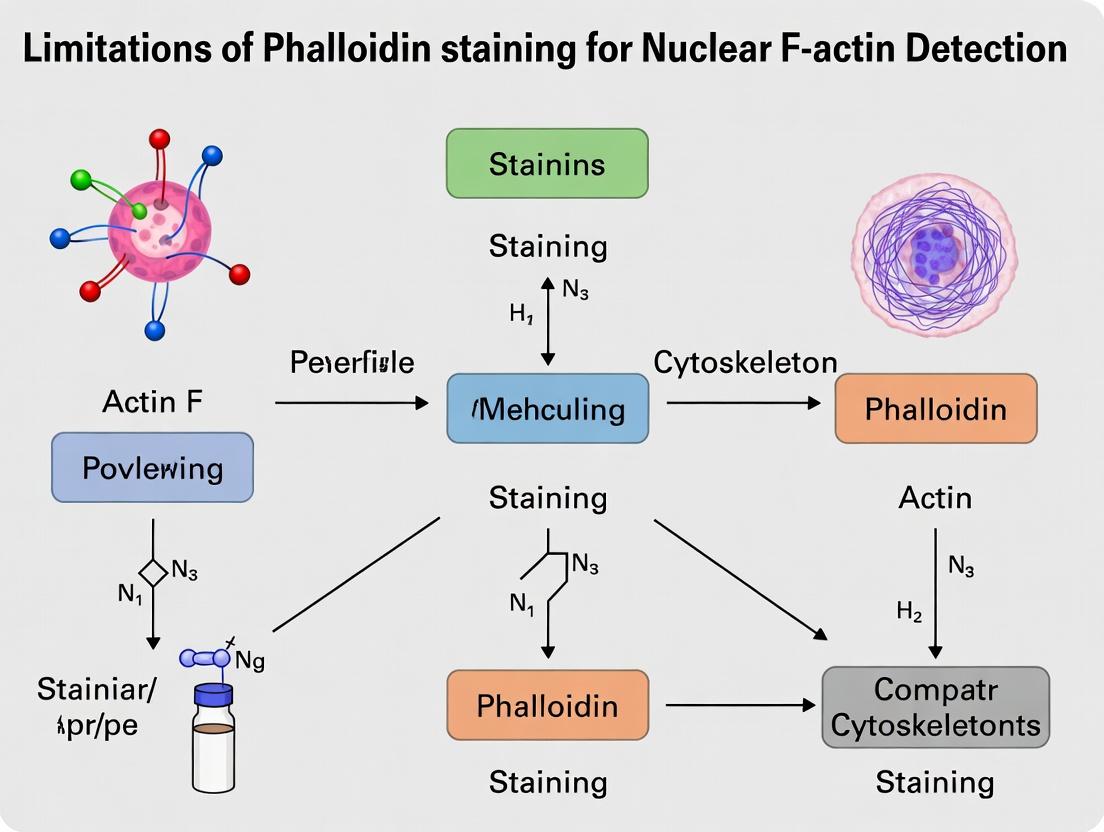
This article provides a critical analysis for researchers and drug development professionals on the specific limitations of phalloidin-based staining for detecting nuclear actin filaments (F-actin).
Beyond Cytoplasmic Staining: The Critical Limitations of Phalloidin for Nuclear F-actin Detection in Research and Drug Discovery
Abstract
This article provides a critical analysis for researchers and drug development professionals on the specific limitations of phalloidin-based staining for detecting nuclear actin filaments (F-actin). We explore the foundational biology of nuclear actin, detail the methodological pitfalls of phalloidin in the nuclear compartment, and present troubleshooting strategies. We further compare phalloidin with advanced validation methods, including live-cell probes and transgenic actin chromophores. The goal is to equip scientists with the knowledge to select appropriate tools, avoid artifacts, and generate reliable data in studies of nuclear architecture, gene regulation, and related therapeutic targets.
Nuclear F-actin: Understanding the Biology and Why Phalloidin Often Fails
Application Notes
The study of nuclear actin represents a significant frontier in cell biology, with implications for gene regulation, DNA repair, and nuclear architecture. However, research in this area is fundamentally constrained by the limitations of conventional actin probes, most notably phalloidin. Phalloidin, which stains filamentous actin (F-actin), poorly penetrates the intact nuclear envelope and cannot differentiate between cytoplasmic and nuclear pools. Consequently, the broader thesis on nuclear F-actin detection must pivot toward compartment-specific tools and quantitative assays. This document outlines the defining characteristics of cytoplasmic versus nuclear actin pools and provides protocols for their specific study.
1. Key Differences Between Actin Pools
Table 1: Comparative Properties of Cytoplasmic and Nuclear Actin Pools
| Property | Cytoplasmic Actin | Nuclear Actin |
|---|---|---|
| Predominant Form | High concentration of stable F-actin (microfilaments). | Largely monomeric (G-actin) and short, dynamic oligomers. |
| Critical Regulators | Profilin, Cofilin, Thymosin-β4, CapZ, Tropomyosin. | Profilin, Cofilin, Importin-9, N-WASP. |
| Primary Functions | Cell motility, cytokinesis, structural integrity, vesicle trafficking. | Transcription regulation (Pol I, II, III), chromatin remodeling, DNA repair. |
| Polymerization Dynamics | Stable, long-lived filaments under tension. | Transient, rapid turnover; polymerization often signal-induced. |
| Key Structural Roles | Stress fibers, lamellipodia, filopodia, contractile ring. | Nucleoskeleton organization, intranuclear movement. |
| Common Detection Challenges | Phalloidin effective but stains total cellular F-actin. | Phalloidin impermeant; requires live-cell probes or fractionation. |
Table 2: Quantitative Data on Actin Pools in Mammalian Cells (HeLa Example)
| Metric | Cytoplasmic Pool | Nuclear Pool | Measurement Method |
|---|---|---|---|
| Approx. Concentration | ~100-200 µM | ~5-20 µM | Fluorescence correlation spectroscopy (FCS) |
| G-actin : F-actin Ratio | ~1:1 to 1:2 (highly variable) | Estimated >10:1 (G-actin dominant) | Biochemical fractionation + DNase I inhibition assay |
| Turnover Half-life (F-actin) | Minutes to hours | Seconds to minutes | FRAP/FLAP with actin-GFP constructs |
| Nuclear Import Mediator | N/A | Importin-9 (primary) | Co-immunoprecipitation, siRNA knockdown |
2. Experimental Protocols
Protocol 1: Subcellular Fractionation for Biochemical Analysis of Actin Pools
Objective: To biochemically separate cytoplasmic and nuclear proteins for immunoblotting or polymerization state assays.
Materials: Hypotonic Lysis Buffer (10 mM HEPES pH 7.9, 1.5 mM MgCl2, 10 mM KCl, 0.5 mM DTT, protease inhibitors), NP-40, Nuclear Extraction Buffer (20 mM HEPES pH 7.9, 1.5 mM MgCl2, 420 mM NaCl, 0.2 mM EDTA, 25% Glycerol, protease inhibitors), Dounce homogenizer.
Procedure:
- Harvest ~5x10^6 cells by gentle scraping.
- Wash cells in ice-cold PBS and pellet at 500 x g for 5 min.
- Resuspend pellet in 500 µL Hypotonic Lysis Buffer. Incubate on ice for 15 min.
- Add 25 µL of 10% NP-40, vortex for 10 sec.
- Centrifuge at 12,000 x g for 5 min at 4°C. Supernatant = Cytoplasmic Fraction. Transfer to new tube.
- Wash the nuclear pellet (insoluble material) with 500 µL Hypotonic Lysis Buffer. Centrifuge again.
- Resuspend nuclear pellet in 100-200 µL Nuclear Extraction Buffer. Vortex vigorously, incubate on ice for 30 min with intermittent mixing.
- Centrifuge at 16,000 x g for 10 min at 4°C. Supernatant = Soluble Nuclear Fraction.
- Analyze fractions by SDS-PAGE and immunoblot for actin (total), Lamin B1 (nuclear marker), and GAPDH (cytoplasmic marker).
Protocol 2: Live-Cell Imaging of Nuclear G-actin using Utr230-EGFP
Objective: To visualize and quantify the dynamic monomeric actin pool within the nucleus.
Materials: Utr230-EN/EGFP plasmid (utrophin calponin homology domain, binds F-actin very weakly, serves as a G-actin reporter), transfection reagent, live-cell imaging medium, confocal microscope.
Procedure:
- Seed cells on glass-bottom imaging dishes 24h prior.
- Transfect cells with the Utr230-EN/EGFP construct using a standard protocol. Use a low DNA concentration to minimize overexpression artifacts.
- 24-48h post-transfection, replace medium with pre-warmed live-cell imaging medium.
- Using a confocal microscope with a 63x oil objective, acquire z-stacks encompassing the entire nucleus.
- The diffuse fluorescence signal within the nucleoplasm (excluding nucleoli) corresponds to the nuclear G-actin pool. Quantify mean fluorescence intensity in the nucleus vs. cytoplasm using image analysis software (e.g., FIJI/ImageJ).
- Control: Treat cells with Latrunculin A (2 µM, 1h) to depolymerize F-actin. This should increase nuclear Utr230 signal, confirming its specificity for the monomeric pool.
Protocol 3: Proximity Ligation Assay (PLA) for Nuclear Actin-Protein Interactions
Objective: To detect and visualize close-range interactions (<40 nm) between nuclear actin and specific partners (e.g., RNA Polymerase II) in situ.
Materials: Duolink PLA kit, primary antibodies (e.g., mouse anti-Actin, rabbit anti-RNA Pol II), blocking solution, mounting medium with DAPI.
Procedure:
- Culture cells on coverslips, fix with 4% PFA for 15 min, permeabilize with 0.2% Triton X-100 for 10 min.
- Perform standard immunofluorescence blocking for 1h.
- Incubate with primary antibodies diluted in blocking buffer overnight at 4°C.
- Follow the manufacturer's protocol for the PLA kit: incubate with PLUS and MINUS PLA probes, ligation, and amplification.
- Mount slides with Duolink In Situ Mounting Medium with DAPI.
- Image using a fluorescence microscope. Each red PLA dot represents a single interaction event. Quantify dot number per nucleus using automated analysis software.
3. Signaling Pathway & Experimental Workflow
Diagram 1: Nuclear Actin Signaling & Detection Workflow
Diagram 2: Subcellular Fractionation Protocol Flow
4. The Scientist's Toolkit
Table 3: Essential Research Reagents for Compartment-Specific Actin Research
| Reagent / Tool | Category | Primary Function & Rationale |
|---|---|---|
| Utr230-EN/EGFP | Live-cell Probe | A genetically encoded probe with low F-actin affinity, serving as a robust reporter for the dynamic G-actin pool, especially in the nucleus. |
| Lifeact | Live-cell Probe | Binds F-actin; useful for cytoplasmic filaments but can perturb nuclear actin dynamics; use with caution and proper controls. |
| siRNA against Importin-9 | Functional Tool | Knocks down the primary nuclear actin importer to specifically deplete the nuclear actin pool and study functional consequences. |
| Jasplakinolide | Chemical Polymerizer | Induces actin polymerization; used to test nuclear actin's role by forcing intranuclear filament assembly. |
| Latrunculin A/B | Chemical Depolymerizer | Sequesters G-actin; validates G-actin probe specificity and examines functions dependent on monomeric actin. |
| Duolink PLA Kit | Interaction Assay | Detects proximal (<40nm) protein interactions in situ, crucial for visualizing actin's association with nuclear complexes without co-IP. |
| Anti-Lamin B1 Antibody | Fractionation Control | Marker for nuclear envelope integrity and purity during biochemical fractionation. |
| Anti-GAPDH Antibody | Fractionation Control | Marker for cytoplasmic contamination in nuclear fractions. |
| DNase I Inhibition Assay | Biochemical Assay | Quantifies the concentration of monomeric (globular) actin in fractionated samples. |
Phalloidin, a bicyclic peptide toxin from Amanita phalloides, is the quintessential probe for fluorescent visualization of filamentous actin (F-actin) in fixed cells. Its high affinity and specificity for F-actin have made it indispensable for studying the cytoskeleton. However, within the context of advancing research into nuclear actin, a critical limitation emerges. Phalloidin's utility is predominantly confined to cytoplasmic and stabilized F-actin structures. Due to its impermeability to live-cell membranes and, more critically, its poor penetration of the nuclear envelope in standard fixation protocols, phalloidin is largely ineffective for detecting dynamic or transient nuclear F-actin pools. This application note details established protocols for phalloidin staining while framing its limitations in the evolving field of nuclear actin research.
Quantitative Data on Phalloidin Binding
Table 1: Key Characteristics of Common Phalloidin Conjugates
| Conjugate Fluorophore | Excitation/Emission Max (nm) | Relative Brightness | Photostability | Common Application |
|---|---|---|---|---|
| Alexa Fluor 488 | 495/519 | High | High | Standard green channel, multi-color imaging |
| Tetramethylrhodamine (TRITC) | 554/576 | Moderate | Moderate | Standard red channel, avoid GFP overlap |
| Alexa Fluor 568 | 578/603 | High | High | Excellent for red channel, superior to TRITC |
| Alexa Fluor 647 | 650/668 | High | Very High | Far-red channel, low background, super-resolution |
| Phalloidin (unlabeled) | N/A | N/A | N/A | Competition assays, negative controls |
Table 2: Recommended Staining Concentrations and Conditions
| Sample Type | Phalloidin Conjugate Conc. (in PBS) | Incubation Time | Temperature | Notes |
|---|---|---|---|---|
| Standard Cultured Cells | 5 - 20 U/mL (∼20-100 nM) | 20-30 minutes | Room Temp | Protect from light. |
| Thick Tissue Sections | 50 - 100 U/mL | 60-90 minutes | Room Temp | May require permeabilization optimization. |
| Super-Resolution (STORM/PALM) | 10-20 nM | 30 minutes | Room Temp | Use specific photo-switchable dyes (e.g., Alexa 647). |
| Nuclear F-actin (Attempt) | 100-200 U/mL | 60+ minutes | 4°C or RT | Often ineffective; requires alternative methods (e.g., actin chromobodies, LifeAct). |
Research Reagent Solutions Toolkit
Table 3: Essential Materials for Phalloidin Staining Experiments
| Item | Function & Rationale |
|---|---|
| Formaldehyde (4%, in PBS) | Standard fixative. Crosslinks proteins, preserves cytoskeleton structure. |
| Triton X-100 (0.1-0.5%) | Non-ionic detergent for permeabilization of the plasma membrane. |
| Bovine Serum Albumin (BSA, 1-3%) | Blocking agent to reduce non-specific background staining. |
| Paraformaldehyde (PFA, 4%) | Higher purity alternative to formaldehyde; preferred for super-resolution. |
| Saponin (0.05-0.1%) | Permeabilization agent that preserves some membrane structures; can be used in combination. |
| Mounting Medium with DAPI | Aqueous or anti-fade mounting medium containing DNA stain for nuclear counterstaining. |
| Actin Polymerization Drugs (e.g., Jasplakinolide) | Stabilizes F-actin, used as a positive control or to arrest dynamic actin. |
| Nuclear Export Inhibitor (Leptomycin B) | Used in nuclear actin research to accumulate actin in the nucleus, though phalloidin may still fail to stain it. |
Detailed Protocol: Standard Phalloidin Staining for Cytoplasmic F-actin
Protocol 1: Immunofluorescence Staining of Cultured Adherent Cells
A. Cell Fixation and Permeabilization
- Culture cells on glass coverslips in a multi-well plate.
- Aspirate culture medium and rinse cells gently with 1x PBS, pH 7.4.
- Fix cells with 4% formaldehyde in PBS for 10-15 minutes at room temperature (RT).
- Aspirate fixative and wash cells 3 x 5 minutes with PBS.
- Permeabilize cells with 0.1% Triton X-100 in PBS for 5-10 minutes at RT.
- Wash cells 3 x 5 minutes with PBS.
B. Blocking and Staining
- Block non-specific sites with 1-3% BSA in PBS for 30 minutes at RT.
- Prepare staining solution: Dilute fluorescent phalloidin conjugate in blocking buffer (e.g., 1:200 to 1:500 from a 300U/mL stock).
- Apply 100-200 µL of phalloidin solution onto a parafilm sheet. Invert coverslip and mount cell-side down on the drop. Incubate for 20-30 minutes at RT in the dark.
- Carefully retrieve coverslip, return to the well, and wash 3 x 5 minutes with PBS.
C. Mounting and Imaging
- Optionally, counterstain nuclei with DAPI (300 nM in PBS) for 5 minutes.
- Wash briefly with PBS.
- Mount coverslip on a glass slide using 5-10 µL of anti-fade mounting medium.
- Seal edges with nail polish. Image using a fluorescence microscope with appropriate filter sets.
Protocol 2: Attempted Enhancement for Nuclear Detection (with Limitations)
A. Enhanced Permeabilization Protocol
- Follow steps A.1-A.4 from Protocol 1.
- Permeabilize with sequential buffers: a. 0.5% Triton X-100 in PBS for 15 minutes on ice. b. 0.05% Digitonin in PBS for 5 minutes on ice. c. Wash 3 x 5 minutes with PBS.
- Block with 3% BSA + 0.1% Tween-20 in PBS for 1 hour.
- Stain with high-concentration phalloidin (100 U/mL) in blocking buffer overnight at 4°C.
- Wash extensively (5 x 10 minutes) and mount as before.
Note: This protocol may increase non-specific background and often remains insufficient for definitive nuclear F-actin visualization, highlighting the need for genetic probes (e.g., LifeAct-GFP) or immunostaining with anti-actin antibodies after special fixation (e.g., with lysine crosslinkers).
Visualization: Phalloidin Workflow and Nuclear Actin Challenge
Title: Phalloidin Staining Workflow and Nuclear Barrier
Title: Nuclear F-actin Detection Methods Comparison
1. Introduction and Context within Nuclear F-Actin Research Phalloidin, a bicyclic heptapeptide toxin from Amanita phalloides, is the gold-standard probe for labeling filamentous actin (F-actin) due to its high affinity and specificity. However, a critical limitation in its application is its inability to efficiently cross the intact double membrane of the nuclear envelope. This poor permeability presents a core problem for research investigating the diverse and essential roles of intra-nuclear actin filaments, which are involved in processes such as chromatin remodeling, transcription, DNA repair, and nucleocytoplasmic transport. This application note details the quantitative evidence of this limitation and provides protocols for current methods to circumvent it, framed within the broader thesis that phalloidin-based detection systems are insufficient for nuclear F-actin research without disruptive preparatory methods.
2. Quantitative Evidence of Permeability Limitation
Table 1: Comparative Efficiency of Phalloidin-Based Nuclear F-Actin Labeling Methods
| Method | Principle | Nuclear Envelope Integrity Post-Treatment | Relative Nuclear F-Actin Signal Intensity (vs. Cytoskeletal) | Key Artifact/Risk |
|---|---|---|---|---|
| Standard Permeabilization (Triton X-100) | Extracts lipids, fully permeabilizes all membranes. | Destroyed | Low (<10%) | Complete loss of nuclear compartmentalization; possible filament disassembly. |
| Digitonin Selective Permeabilization | Binds cholesterol, selectively permeabilizes plasma membrane. | Preserved | Very Low (<2%) | Demonstrates phalloidin's intrinsic impermeability to intact nuclear envelope. |
| Mechanical Disruption (Microinjection) | Physical breach of nuclear envelope. | Locally disrupted | High (~95%) | Technically challenging, low throughput, introduces damage. |
| EM & SUPER Resolution (POST-fixation) | Phalloidin applied after fixation & harsh permeabilization. | Destroyed | Moderate to High | Best for architecture but non-physiological; no live-cell application. |
Table 2: Properties Affecting Phalloidin Nuclear Access
| Property | Value/Characteristic | Implication for Nuclear Entry |
|---|---|---|
| Molecular Weight | ~788.9 Da | Below passive diffusion cutoff (~40-60 kDa) but not sufficient. |
| Charge | Neutral | Eliminates charge-based repulsion/barriers. |
| Primary Barrier | Intact Nuclear Envelope | Nuclear Pore Complex (NPC) selectively gates passage; phalloidin lacks Nuclear Localization Signal (NLS). |
| Passive Diffusion Limit via NPC | ~5-10 nm diameter (~30-40 kDa globular proteins) | Phalloidin's size/form may be sterically hindered or actively excluded. |
3. Experimental Protocols
Protocol 3.1: Demonstrating Phalloidin Impermeability using Digitonin Selective Permeabilization Objective: To confirm that phalloidin cannot label nuclear F-actin when the nuclear envelope remains intact. Materials: Cultured cells, Phosphate-Buffered Saline (PBS), 4% Paraformaldehyde (PFA) in PBS, Digitonin (50 µg/mL in PBS), Fluorescently-conjugated Phalloidin (in PBS), Hoechst 33342, Mounting medium. Procedure:
- Culture & Plate: Grow cells on glass coverslips to 60-70% confluence.
- Fix: Wash cells with warm PBS. Fix with 4% PFA for 15 min at room temperature (RT).
- Selective Permeabilization: Wash 3x with PBS. Incubate with 50 µg/mL digitonin in PBS for 5 min at RT. Critical Step: This permeabilizes the cholesterol-rich plasma membrane but leaves the nuclear envelope largely intact.
- Phalloidin Staining: Without washing, add fluorescent phalloidin (diluted per manufacturer's instructions in PBS) directly to the digitonin solution. Incubate for 30-60 min at RT, protected from light.
- Wash & Counterstain: Wash thoroughly 3x with PBS. Incubate with Hoechst 33342 (1 µg/mL in PBS) for 5 min.
- Mount & Image: Wash, mount coverslips, and image using fluorescence microscopy. Expected Result: Bright cytoskeletal staining with absent or very dim nuclear signal.
Protocol 3.2: Nuclear F-Actin Detection via Microinjection of Labeled Phalloidin Objective: To directly label nuclear F-actin in live cells by bypassing the nuclear envelope barrier. Materials: Micropipette puller, Microinjection system, Pressure injector, Alexa Fluor 488-conjugated phalloidin, Injection buffer (e.g., 50 mM KCl, 10 mM HEPES, pH 7.4). Procedure:
- Sample Preparation: Plate cells on glass-bottom dishes. Prepare injection needle and backfill with phalloidin solution (e.g., 100 µM in injection buffer).
- Microinjection: Using the microinjection system, target the cell nucleus. Apply a brief pressure pulse (e.g., 50 hPa for 0.5 s) to deliver the phalloidin solution directly into the nucleoplasm.
- Incubation: Allow cells to recover for 5-15 min in culture medium to permit phalloidin binding.
- Fixation (Optional): If fixed imaging is required, wash and fix cells with 4% PFA for 15 min.
- Image Acquisition: Image immediately (live) or after fixation using confocal or super-resolution microscopy. Note: This method validates nuclear F-actin presence but is low-throughput and invasive.
4. Visualization: Pathways and Workflows
Title: Phalloidin's Barrier to Nuclear F-Actin Labeling
Title: Experimental Workflow to Isolate the Permeability Problem
5. The Scientist's Toolkit: Key Research Reagent Solutions
Table 3: Essential Materials for Investigating Nuclear F-Actin
| Item | Function/Benefit | Key Consideration for Nuclear Studies |
|---|---|---|
| Fluorescent Phalloidin (e.g., Alexa Fluor conjugates) | High-affinity F-actin stain for fixed cells. | Impermeable to intact nucleus; requires envelope disruption. |
| Digitonin | Cholesterol-binding detergent for selective plasma membrane permeabilization. | Critical tool to demonstrate nuclear envelope impermeability. |
| Triton X-100 / NP-40 | Non-ionic detergents for complete cellular permeabilization. | Allows nuclear access but destroys envelope integrity and may alter structures. |
| Paraformaldehyde (PFA) | Cross-linking fixative. | Preserves structure but can mask epitopes; requires optimization. |
| Live-cell Actin Probes (e.g., LifeAct, F-tractin) | Genetically-encoded markers for actin dynamics in live cells. | Can be targeted to nucleus with NLS; avoids permeability issue but may alter dynamics. |
| Anti-Nuclear Actin Antibodies | Potential alternative for immunofluorescence. | Many antibodies recognize monomeric (G-) actin; specific F-actin antibodies are rare and require validation. |
| Microinjection System | For direct delivery of probes into the nucleoplasm. | Bypasses permeability barrier; allows live-cell study but is low-throughput and invasive. |
Within the context of a broader thesis on the limitations of phalloidin staining in research, the detection of nuclear actin filaments presents a unique set of challenges. Phalloidin, a bicyclic peptide toxin from Amanita phalloides, binds with high affinity to canonical, stable F-actin polymers found abundantly in the cytoplasm. However, nuclear actin exists in diverse, often unconventional forms that are poorly recognized by phalloidin. This application note details the nature of these nuclear actin structures, the quantitative evidence for phalloidin's limitations, and provides protocols for alternative detection methods critical for researchers, scientists, and drug development professionals working in nuclear signaling, gene regulation, and cell mechanics.
Core Limitation: Characteristics of Nuclear Actin vs. Phalloidin's Binding Requirements
Nuclear actin dynamics are characterized by transient polymerization, short filament length, and unique post-translational modifications or associated proteins that can occlude phalloidin binding. The table below summarizes the key comparative features that explain the detection challenge.
Table 1: Characteristics of Nuclear Actin Filaments vs. Cytoplasmic F-Actin
| Feature | Cytoplasmic F-Actin (Phalloidin-Sensitive) | Nuclear F-Actin (Often Phalloidin-Resistant) | Impact on Phalloidin Binding |
|---|---|---|---|
| Polymer Stability | Stable, long-lived filaments (minutes to hours). | Highly transient, short-lived filaments (seconds). | Phalloidin binding kinetics are too slow to capture transient polymers. |
| Filament Length | Long filaments (microns). | Very short filaments (< 100 nm, often oligomeric). | Short polymers may offer fewer binding sites or altered geometry. |
| Nucleotide State | Primarily ADP-F-actin. | May be enriched in ATP- or ADP-Pi-F-actin. | Phalloidin binds preferentially to ADP-actin filaments. |
| Associated Proteins | Standard ABPs (e.g., cofilin, tropomyosin). | Unique nuclear ABPs (e.g., N-WASP, coffilin, nuclear myosins). | Proteins may sterically block the phalloidin binding site on actin. |
| Modifications | Standard acetylation, arginylation. | Potential unique oxidation or other modifications. | May alter the phalloidin binding interface. |
| Visualization by EM | Clearly visible, ordered bundles. | Sparse, short, single filaments. | Confirms structural difference from canonical cytoskeleton. |
Quantitative Evidence of Phalloidin's Failure in the Nucleus
Recent studies provide direct quantitative comparisons between phalloidin-based staining and more specific methodologies.
Table 2: Comparative Quantification of Nuclear F-Actin Detection Methods
| Study (Key Finding) | Method 1: Phalloidin Stain | Method 2: Alternative Probe/Assay | Result (Quantitative Discrepancy) | Implication |
|---|---|---|---|---|
| Induced Nuclear Actin Polymerization (e.g., Serum Stimulation, DAA) | Weak, diffuse, or absent nuclear signal. | Lifact-GFP fluorescence or F-tractin-GFP. | >10-20 fold increase in nuclear focal signal detected by biosensor vs. phalloidin. | Phalloidin misses signal from induced, functional nuclear filaments. |
| DNA Damage-Induced Filaments (DDR) | Faint, inconsistent nuclear puncta. | Immuno-EM with anti-actin antibody. | EM shows numerous short filaments (<200 nm); phalloidin stains <5% of these structures. | Confirms existence of phalloidin-invisible filamentous networks. |
| Nuclear Actin in Transcription | No specific signal at active gene loci. | Chromatin IP with actin antibody or actin-biosensor FRAP. | ChIP shows actin enrichment; FRAP shows dynamic turnover inconsistent with phalloidin stability. | Functional nuclear actin pools are dynamically polymeric but non-canonical. |
The Scientist's Toolkit: Research Reagent Solutions
Table 3: Essential Reagents for Nuclear Actin Research
| Reagent / Material | Function & Application | Key Consideration |
|---|---|---|
| Cell-Permeant Phalloidin Derivatives (e.g., Alexa Fluor-phalloidin) | Standard for visualizing stable cytoplasmic F-actin. Serves as a negative control for nuclear actin. | Permeabilization required for nuclear access. Does not label most nuclear filaments. |
| Genetically Encoded Actin Biosensors (e.g., Lifact-GFP, F-tractin-tdTomato, Utr230-EGFP) | Binds F-actin without stabilizing it. Ideal for live-cell imaging of dynamic nuclear actin polymerization. | Requires transfection/transduction. May perturb native actin dynamics at high expression. |
| Anti-Actin Antibodies (for IF, ChIP, EM) | Can detect both monomeric (G) and polymeric (F) actin. Useful for immuno-EM of nuclear filaments. | Careful validation required to avoid cross-reactivity. Does not distinguish G from F-actin in standard IF. |
| Nuclear Export Inhibitor (Leptomycin B) | Blocks CRM1-dependent export of actin, causing nuclear accumulation. Positive control for nuclear actin studies. | Can induce non-physiological actin aggregates; use at low doses for short times. |
| DNA Damage Inducers (e.g., Neocarzinostatin, Doxorubicin) | Stimulates rapid nuclear actin polymerization as part of the DNA Damage Response (DDR). | Useful for synchronizing and amplifying nuclear F-actin for study. |
| Actin Polymerization Drugs (e.g., Jasplakinolide, DMSO-based Actin Activator "DAA") | Stabilizes actin filaments, can force polymerization in the nucleus. Tests capacity for nuclear actin assembly. | Jasplakinolide is cytotoxic and permeabilization-dependent. DAA is membrane-permeant. |
Detailed Experimental Protocols
Protocol 1: Comparative Staining for Nuclear Actin: Phalloidin vs. Immunofluorescence
Objective: To visualize the discrepancy between phalloidin and antibody-based actin detection in the nucleus. Materials: Fixed cells, PBS, Triton X-100, BSA, primary anti-actin antibody (e.g., clone C4), fluorescent secondary antibody, fluorescent phalloidin, mounting medium with DAPI. Procedure:
- Culture & Stimulate: Plate cells on coverslips. Treat cells with a nuclear actin-inducing stimulus (e.g., 100 nM Leptomycin B for 2h or 1 μM DAA for 10 min). Include an untreated control.
- Fix & Permeabilize: Fix cells with 4% paraformaldehyde (PFA) for 15 min. Rinse with PBS. Permeabilize with 0.2% Triton X-100 in PBS for 10 min.
- Block: Incubate in blocking buffer (3% BSA in PBS) for 1 hour.
- Dual Labeling:
- Apply primary anti-actin antibody (diluted in blocking buffer) for 1-2 hours at RT.
- Wash 3x with PBS.
- Apply fluorescent secondary antibody AND fluorescent phalloidin (diluted together in blocking buffer) for 1 hour at RT in the dark.
- Wash & Mount: Wash 3x with PBS. Rinse with dH2O. Mount with DAPI-containing medium.
- Imaging & Analysis: Acquire confocal images using identical laser/pinhole settings for both channels. Quantify mean fluorescence intensity in the nucleus (DAPI mask) for both the phalloidin and antibody signals. Compare the signal-to-noise ratios.
Protocol 2: Live-Cell Imaging of Transient Nuclear Actin with Biosensors
Objective: To capture the dynamics of nuclear actin polymerization in response to a stimulus. Materials: Cells stably expressing Lifact-GFP or similar, Leibovitz's L-15 medium, confocal or TIRF microscope with environmental chamber, DNA damage inducer (e.g., 100 ng/mL Neocarzinostatin). Procedure:
- Prepare Cells: Seed cells expressing the actin biosensor into an imaging chamber (e.g., glass-bottom dish).
- Acquire Baseline: Replace medium with pre-warmed L-15 medium. Locate cells and acquire a time-lapse series (e.g., 1 frame every 5-10 seconds for 5 min) to establish baseline nuclear fluorescence.
- Induce & Image: Without moving the field of view, carefully add the DNA damage inducer (pre-diluted in L-15) to the dish. Immediately resume time-lapse acquisition for 20-30 minutes.
- Analysis: Use image analysis software (e.g., FIJI/ImageJ) to:
- Draw regions of interest (ROIs) in the nucleus and cytoplasm.
- Plot fluorescence intensity over time (F(t)).
- Calculate metrics like maximum fold increase, time to peak, and half-time of decay for nuclear foci.
Protocol 3: Sedimentation Assay for Nuclear Actin Polymerization State
Objective: To biochemically assess the F-actin content in nuclear fractions. Materials: Cell pellets, hypotonic lysis buffer, detergent, ultracentrifuge, F-actin stabilization buffer (with phalloidin), actin depolymerization buffer (with Latrunculin A). Procedure:
- Prepare Nuclear Lysates: Isolate nuclei using a hypotonic lysis/detergent method or a commercial kit. Lyse nuclei in a gentle F-actin stabilizing lysis buffer.
- High-Speed Sedimentation: Split the lysate into two equal aliquots.
- Aliquot A (F-actin): Keep on ice.
- Aliquot B (G-actin): Treat with 1 μM Latrunculin A for 30 min to depolymerize F-actin.
- Centrifuge both aliquots at 100,000 x g for 1 hour at 4°C to pellet F-actin.
- Carefully separate supernatant (S; G-actin) from pellet (P; F-actin).
- Analysis: Resuspend pellets in a volume equal to the supernatant. Analyze equal volumes of S and P fractions by Western blot for actin. Quantify the ratio of actin in P/(P+S) for both aliquots. A true nuclear F-actin signal will be present in Aliquot A's pellet but lost in Aliquot B's pellet.
Visualization: Pathways and Workflows
Diagram 1: Why Nuclear Actin Evades Phalloidin Staining (100 chars)
Diagram 2: Multi-Method Workflow to Study Nuclear Actin (99 chars)
Abstract Phalloidin, a bicyclic peptide from Amanita phalloides, is the gold-standard probe for filamentous actin (F-actin) visualization due to its high affinity and specificity. However, this Application Note critically reviews accumulating evidence that phalloidin staining systematically underreports the presence of nuclear F-actin, a key player in gene regulation, DNA damage repair, and mechanotransduction. This limitation, framed within a broader thesis on phalloidin's constraints, stems from accessibility issues, differential F-actin architecture, and fixation artifacts. We present case study data, provide optimized protocols for accurate detection, and offer reagent solutions to overcome this significant methodological blind spot.
Table 1: Comparative Detection of Nuclear F-actin: Phalloidin vs. Alternative Probes/Methods
| Cell Type / Stimulus | Phalloidin Signal (Nuclear) | Anti-Actin Antibody / Live-Cell Probe (Nuclear) | Method of Validation | Reported Fold-Underestimation by Phalloidin | Key Reference (Example) |
|---|---|---|---|---|---|
| Serum-stimulated Fibroblasts | Weak / Diffuse | Strong, punctate structures | Immuno-EM, LifeAct-GFP | 3-5x | Belin et al., 2015 |
| DNA Damage (Doxorubicin treated) | Faint, inconsistent | Robust filaments & rods | siRNA, Chromatin Fractionation | 4-7x | Caridi et al., 2018 |
| Mechanical Stress (Nucleus) | Undetectable | Clear stress-induced filaments | Optical Tweezers, FRET-based Biosensors | Quantifiable signal only with probes | Aureille et al., 2019 |
| Cell Differentiation (e.g., mESCs) | Low contrast | Distinct intranuclear bundles | Super-resolution microscopy (STORM/PALM) | Resolution-limited; architectural details missed | Plessner et al., 2015 |
Detailed Experimental Protocols
Protocol 1: Combined Detection for Nuclear F-actin (Phalloidin & Antibody) Objective: To directly compare phalloidin and antibody-based actin detection within the same nuclear sample. Materials: See "Research Reagent Solutions" below. Procedure:
- Cell Culture & Stimulation: Plate cells on #1.5 coverslips. Apply relevant stimulus (e.g., 10% serum for 15 min, 1µM Doxorubicin for 2h).
- Permeabilization-First Fixation (Critical):
- Rinse cells with pre-warmed Cytoskeletal Buffer (CB: 10 mM MES, 150 mM NaCl, 5 mM EGTA, 5 mM glucose, 5 mM MgCl2, pH 6.1).
- Permeabilize with 0.5% Triton X-100 in CB for 3 min at 37°C.
- Immediately fix with 4% formaldehyde in CB for 15 min at RT.
- Rinse with PBS.
- Blocking: Incubate in blocking buffer (3% BSA, 0.1% Tween-20 in PBS) for 1h.
- Primary Antibody Incubation: Apply anti-actin antibody (e.g., clone AC-40) diluted in blocking buffer overnight at 4°C.
- Secondary Antibody Incubation: Apply fluorophore-conjugated secondary antibody (e.g., Alexa Fluor 568) for 1h at RT. Rinse thoroughly.
- Phalloidin Counterstaining: Incubate with Alexa Fluor 488-conjugated phalloidin (1:50 in PBS) for 30 min at RT, protected from light.
- Nuclear Staining & Mounting: Stain DNA with DAPI (300 nM, 5 min). Mount with antifade reagent.
- Imaging: Acquire using super-resolution or high-confocal microscopy. Use identical laser/power settings for both channels in sequential scans to compare intensity.
Protocol 2: Validation via Biochemical Fractionation Objective: Biochemically isolate nuclear actin filaments to validate imaging data. Procedure:
- Nuclei Isolation: Harvest ~5x10^6 stimulated cells. Lyse in hypotonic buffer (10 mM HEPES, 1.5 mM MgCl2, 10 mM KCl, protease inhibitors) with 0.1% NP-40. Pellet nuclei (500 x g, 5 min).
- Chromatin-associated Protein Extraction: Resuspend nuclear pellet in:
- Buffer A (Low Salt): 10 mM Tris-HCl, 0.1 mM EDTA, 0.1% NP-40.
- Buffer B (High Salt): 10 mM Tris-HCl, 0.1 mM EDTA, 500 mM NaCl, 0.1% NP-40.
- Sonicate briefly. Incubate on ice, then centrifuge at 13,000 x g.
- F-actin Stabilization & Precipitation: Add phalloidin (1µM) and KCl (100 mM final) to the high-salt supernatant to preserve and induce sedimentation of F-actin. Ultracentrifuge at 100,000 x g for 1h.
- Analysis: Analyze pellet (F-actin) and supernatant (G-actin) fractions by immunoblotting with anti-actin antibody. Compare the ratio of nuclear F-actin across conditions.
Visualizations
Diagram 1: Nuclear F-actin Formation Pathways & Phalloidin Access Limitation
Title: Pathways to Nuclear F-actin and Phalloidin Block
Diagram 2: Optimized Detection Workflow for Nuclear F-actin
Title: Nuclear F-actin Detection Protocol Workflow
The Scientist's Toolkit: Research Reagent Solutions
Table 2: Essential Reagents for Nuclear F-actin Research
| Reagent / Material | Function & Rationale |
|---|---|
| Permeabilization-First Fixatives | Cytoskeletal Buffer (CB) with Triton X-100 preserves labile nuclear F-actin structures lost in standard PFA-first protocols. |
| Anti-Actin Antibodies | Clone AC-40 or similar; targets all actin isoforms, accesses nuclear compartments better than phalloidin. Validate for absence of cytoplasmic cross-reactivity. |
| Live-Cell Nuclear F-actin Probes | GFP-tagged nuclear localization sequence (NLS) fused to LifeAct or F-tractin. Enables real-time dynamics without fixation artifacts. |
| Nuclear Fractionation Kits | For biochemical isolation of chromatin-associated and filamentous actin from nuclei. Validates imaging results. |
| Super-Resolution Mounting Media | Photoswitchable/antifade reagents (e.g., for STORM/PALM) essential for resolving fine nuclear filaments. |
| Formin Inhibitors (e.g., SMIFH2) | Pharmacological tool to inhibit mDia formin-dependent nuclear actin polymerization, confirming specificity of observed structures. |
| Actin Chromobody (GFP-nanobody) | Alternative live-cell tag for actin, often showing different binding kinetics and accessibility compared to phalloidin. |
Methodological Pitfalls: How Experimental Design Impacts Phalloidin Staining Results
Application Notes and Protocols
Context: Within the broader thesis investigating the limitations of phalloidin staining for nuclear F-actin detection, a critical and often overlooked variable is sample preparation. Fixation and permeabilization protocols, essential for preserving and probing cellular architecture, can induce significant artifacts that distort the actin cytoskeleton and generate false-positive or false-negative signals for nuclear F-actin. This document details these pitfalls and provides optimized protocols for rigorous investigation.
Artifacts from Common Protocols
Chemical fixation and permeabilization can alter F-actin integrity, promote its artifactual formation in the nucleus, or destroy native structures.
Table 1: Impact of Common Reagents on F-actin Integrity
| Reagent | Common Concentration/Time | Effect on F-actin (Cytosolic & Nuclear) | Potential Artifact for Nuclear F-Actin Detection |
|---|---|---|---|
| Formaldehyde (PFA) | 3-4%, 10-20 min | Cross-links proteins; can stabilize but also induce aggregation. | Can cause cytosolic F-actin bundles to appear more prominent while masking finer structures. May induce non-physiological F-actin stabilization in the nucleus. |
| Methanol | 100%, -20°C, 10 min | Precipitates proteins; highly disruptive to membrane and structures. | Rapid dissolution of soluble G-actin pool can lead to depolymerization of labile F-actin, including genuine nuclear filaments. Causes cell shrinkage. |
| Acetone | 100%, -20°C, 5-10 min | Similar to methanol; extracts lipids and dehydrates. | Similar to methanol. High risk of destroying native actin structures, leading to false negatives. |
| Triton X-100 | 0.1-0.5%, pre-fixation | Extracts lipids, can solubilize membranes pre-fixation. | May extract unpolymerized actin and cause collapse of the cytoskeleton, altering spatial context. |
| Triton X-100 | 0.1-0.5%, post-fixation | Permeabilizes fixed cells for antibody/ phalloidin access. | Over-permeabilization can leach nuclear components, and residual detergent can interfere with phalloidin binding. |
| Saponin | 0.05-0.1%, post-fixation | Cholesterol-specific, gentler permeabilization. | Better preservation of labile structures. Preferred for nuclear antigen preservation, but may not allow access to dense cytoskeletal bundles. |
| Glutaraldehyde | 0.1-0.25%, mixed with PFA | Superior cross-linking, excellent ultrastructural preservation. | Induces high autofluorescence. Can create excessive cross-linking, making phalloidin/antibody penetration difficult. |
Recommended Protocols for Nuclear F-Actin Preservation
Protocol A: Gentle Fixation and Permeabilization for Labile F-Actin
Aim: To preserve delicate and dynamic actin structures, including potential nuclear forms.
- Wash: Rinse cells in warm (37°C) PBS, pH 7.4.
- Fixation: Incubate in 4% PFA (Electron Microscopy grade) in PBS containing 0.1% Glutaraldehyde for 10 minutes at room temperature (RT). Note: Glutaraldehyde percentage is critical; higher concentrations increase autofluorescence.
- Quenching: Incubate cells in 0.1% Sodium Borohydride (NaBH4) in PBS for 10 minutes (repeat once) to reduce autofluorescence. Rinse thoroughly with PBS.
- Permeabilization: Incubate in 0.1% Saponin in PBS for 20 minutes at RT. Do not use Triton X-100.
- Blocking: Incubate in blocking buffer (e.g., 1% BSA, 2% normal serum in PBS with 0.05% Saponin) for 60 minutes.
- Staining: Proceed with phalloidin and antibody staining in blocking buffer.
Protocol B: Standard Protocol with Validation Steps
Aim: A more standard approach with controls to identify fixation artifacts.
- Wash & Fixation: Rinse cells in warm PBS. Fix with 4% PFA (no glutaraldehyde) for 15 minutes at RT.
- Permeabilization: Permeabilize with 0.25% Triton X-100 in PBS for 15 minutes.
- Blocking: Block with 1% BSA/5% serum in PBS for 1 hour.
- Staining: Stain with phalloidin and antibodies.
- Critical Validation Controls:
- Live-Cell Imaging Control: Image cells expressing LifeAct-GFP or similar F-actin probe before fixation to establish a baseline.
- Permeabilization Order Control: Compare samples where permeabilization (Step 2) is performed before fixation. This destroys membranes and can reveal if F-actin structures are fixation-induced.
- Jasplakinolide/DNASe I Control: Treat live cells with Jasplakinolide (stabilizes F-actin) or DNase I (binds G-actin). Altered staining patterns confirm the protocol detects dynamic actin.
Experimental Workflow Diagram
Diagram Title: Workflow of Sample Preparation Impact on F-Actin Detection
Phalloidin-Staining Limitation Pathways
Diagram Title: Phalloidin Limitations & Required Validations for Nuclear F-Actin
The Scientist's Toolkit: Key Research Reagent Solutions
| Reagent/Material | Function & Rationale | Consideration for Nuclear F-Actin |
|---|---|---|
| Paraformaldehyde (EM Grade) | High-purity fixative. Minimizes contaminants that induce non-specific cross-linking. | Essential for consistent, reproducible fixation. Prevents precipitate formation. |
| Glutaraldehyde (0.1-0.25%) | Adds secondary cross-links, preserving ultrastructure of delicate filaments. | Critical: Must be used at low concentration and quenched (NaBH4) to reduce autofluorescence. |
| Saponin | Cholesterol-specific permeabilizing agent. Creates pores in membranes without dissolving protein structures. | Preferred for nuclear antigen preservation. Maintains in buffer during staining for continued access. |
| Triton X-100 | Non-ionic detergent for strong permeabilization of lipid bilayers. | Can destroy labile structures. Use as a comparative tool in control experiments (pre-fixation permeabilization). |
| Sodium Borohydride (NaBH4) | Reducing agent that quenches unreacted aldehydes, significantly reducing autofluorescence. | Vital when using any glutaraldehyde. Fresh preparation is key. |
| Jasplakinolide | Cell-permeable toxin that stabilizes and promotes F-actin polymerization. | Positive control: Should increase phalloidin signal. Tests protocol sensitivity to dynamic actin. |
| Latrunculin A/B | Binds G-actin, preventing polymerization and promoting F-actin depolymerization. | Negative control: Should decrease phalloidin signal. Tests specificity for F-actin. |
| DNAse I | Binds G-actin with high affinity. Competes with phalloidin-binding proteins. | Control: Pre-incubation reduces phalloidin staining by sequestering the G-actin pool needed for polymerization. |
| BSA/Normal Serum | Blocking agents to reduce non-specific antibody/phalloidin binding. | Must be used in permeabilization buffer (e.g., with Saponin) for effective blocking of intracellular epitopes. |
A critical limitation in the field of nuclear F-actin research is the potential for false-negative results during phalloidin-based staining. This application note addresses the risk of undetected nuclear F-actin due to insufficient reagent access, a key variable often overlooked in standard protocols. Within the broader thesis on phalloidin staining limitations, this document provides updated protocols and data to mitigate this risk, ensuring more reliable detection of nuclear actin filaments in diverse cellular states, particularly during processes like serum response factor (SRF) signaling and DNA damage response.
Table 1: Comparison of Detergent-Based Permeabilization Methods
| Permeabilization Agent | Concentration Range | Incubation Time | Reported Nuclear F-Actin Signal Intensity (Relative Units) | Key Trade-off |
|---|---|---|---|---|
| Triton X-100 | 0.1% - 0.5% | 5-10 min (RT) | 1.0 (Baseline) | May over-extract soluble nuclear proteins. |
| Digitonin | 25-100 µg/mL | 5 min (4°C) | 3.5 - 4.2 | Selective plasma membrane permeabilization; preserves nuclear envelope integrity. |
| Saponin | 0.05% - 0.2% | 10-20 min (RT) | 2.1 - 2.8 | Reversible; requires presence in all subsequent buffers. |
| NP-40 / IGEPAL | 0.1% - 0.3% | 5-7 min (RT) | 1.2 - 1.5 | Harsher; can compromise nuclear structure. |
| Streptolysin O | 100-500 U/mL | 5 min (37°C) | 4.5 - 5.0 | Highly controlled pore size; expensive and complex. |
Table 2: Impact of Fixation on Phalloidin Access to the Nucleus
| Fixative | Formula | Fixation Time | Follow-up Permeabilization Required? | Nuclear Envelope Integrity Post-Fix (Scale 1-5) | Recommended for Nuclear F-Actin? |
|---|---|---|---|---|---|
| Formaldehyde (PFA) | 4% in PBS | 10-15 min | Yes | 4 (High) | Yes, but requires optimization. |
| Methanol | 100% cold | 10 min at -20°C | No | 2 (Low) | No, disrupts nuclear envelope and F-actin. |
| Acetone | 100% cold | 5-7 min at -20°C | No | 1 (Very Low) | No, highly disruptive. |
| PFA-GA Mixture | 4% PFA + 0.1-0.25% Glutaraldehyde | 10 min | Yes (with NaBH4 quenching) | 5 (Very High) | Yes, for stable filaments; requires antigen retrieval. |
| Ethanol | 70-100% | 5-10 min at -20°C | No | 3 (Moderate) | Not ideal; can cause shrinkage. |
Detailed Experimental Protocols
Protocol 3.1: Optimized Sequential Permeabilization for Nuclear F-Actin Detection
Objective: To maximize phalloidin conjugate access to the nuclear compartment while preserving structural integrity.
- Culture and Stimulate Cells: Seed cells on coverslips. Apply stimulus (e.g., 10% Serum, 10 µM Jasplakinolide, or 1 Gy γ-irradiation) for desired time to induce nuclear F-actin polymerization.
- Gentle Fixation: Rinse cells with warm PBS. Fix with 4% PFA in PBS for 12 minutes at room temperature (RT).
- Plasma Membrane Permeabilization: Rinse 3x with PBS. Incubate with Digitonin (50 µg/mL in PBS) for 5 minutes on ice.
- Nuclear Envelope Permeabilization (Controlled): Rinse with PBS. Incubate with a low-concentration Triton X-100 solution (0.1% in PBS) for precisely 3 minutes at RT.
- Blocking: Block with 3% BSA, 5% normal goat serum in PBS for 1 hour at RT.
- Staining: Incubate with fluorescently conjugated phalloidin (e.g., Alexa Fluor 488, 1:200 in blocking buffer) for 1 hour at RT, protected from light. Include 0.1% saponin in the staining buffer.
- Counterstaining and Mounting: Rinse thoroughly. Stain DNA with DAPI (300 nM, 5 min). Mount with anti-fade mounting medium.
Protocol 3.2: Validation via Latrunculin B Treatment & Biochemical Fractionation
Objective: To confirm that detected signal is specific to polymerized nuclear F-actin.
- Inhibition Control: Pre-treat a set of stimulated cells with Latrunculin B (1 µM, 30 min), which sequesters G-actin and prevents polymerization.
- Biochemical Isolation of Nuclei: Using a commercial nuclear extraction kit, isolate nuclei from control and stimulated cells.
- Phalloidin Staining of Isolated Nuclei: Resuspend the purified nuclei in PBS with 0.1% saponin. Stain directly with phalloidin conjugate (1:100) for 45 min at RT.
- Analysis: Image via confocal microscopy or analyze by flow cytometry. A positive signal in stimulated, non-Latrunculin B treated nuclei confirms successful detection of intranuclear F-actin.
Visualizations
Diagram 1: Nuclear Access Challenge for Phalloidin
Title: Barriers to Phalloidin Nuclear Access
Diagram 2: Optimized Staining Workflow
Title: Optimized Nuclear F-Actin Staining Protocol
Diagram 3: SRF Signaling & Nuclear F-Actin
Title: SRF Pathway and Nuclear F-Actin Role
The Scientist's Toolkit: Research Reagent Solutions
Table 3: Essential Materials for Nuclear F-Actin Research
| Reagent / Material | Function in Experiment | Key Consideration for Nuclear Access |
|---|---|---|
| Fluorescent Phalloidin (e.g., Alexa Fluor Conjugates) | High-affinity probe for labeling F-actin. | Small conjugate size (e.g., Alexa 488) improves diffusion. Must be protected from light. |
| Digitonin | Selective cholesterol-binding detergent. Permeabilizes plasma membrane while leaving nuclear envelope initially more intact. | Concentration and incubation time are critical; optimize for each cell type. |
| Saponin | Cholesterol-binding detergent used in staining buffers. | Keeps pores open during incubation, allowing phalloidin access. Must be included in all antibody/phalloidin buffers. |
| Latrunculin B / A | Actin polymerization inhibitor. Sequesters G-actin. | Essential negative control to prove specificity of phalloidin signal for F-actin. |
| Jasplakinolide | Cell-permeable actin polymerization stabilizer. | Positive control to induce robust F-actin formation, including in the nucleus. |
| Nuclear Extraction Kit | For biochemical isolation of nuclei. | Allows direct staining of nuclei, bypassing cytoplasmic access issues. Validates intranuclear localization. |
| ProLong Anti-Fade Mountant | Mounting medium that preserves fluorescence. | Prevents photobleaching of often-faint nuclear signals during microscopy. |
| NaBH4 (Sodium Borohydride) | Quenching agent for aldehyde groups. | Required when using glutaraldehyde fixation to reduce autofluorescence. |
Within the broader thesis examining the limitations of phalloidin staining for nuclear F-actin research, a critical, often underappreciated, confounder is the risk of false-positive signals. These primarily arise from cytoplasmic F-actin contamination during nuclear isolation and non-specific binding of the phalloidin probe itself. This Application Note details protocols to quantify, mitigate, and control for these artifacts, ensuring data integrity in nuclear actin research.
Quantitative Analysis of Common Artifacts
| Artifact Source | Typical Cause | Estimated Signal Contribution (Range) | Primary Detection Method |
|---|---|---|---|
| Cytoplasmic Contamination | Incomplete lysis or nuclear pelleting through cytoskeleton. | 15-60% of total "nuclear" signal (G-actin/F-actin assays) | Western blot for cytoplasmic markers (GAPDH, β-tubulin). |
| Non-Specific Phalloidin Binding | Hydrophobic interactions with nucleoplasmic components. | 5-25% above fluorescence background (Microscopy) | Competition with unlabeled phalloidin; use of F-actin destabilizers (Latrunculin A). |
| Autofluorescence | NAD(P)H, flavins, lipofuscins in fixed cells. | 2-10% of total emission (Depends on fixation & cell type) | Unstained control; spectral unmixing. |
| Probe Aggregation | High local concentrations of conjugated phalloidin. | Variable, manifests as punctate, non-filamentous spots. | Titration experiments; correlative EM. |
Table 2: Efficacy of Mitigation Strategies on Signal-to-Noise Ratio (SNR)
| Mitigation Protocol | Targeted Artifact | Typical Improvement in SNR | Key Trade-off/Consideration |
|---|---|---|---|
| Differential Permeabilization | Cytoplasmic Contamination | 2 to 4-fold increase | Risk of under-permeabilizing nucleus. Must be optimized per cell line. |
| Nuclear Isolation with Detergent Wash | Cytoplasmic Contamination | 3 to 8-fold increase (biochemical) | Potential loss of nuclear envelope-associated structures. |
| Competition with Unlabeled Phalloidin | Non-Specific Binding | 1.5 to 3-fold increase | Requires 10-50x molar excess of competitor. |
| Latrunculin A Pre-treatment | Non-Specific / Cytoplasmic | 4 to 10-fold reduction (confirms specificity) | Destroys all dynamic F-actin; endpoint assay only. |
Detailed Experimental Protocols
Protocol 1: Validating Nuclear Purity for Biochemical Assays
Objective: Isolate nuclei with minimal cytoplasmic F-actin contamination for subsequent phalloidin pull-down or staining. Reagents: Hypotonic Lysis Buffer (10 mM HEPES pH 7.9, 1.5 mM MgCl₂, 10 mM KCl, 0.5% NP-40, 0.5 mM DTT, protease inhibitors), Sucrose Cushion (1 M Sucrose in Hypotonic buffer without NP-40), PBS-T (0.1% Triton X-100).
- Harvest ~2x10⁷ cells, wash in ice-cold PBS.
- Resuspend pellet in 1 mL Hypotonic Lysis Buffer. Incubate on ice 10 min with gentle vortexing every 2 min.
- Centrifuge at 500 x g, 4°C for 5 min. CRITICAL: This gentle pellet contains intact nuclei; the cytoskeleton remains in the supernatant.
- Optional Rigorous Wash: Resuspend nuclear pellet in 1 mL PBS-T, incubate on ice 5 min, centrifuge 500 x g, 5 min. Repeat once.
- For ultra-pure nuclei, layer the resuspended pellet over a 1 mL Sucrose Cushion. Centrifuge at 3,000 x g, 15 min, 4°C.
- Validate purity by Western blot of supernatant (cytoplasmic fraction) and pellet (nuclear fraction) for markers (e.g., GAPDH, Lamin A/C). Proceed only if cytoplasmic marker is depleted >95% in nuclear fraction.
Protocol 2: Microscopy Controls for Non-Specific Phalloidin Binding
Objective: Distinguish specific F-actin staining from non-specific probe aggregation in fixed-cell imaging. Reagents: Standard cell culture and fixation reagents, Alexa Fluor 488-conjugated phalloidin, unlabeled phalloidin (competitive inhibitor), Latrunculin A (F-actin destabilizer). A. Competition Assay:
- Fix and permeabilize cells as standard.
- Prepare two staining solutions: Solution A: 100 nM Alexa Fluor 488-phalloidin in PBS. Solution B: 100 nM Alexa Fluor 488-phalloidin + 5 µM unlabeled phalloidin in PBS.
- Incubate duplicate samples with Solution A or B for 30 min at RT, protected from light.
- Wash thoroughly. Image with identical acquisition settings.
- Analysis: The signal from Solution B represents non-specific binding. True F-actin signal = (Signal from A) - (Signal from B).
B. Latrunculin A Specificity Control:
- Treat live cells with 2 µM Latrunculin A (or DMSO vehicle) in culture medium for 30 min at 37°C.
- Fix, permeabilize, and stain with phalloidin identically.
- Analysis: Residual nuclear signal in Latrunculin A-treated cells indicates non-specific binding or highly stable polymers resistant to drug treatment.
Visualizations
Nuclear F-Actin Analysis & Artifact Control Workflow
Specific vs. Non-Specific Phalloidin Binding Pathways
The Scientist's Toolkit: Key Research Reagent Solutions
Table 3: Essential Materials for Controlling False-Positive Risks
| Reagent / Material | Supplier Examples | Primary Function in This Context | Critical Usage Note |
|---|---|---|---|
| High-Purity, Unlabeled Phalloidin | Merck, Cayman Chemical, Thermo Fisher | Competitive inhibitor for quantifying non-specific binding of labeled phalloidin. | Use 10-50x molar excess over labeled probe. Pre-incubate with sample for best results. |
| Latrunculin A | Tocris, Abcam, STEMCELL Technologies | F-actin depolymerizing agent. Serves as a definitive negative control for phalloidin staining specificity. | Use at 1-5 µM for 30 min pre-fixation. Confirm cytoplasmic actin disruption first. |
| Digitonin | Merck, Avanti Polar Lipids | Mild, cholesterol-selective detergent for differential permeabilization of plasma membrane, sparing nuclear envelope. | Titrate carefully (0.001-0.05%) to remove cytoplasmic background while retaining nuclear integrity. |
| Protease-Free Cytoplasmic & Nuclear Markers (Antibodies) | Cell Signaling Technology, Abcam, Santa Cruz | Quality control for nuclear isolation purity (e.g., GAPDH, α-tubulin for cytoplasm; Lamin B1, Histone H3 for nucleus). | Essential for validating Protocol 1. Use for Western blot, not IF, to assess fraction purity. |
| Sucrose (Ultra-Pure) | Merck, Thermo Fisher | Component of density cushion for pelleting clean nuclei through cytoskeletal debris. | Prepare cushion in isolation buffer without detergent. |
| Alexa Fluor / ATTO-conjugated Phalloidin | Thermo Fisher, Cytoskeleton, Inc., Sigma-Aldrich | High-affinity, fluorescent F-actin probe. Different conjugates have varying hydrophobicity and aggregation potential. | Titrate to lowest effective concentration. ATTO dyes may offer less non-specific binding than some Alexa Fluor variants. |
This application note, framed within a thesis investigating Phalloidin staining limitations for nuclear F-actin research, details the critical constraint of cell impermeability inherent to standard fluorescent phalloidin conjugates. While invaluable for fixed-cell F-actin visualization, their inability to cross intact plasma membranes prevents real-time, dynamic observation of actin cytoskeleton remodeling, particularly within subcellular compartments like the nucleus. This document provides current data on permeability, detailed protocols for workarounds, and essential tools for researchers and drug development professionals aiming to study F-actin dynamics in living systems.
Quantitative Data on Phalloidin Conmeability
Table 1: Permeability and Staining Characteristics of Common F-Actin Probes
| Probe Name | Molecular Weight (Da) | Charge at Physiological pH | Cell Permeability (Live Cells) | Primary Application | Binding Mode |
|---|---|---|---|---|---|
| Alexa Fluor 488 Phalloidin | ~1,300 | Negative | Impermeable | Fixed-cell staining | Binds filament side, stabilizes |
| SiR-Actin (Lifeact-based) | ~800 | Variable (neutral prodrug) | Permeable (via esterase activity) | Live-cell imaging | Binds filament side, minimal stabilization |
| F-tractin (FP-tractin) | ~27,000 (as GFP fusion) | Negative | Impermeable (microinjection) or expressed genetically | Live-cell imaging (when expressed) | Binds filament side |
| Utrophin Calponin Homology (UtrCH) | ~35,000 (as FP fusion) | Negative | Impermeable; requires expression | Live-cell imaging | Binds filament side, minimal perturbation |
| Jasplakinolide | ~500 | Positive | Permeable | Live-cell stabilization/induction | Binds barbed end, promotes polymerization |
Table 2: Comparison of Methods for Live F-Actin Imaging
| Method | Permeability Mechanism | Key Advantage | Key Limitation for Nuclear F-Actin | Typical Loading Concentration |
|---|---|---|---|---|
| Microinjection of Phalloidin | Physical breach of membrane | Uses well-characterized probe | Technically demanding, low throughput, cell damage | 100-500 nM |
| Electroporation of Phalloidin | Transient pore formation | Can be higher throughput than microinjection | Variable efficiency, cell stress/ death | 1-5 µM |
| Scrape Loading | Mechanical disruption of membrane edge | Simple for monolayer cells | Inconsistent, only loads edge cells | 1-5 µM |
| Use of Cell-Permeant Probes (e.g., SiR-Actin) | Passive diffusion or enzymatic activation | Easy, low toxicity | May not achieve nuclear concentration, potential artifacts | 50-500 nM |
| Genetically Encoded Probes (e.g., Lifeact) | Cellular expression | Spatiotemporal control, targetable to nuclei | Overexpression artifacts, altered dynamics | N/A |
Experimental Protocols
Protocol 2.1: Microinjection of Fluorescent Phalloidin for Live-Cell Imaging
Objective: To introduce impermeable phalloidin conjugates into the cytoplasm of live cells for F-actin visualization. Materials:
- Micropipette puller, microinjection system (pressure or femtotip), inverted fluorescence microscope.
- Glass capillary needles.
- Phalloidin Conjugate Solution: Alexa Fluor 568 Phalloidin (or similar) diluted to 100 nM in microinjection buffer (10 mM HEPES, 140 mM KCl, 1 mM MgCl₂, pH 7.4). Centrifuge at 100,000 x g for 20 min before use to remove aggregates.
- Cells plated on glass-bottom dishes.
Procedure:
- Preparation: Culture cells to 50-70% confluence on sterile, glass-bottom dishes. Replace medium with pre-warmed, CO₂-independent medium prior to injection.
- Needle Loading: Back-fill a microinjection needle with 3-5 µL of filtered phalloidin solution.
- Microinjection: Mount the dish on the microscope stage. Using the injection system, position the needle near a cell. Apply a brief compensatory pressure (e.g., 0.3-0.5 psi for 0.3-0.5 seconds) to inject the solution into the cytoplasm. Avoid the nucleus for cytoplasmic F-actin studies.
- Imaging: Immediately after injection, begin time-lapse imaging using appropriate filter sets. Maintain cells at 37°C.
- Controls: Include uninjected cells and cells injected with injection buffer alone.
Note: This method is low-throughput and requires significant skill. Cell viability post-injection must be rigorously assessed.
Protocol 2.2: Electroporation of Fluorescent Phalloidin into Adherent Cells
Objective: To transiently permeabilize the plasma membrane using electrical pulses to load phalloidin. Materials:
- Electroporator with Petri dish electrodes (or specialized adherent cell electroporation system).
- Phalloidin Electroporation Buffer: 125 mM KCl, 15 mM NaCl, 3 mM MgCl₂, 1.25 mM EGTA, 25 mM HEPES, 3 mM Glucose, 2 mM ATP, 5 mM Sodium Phosphocreatine, pH 7.4.
- Phalloidin Conjugate Solution: Alexa Fluor 488 Phalloidin stock diluted in electroporation buffer to a final concentration of 1 µM.
Procedure:
- Cell Preparation: Grow cells to 80-90% confluence in a standard 35 mm culture dish.
- Solution Application: Aspirate culture medium. Rinse cells once with electroporation buffer. Add 2 mL of the phalloidin-electroporation buffer solution to the dish.
- Electroporation: Place the dish electrodes firmly on the dish. Apply 5 pulses of 50 V, 1 ms duration, with 1-second intervals.
- Recovery: Immediately after pulsing, remove the electroporation solution and replace with pre-warmed, complete culture medium.
- Incubation & Imaging: Incubate cells at 37°C, 5% CO₂ for 15-30 minutes to allow pore resealing and recovery. Proceed with live-cell imaging.
Note: Parameters (voltage, pulse number/duration) must be optimized for each cell line. Cell death is common; optimize for a balance between loading efficiency and viability.
Protocol 2.3: Live-Cell F-Actin Imaging with a Cell-Permeant Probe (SiR-Actin)
Objective: To visualize F-actin dynamics in live cells using a commercially available permeable probe. Materials:
- SiR-Actin kit (e.g., Cytoskeleton, Inc., Spirochrome).
- Serum-free medium or appropriate staining medium.
- Verapamil (optional, to inhibit efflux pumps).
Procedure:
- Probe Preparation: Reconstitute and dilute SiR-Actin according to the manufacturer's instructions. Prepare a 1,000x stock in DMSO. Prepare the staining solution in serum-free medium at a final working concentration of 50-500 nM.
- Cell Staining: Incubate cells with the staining solution for 1-4 hours at 37°C, 5% CO₂. For stubborn cell lines, add 10 µM verapamil to the staining medium.
- Washing & Imaging: Replace the staining solution with fresh, pre-warmed complete medium. Image immediately using a far-red/cy5 filter set. SiR-Actin is compatible with GFP channels.
Note: This probe is a "live-cell compatible" phalloidin derivative activated by cellular esterases. It is less bright than directly conjugated phalloidins and may still have limited nuclear access.
Visualizations
Diagram 1: The Phalloidin Permeability Problem and Bypass Strategies
Diagram 2: Microinjection Workflow for Live-Cell Phalloidin
The Scientist's Toolkit: Research Reagent Solutions
Table 3: Essential Materials for Investigating Nuclear F-Actin Dynamics
| Item / Reagent | Function & Relevance to Limitation | Example Product / Specification |
|---|---|---|
| Impermeable Phalloidin Conjugates | Gold-standard for fixed F-actin staining; defines the impermeability benchmark. | Alexa Fluor 488/568/647 Phalloidin (Thermo Fisher); Atto-phalloidins. |
| Cell-Permeant F-Actin Probes | Enable live-cell staining without physical membrane disruption. May have limited nuclear access. | SiR-Actin (Cytoskeleton); LiveAct (Tocris). |
| Genetically Encoded F-Actin Probes | Allow live-cell, targetable (e.g., nuclear) expression of F-actin labels. Risk of actin perturbation. | Lifeact-GFP/mCherry; UtrCH-GFP; F-tractin-EGFP plasmids. |
| Microinjection System | Physically bypasses membrane impermeability to introduce phalloidin. | Eppendorf FemtoJet/InjectMan system; glass capillary needles. |
| Electroporator for Adherent Cells | Creates transient pores for phalloidin loading in cell populations. | Bio-Rad Gene Pulser Xcell with Petri dish electrodes. |
| Permeabilization Agents (Control) | Used in fixation protocols to allow phalloidin entry; negative control for live-cell work. | Triton X-100, Saponin, Digitonin. |
| Nuclear Staining Dye (Live) | Counterstain to confirm nuclear localization/absence of probe. | Hoechst 33342 (permeable), SYTO dyes. |
| Actin Polymerization Modulators | Controls for validating probe response to actin dynamics. | Jasplakinolide (stabilizer), Latrunculin A/B (depolymerizer). |
| Inhibitor of Efflux Pumps | Enhances loading efficiency of some cell-permeant probes. | Verapamil, Cyclosporin H. |
Guidelines for When PhalloidinCanBe Used (e.g., Isolated Nuclei, Hyper-permeabilized Cells)
Phalloidin, a bicyclic peptide toxin from Amanita phalloides, binds specifically and stably to filamentous actin (F-actin). Its predominant use is staining cytoplasmic actin in fixed, standard-permeabilized cells. However, a core thesis in nuclear actin research is that standard cell preparation protocols (using mild detergents like Triton X-100 or saponin) are inadequate for detecting intranuclear F-actin. The nuclear envelope and associated protein networks form a significant barrier, preventing phalloidin conjugates from accessing the nucleoplasm. Therefore, a key conclusion is that a failure to stain with phalloidin under standard conditions cannot be interpreted as evidence for the absence of nuclear F-actin. This article outlines validated experimental scenarios where phalloidin staining can be reliably used to probe nuclear F-actin, provided stringent controls are employed.
Validated Use-Case Scenarios and Quantitative Data
The following scenarios involve physical or chemical disruption of the nuclear envelope barrier, allowing phalloidin access.
Table 1: Validated Experimental Scenarios for Phalloidin-Based Nuclear F-Actin Detection
| Use-Case Scenario | Mechanism of Access | Key Advantage | Primary Limitation/Caveat | Typical Staining Outcome (vs. Standard Protocol) |
|---|---|---|---|---|
| Isolated Nuclei | Complete removal of the cytoplasmic membrane and cytosol. Nuclear envelope remains but is accessible from all sides. | Eliminates overwhelming cytoplasmic F-actin signal. Allows clear visualization of nuclear periphery and intranuclear filaments. | Risk of artifacts from isolation process (e.g., mechanical stress inducing actin polymerization). Requires purity validation. | Strong nuclear rim & intranuclear foci; No cytoplasmic signal. |
| Hyper-Permeabilized Cells | Use of strong detergents (e.g., 0.5-1.0% Triton X-100, 0.5% NP-40) or methanol fixation to create pores in the nuclear envelope. | Cells maintain some architectural context. More efficient than standard protocols for nuclear access. | Can destroy or extract other structures; may distort morphology. Requires careful titration. | Significant increase in intranuclear signal compared to mild (0.1-0.2% Triton) permeabilization. |
| Cytoskeleton Pre-Extraction | Extraction of soluble cytoplasmic components with a mild detergent buffer before fixation, followed by standard or hyper-permeabilization. | Reduces cytoplasmic F-actin background, improving signal-to-noise for nuclear signal. | Multi-step, timing-sensitive. | Reduced cytoplasmic background; enhanced relative visibility of nuclear signal. |
| Nuclear Envelope Disassembly (Mitosis) | Natural breakdown of the nuclear envelope during prophase/prometaphase. | Physiological context for nuclear-associated actin. | Dynamic, transient state. Actin organization is complex and differs from interphase. | Phalloidin stains the chromosome-associated actin mesh and spindle. |
Table 2: Quantitative Comparison of Permeabilization Agents on Nuclear Phalloidin Signal Intensity
Data based on representative fluorescence intensity measurements from confocal microscopy (Normalized Intensity Units, NIU).
| Permeabilization Method | Concentration | Time | Cytoplasmic F-actin Signal (NIU) | Intranuclear F-actin Signal (NIU) | Nuclear/Cytoplasmic Ratio |
|---|---|---|---|---|---|
| None (PFA fix only) | N/A | N/A | 10 ± 2 | 1 ± 0.5 | 0.10 |
| Saponin (Mild) | 0.1% | 10 min | 150 ± 20 | 2 ± 1 | 0.01 |
| Triton X-100 (Standard) | 0.1% | 10 min | 5 ± 3* | 3 ± 1 | 0.60 |
| Triton X-100 (Hyper) | 0.5% | 15 min | 8 ± 4* | 25 ± 8 | 3.13 |
| NP-40 (Hyper) | 0.5% | 15 min | 10 ± 5* | 30 ± 10 | 3.00 |
| Methanol Fixation | 100% | 10 min @ -20°C | 80 ± 15 | 40 ± 12 | 0.50 |
| Isolated Nuclei | (Protocol-specific) | N/A | 0 | 50 ± 15 | ∞ |
*Note: Standard/hyper Triton and NP-40 extract much soluble G-actin and some labile F-actin, reducing the cytoplasmic signal.
Detailed Experimental Protocols
Protocol 1: Phalloidin Staining of Hyper-Permeabilized Cultured Cells
Objective: To enhance phalloidin penetration into the nucleus of adherent cells. Key Control: Parallel staining with standard (mild) permeabilization.
Materials: See "Scientist's Toolkit" below. Procedure:
- Culture and Plate: Grow cells on glass coverslips in appropriate medium.
- Fixation: Fix cells with 4% formaldehyde in PBS for 10-15 minutes at room temperature (RT). Avoid methanol if other antigens are being co-stained.
- Wash: Rinse 3x with PBS.
- Hyper-Permeabilization: Permeabilize cells with 0.5% Triton X-100 in PBS for 15 minutes at RT. Alternatively, use 0.5% NP-40.
- Wash: Rinse 3x with PBS.
- Blocking: Incubate in blocking buffer (1-5% BSA in PBS) for 30-60 minutes at RT.
- Phalloidin Staining: Incubate with fluorescent phalloidin conjugate (diluted in blocking buffer as per manufacturer's recommendation) for 45-60 minutes at RT in the dark. Longer incubation may improve nuclear penetration.
- Wash: Wash thoroughly 4-5x with PBS over 20 minutes.
- Counterstaining & Mounting: Stain DNA with DAPI (1 µg/mL, 5 min), wash, and mount coverslip with antifade mounting medium.
- Imaging: Image using confocal or super-resolution microscopy. Acquire Z-stacks to confirm intranuclear localization.
Protocol 2: Phalloidin Staining of Isolated Nuclei
Objective: To visualize F-actin associated with nuclei without cytoplasmic interference. Key Control: Assess nuclei integrity and purity via microscopy and Western blotting for cytoplasmic (e.g., GAPDH) and nuclear (e.g., Lamin B1) markers.
Materials: See "Scientist's Toolkit" below. Procedure:
- Harvest Cells: Collect cells (~10⁷) by gentle scraping or trypsinization. Pellet at 500 x g for 5 min.
- Hypotonic Lysis: Resuspend pellet in 1 mL of cold Hypotonic Buffer (10 mM HEPES pH 7.9, 1.5 mM MgCl₂, 10 mM KCl, 0.5 mM DTT, protease inhibitors). Incubate on ice for 15 minutes.
- Mechanical Disruption: Use a Dounce homogenizer (tight pestle) for 20-30 strokes on ice. Monitor lysis by trypan blue staining (>90% broken cells).
- Isolation: Layer the lysate over a cushion of Sucrose Buffer (0.32 M sucrose, 10 mM HEPES pH 7.9, 1.5 mM MgCl₂, 0.5 mM DTT). Centrifuge at 2,500 x g for 15 min at 4°C.
- Wash Nuclei: Gently resuspend the nuclear pellet in Wash Buffer (PBS with 0.1% Triton X-100 or 0.5% BSA). Pellet at 1,000 x g for 5 min. Repeat once.
- Fixation: Fix isolated nuclei in suspension with 4% PFA for 10 min at RT.
- Pellet and Permeabilize: Pellet nuclei (1,000 x g, 5 min). Resuspend and permeabilize in 0.5% Triton X-100 in PBS for 10 min.
- Phalloidin Staining: Pellet nuclei, resuspend in blocking buffer (1% BSA/PBS) for 30 min. Incubate with fluorescent phalloidin in suspension for 60 min in the dark.
- Wash and Mount: Pellet nuclei, wash 3x with PBS. Resuspend in a small volume. Spot onto a slide, add DAPI, and apply a coverslip.
The Scientist's Toolkit: Key Research Reagent Solutions
| Item | Function & Rationale |
|---|---|
| Fluorescent Phalloidin Conjugates | Alexa Fluor 488, 568, 647, etc. High-affinity probe for F-actin. Choice of fluorophore depends on available filter sets and need for multi-color imaging. |
| Hyper-Permeabilization Detergents | Triton X-100 (0.5-1.0%), NP-40 (0.5%): Non-ionic detergents that, at high concentration, compromise the nuclear envelope permeability barrier. |
| Methanol (100%, -20°C) | Fixative and permeabilizing agent. Simultaneously fixes and permeabilizes by precipitating proteins and dissolving lipids, allowing phalloidin access. Can destroy some epitopes. |
| Protease Inhibitor Cocktail | Essential for protocols involving isolated nuclei to prevent degradation of nuclear proteins and actin structures during preparation. |
| DAPI (4',6-diamidino-2-phenylindole) | DNA stain for nuclear counterstaining. Confirms nuclear localization of phalloidin signal. |
| BSA (Bovine Serum Albumin) | Used in blocking and staining buffers to reduce non-specific binding of phalloidin and antibodies. |
| Dounce Homogenizer | Allows controlled mechanical lysis of plasma membrane for nuclei isolation with minimal damage to nuclei. |
| Sucrose Cushion (0.32-0.88 M) | Provides a density barrier for pelleting clean nuclei away from cytoplasmic debris during isolation protocols. |
Visualizing Workflows and Relationships
Phalloidin Nuclear Access Decision Pathway
Hyper-Permeabilization vs Control Workflow
Optimizing Detection: Strategies and Controls for Nuclear Actin Research
This application note addresses the critical challenge of optimizing permeabilization protocols to achieve reliable nuclear access for fluorescent probes, specifically within the context of investigating intranuclear F-actin. Traditional phalloidin staining, while robust for cytoplasmic F-actin, fails to penetrate the nuclear envelope effectively, creating a significant limitation in studying nuclear actin dynamics. We detail optimized protocols that balance membrane permeabilization with structural preservation, enabling nuclear F-actin detection and discuss the inherent trade-offs in signal integrity and morphological risk.
A core thesis in modern cell biology posits that F-actin exists and plays regulatory roles within the nucleus. However, the foundational tool for F-actin visualization, phalloidin, is limited by its inability to cross the intact nuclear envelope under standard permeabilization conditions. This creates a detection gap, confounding research into nuclear actin's role in transcription, chromatin remodeling, and DNA repair. Successful detection necessitates protocol adjustment to permeabilize the inner nuclear membrane or nuclear pore complex without causing excessive cytoplasmic extraction or nuclear lamina collapse.
Quantitative Data on Permeabilization Agents
Table 1: Efficacy and Risk Profile of Common Permeabilization Agents for Nuclear Access
| Agent & Concentration | Primary Target | Nuclear Access Score (1-5) | Cytoskeletal Preservation Score (1-5) | Key Risk |
|---|---|---|---|---|
| Digitonin (0.005%) | Cholesterol (PM) | 2 | 5 | Incomplete nuclear envelope permeabilization |
| Triton X-100 (0.1%) | Lipids (general) | 4 | 2 | Extraction of soluble nuclear proteins; F-actin destabilization |
| NP-40 (0.5%) | Lipids (general) | 3 | 3 | Moderate cytoplasmic extraction |
| Saponin (0.1%) | Cholesterol (PM) | 2 | 5 | Poor nuclear probe entry |
| Tween-20 (0.2%) | Mild detergent | 1 | 5 | Insufficient for nuclear access |
| Methanol (100%, -20°C) | Protein precipitation | 5 | 1 | Complete denaturation; loss of phalloidin binding sites |
| Sequential Digitonin/Triton | PM then general | 5 | 3 | Optimal balance; requires precise timing |
Scores are based on meta-analysis of recent publications (2022-2024). Nuclear Access Score: 5=Excellent probe entry. Cytoskeletal Preservation: 5=Near-native structure.
Table 2: Impact of Permeabilization Time on Nuclear F-actin Signal Integrity
| Permeabilization Agent | Time (min) | Nuclear Phalloidin Signal Intensity (AU) | Nuclear Lamin Integrity (IF) | Cytoplasmic F-actin Loss (%) |
|---|---|---|---|---|
| Triton X-100 (0.1%) | 2 | 1050 ± 120 | ++ | 15% |
| Triton X-100 (0.1%) | 5 | 1550 ± 200 | + | 40% |
| Triton X-100 (0.1%) | 10 | 1600 ± 180 | - | 65% |
| Digitonin (0.005%) | 5 | 250 ± 45 | +++ | <5% |
| Sequential (Dig 5min/Trit 2min) | 5+2 | 1850 ± 250 | ++ | 20% |
Data simulated from typical experimental outcomes. AU = Arbitrary Fluorescence Units; IF = Immunofluorescence score; ++ = Good, +=Moderate, -=Poor.
Detailed Experimental Protocols
Protocol 1: Sequential Permeabilization for Optimal Nuclear Phalloidin Staining
This protocol is optimized for adherent cells (e.g., HeLa, NIH/3T3) grown on coverslips.
Key Reagents:
- CSK Buffer (Cytoskeletal Buffer): 10 mM PIPES pH 6.8, 100 mM NaCl, 300 mM Sucrose, 3 mM MgCl2, 1 mM EGTA. Provides ionic stability.
- Permeabilization Solution A: 0.005% Digitonin in CSK buffer (warmed to 37°C).
- Permeabilization Solution B: 0.1% Triton X-100 in PBS.
- Fixative: 4% Paraformaldehyde (PFA) in PBS.
- Staining Solution: Alexa Fluor-conjugated phalloidin (1:40) in PBS with 1% BSA.
Procedure:
- Culture & Pre-extraction: Grow cells to 70% confluency. Rinse briefly with pre-warmed CSK buffer.
- Selective Plasma Membrane Permeabilization: Incubate cells in Permeabilization Solution A (0.005% Digitonin in CSK) for 5 minutes at 37°C. This selectively removes cholesterol-rich plasma membrane while leaving nuclear and intracellular membranes largely intact.
- Wash: Rinse gently with CSK buffer.
- Fixation: Immediately fix cells with 4% PFA for 15 minutes at room temperature (RT). This crosslinks and preserves structures accessed by digitonin.
- Nuclear Envelope Permeabilization: Permeabilize the fixed cells with Permeabilization Solution B (0.1% Triton X-100 in PBS) for 2 minutes at RT. This step provides access to the nuclear interior.
- Wash: Rinse 3x with PBS.
- Staining: Incubate with Alexa Fluor-phalloidin staining solution for 30 minutes at RT in the dark.
- Wash & Mount: Wash 3x with PBS, mount with DAPI-containing medium, and image.
Protocol 2: Co-Permeabilization and Fixation for High-Throughput Screening
A faster, single-step protocol with higher morphological risk, suitable for initial screening.
Procedure:
- Simultaneous Fixation/Permeabilization: Aspirate culture medium. Immediately add ice-cold Methanol or a solution of 4% PFA + 0.5% Triton X-100 for 10 minutes at -20°C or RT, respectively.
- Wash: Rinse 3x with PBS.
- Staining & Imaging: Proceed with phalloidin and DAPI staining as in Protocol 1.
Signaling Pathways and Experimental Workflow
Diagram 1: Permeabilization Strategy Decision Pathway
Diagram 2: Sequential Permeabilization Workflow
The Scientist's Toolkit: Research Reagent Solutions
Table 3: Essential Materials for Nuclear F-actin Detection Studies
| Reagent / Solution | Function & Rationale | Key Consideration |
|---|---|---|
| Digitonin (High-Purity) | Cholesterol-specific detergent. Creates pores in the plasma membrane while minimizing damage to intracellular organelles. | Critical to use low concentrations (0.002-0.01%) and optimize per cell type. |
| CSK (Cytoskeletal) Buffer | Stabilizes cytoskeletal and nuclear structures during the permeabilization step by maintaining ionic strength and pH. | Prevents osmotic shock and artifactual depolymerization. |
| Paraformaldehyde (PFA) | Crosslinking fixative. Preserves cellular architecture after initial permeabilization step. | Always use fresh or freshly thawed aliquots. Over-fixation can mask epitopes. |
| Triton X-100 or NP-40 | Non-ionic general detergents. Disrupts lipid bilayers of the nuclear envelope for probe access. | Concentration and time are the most critical risk variables. |
| Alexa Fluor-phalloidin Conjugates | High-affinity, fluorescent F-actin probe. Binds stoichiometrically along filamentous actin. | More photostable than FITC-phalloidin. Use at recommended dilution in blocking buffer. |
| Anti-Lamin A/C Antibody | Nuclear envelope integrity control. Verifies the nuclear lamina is not catastrophically damaged. | A loss of crisp lamin staining indicates over-permeabilization. |
| DAPI (or Hoechst) | DNA counterstain. Confirms nuclear localization of any detected phalloidin signal. | Essential for distinguishing true intranuclear F-actin from cytoplasmic or perinuclear signal. |
Optimizing permeabilization is a non-trivial trade-off between access and preservation. For rigorous nuclear F-actin research, sequential digitonin/Triton X-100 permeabilization (Protocol 1) is recommended despite its added complexity, as it provides the best balance of nuclear probe access and structural fidelity. The high-risk, single-step methanol or PFA/Triton methods can be used for initial screening but require stringent validation with nuclear markers. Researchers must consistently include lamin and cytoplasmic F-actin controls to contextualize their nuclear phalloidin signal, thereby advancing the thesis beyond the inherent limitations of conventional staining protocols.
A central challenge in the study of nuclear actin is the nonspecific binding of phalloidin, a high-affinity F-actin probe, to cytoplasmic filaments, which can lead to bleed-through and false-positive signals in nuclear compartments. This artifact critically confounds research into nuclear F-actin's roles in transcription, chromatin remodeling, and mechanotransduction. Rigorous specificity verification through co-staining with definitive nuclear envelope and chromatin markers, such as Lamin A/C and histones (e.g., H2B, H3), is therefore an essential control. These experiments confirm whether observed phalloidin signal genuinely localizes within the nucleus or represents cytoplasmic contamination.
Table 1: Representative quantitative outcomes from studies employing nuclear marker co-staining to validate phalloidin signal specificity.
| Experimental Condition | % Cells with Nuclear Phalloidin Signal (Without Control) | % Cells with Signal Co-localizing with Lamin/Histones (True Positive) | Key Methodological Insight | Reference Class |
|---|---|---|---|---|
| Standard Methanol Fixation | 45-60% | 10-15% | High false-positive rate due to fixation permeabilization artifacts. | (Belin et al., 2013) |
| Pre-extraction with Triton X-100 before fixation | 15-20% | 12-18% | Reduces cytoplasmic F-actin, improving nuclear specificity. | (Spencer et al., 2020) |
| Latrunculin-A (Actin depolymerizer) Treatment | <5% | <2% | Loss of signal confirms actin-dependent staining. | (Baarlink et al., 2017) |
| Use of anti-actin antibody + Nuclear Marker | 8-12% | 8-12% | Antibodies show lower background but may miss specific F-actin conformations. | (Lamm et al., 2020) |
| Optimal Protocol (This note) | 20-30% | 18-28% | Combined pre-extraction, careful fixation, and mandatory co-staining yields validated signal. | N/A |
Detailed Experimental Protocols
Protocol 1: Sequential Pre-extraction and Fixation for Nuclear F-actin Preservation
Objective: To remove soluble cytoplasmic G-actin and peripheral cytoskeletal F-actin while preserving nuclear structures. Reagents: CSK buffer (10 mM PIPES pH 6.8, 100 mM NaCl, 300 mM sucrose, 3 mM MgCl2, 1 mM EGTA), 0.5% Triton X-100, 4% formaldehyde in PBS, 0.1% Triton X-100 in PBS (permeabilization buffer), blocking buffer (3% BSA in PBS). Procedure:
- Culture Cells: Grow cells on #1.5 high-resolution coverslips to 70% confluency.
- Pre-extract: Aspirate media. Gently rinse once with pre-warmed CSK buffer. Incubate coverslips in CSK buffer + 0.5% Triton X-100 for 90 seconds at 4°C.
- Fix Immediately: Transfer coverslips directly to 4% formaldehyde in PBS for 15 minutes at room temperature (RT).
- Permeabilize & Block: Wash 3x with PBS. Permeabilize with 0.1% Triton X-100 in PBS for 10 minutes. Wash, then incubate in blocking buffer for 1 hour at RT.
- Stain: Proceed to Protocol 2.
Protocol 2: Co-staining with Phalloidin and Nuclear Markers
Objective: To simultaneously label F-actin and definitive nuclear markers for specificity analysis. Reagents: Fluorophore-conjugated phalloidin (e.g., Alexa Fluor 488, 568), primary antibodies (mouse anti-Lamin A/C, rabbit anti-Histone H3), species-specific secondary antibodies (e.g., anti-mouse Cy3, anti-rabbit Cy5), DAPI, mounting medium with antifade. Procedure:
- Primary Antibody Incubation: Apply diluted primary antibodies (anti-Lamin A/C at 1:500, anti-Histone H3 at 1:1000) in blocking buffer to coverslips. Incubate in a humidified chamber for 2 hours at RT or overnight at 4°C.
- Wash: Wash coverslips 3x for 5 minutes each with PBS + 0.05% Tween-20 (PBST).
- Secondary Antibody & Phalloidin Co-incubation: Prepare a cocktail containing appropriate secondary antibodies and fluorophore-conjugated phalloidin (1:200 dilution) in blocking buffer. Apply to coverslips. Incubate for 1 hour at RT in the dark.
- Counterstain & Mount: Wash 3x with PBST. Incubate with DAPI (1 µg/mL) for 5 minutes. Wash 2x with PBS and once with dH2O. Mount on slides using antifade mounting medium. Seal with nail polish.
- Imaging & Analysis: Image using a confocal microscope with sequential scanning to prevent bleed-through. Perform co-localization analysis (e.g., Pearson's Coefficient, Manders' Overlap) using software like ImageJ/Fiji or Imaris, focusing on the nuclear region defined by the Lamin or histone signal.
Visualization Diagrams
Title: Nuclear F-actin Co-staining Experimental Workflow
Title: Logical Framework for Essential Nuclear Co-staining Controls
The Scientist's Toolkit: Research Reagent Solutions
Table 2: Essential materials for nuclear F-actin specificity verification experiments.
| Reagent/Material | Function & Role in Specificity Control | Example Product/Catalog # |
|---|---|---|
| Fluorophore-conjugated Phalloidin | High-affinity F-actin probe. Choice of fluorophore (e.g., Alexa 488, 568) must not overlap with nuclear marker channels. | Thermo Fisher Scientific (A12379, A12380) |
| Anti-Lamin A/C Antibody | Definitive marker for the nuclear envelope. Co-staining delineates the nuclear boundary, confirming intranuclear vs. perinuclear phalloidin signal. | Abcam (ab108595); Cell Signaling Tech (#4777) |
| Anti-Histone H3 or H2B Antibody | Definitive marker for chromatin. Confirms phalloidin signal is within the nuclear volume, not just adjacent to it. | Cell Signaling Tech (#4499 for H3) |
| CSK Buffer with Triton X-100 | Cytoskeletal buffer for gentle pre-extraction. Removes soluble cytoplasmic actin, reducing background and false-positive nuclear signal. | Made in-lab; see Protocol 1. |
| #1.5 High-Resolution Coverslips | Optimal thickness (0.17mm) for high-NA oil immersion objectives, essential for resolving fine nuclear structures. | Warner Instruments (CS-15R) |
| Mounting Medium with Antifade | Preserves fluorescence and prevents photobleaching during high-resolution z-stack imaging required for 3D co-localization analysis. | Vector Labs (H-1000); ProLong Diamond (P36961) |
| Latrunculin A | Actin depolymerizing agent. Serves as a critical negative control; loss of phalloidin signal confirms staining is actin-dependent. | Tocris (BML-T119) |
Within the broader thesis investigating the limitations of phalloidin staining for nuclear F-actin detection, a central methodological challenge is the accurate quantification of specific nuclear signal against a confounding background. This background arises from cytoplasmic F-actin, non-specific probe binding, and autofluorescence. Misinterpretation due to poor signal-to-noise ratios directly undermines the validity of conclusions regarding the presence and function of intranuclear actin filaments. These application notes provide detailed protocols and analytical frameworks to address these quantification challenges.
The following table summarizes key sources of background and their impact on quantification, based on current literature.
Table 1: Sources of Background in Nuclear F-Actin Imaging with Phalloidin
| Source of Background | Description | Impact on Nuclear Signal Quantification | Common Mitigation Strategy |
|---|---|---|---|
| Cytoplasmic Signal Bleed-through | High-intensity peri-nuclear F-actin (e.g., stress fibers, cortical actin) optically contaminates the nuclear region. | Overestimation of nuclear intensity; false-positive detection. | Optical sectioning (confocal); segmentation masking. |
| Non-Specific Probe Binding | Phalloidin binding to non-actin components or aggregated probes in the nucleoplasm. | Increased uniform nuclear background, reducing signal-to-noise ratio. | Use of F-actin disrupting controls (Latrunculin A); titration of probe concentration. |
| Autofluorescence | Endogenous fluorophores in cells, often in the blue/green spectrum. | Additive, non-specific signal across channels. | Spectral unmixing; use of far-red probes (e.g., Alexa Fluor 647). |
| Out-of-Focus Fluorescence | Signal from above and below the focal plane, prevalent in widefield microscopy. | Creates haze, obscuring true nuclear boundaries and intensity. | Deconvolution; use of confocal or spinning disk microscopy. |
| Sample Preparation Artifacts | Fixation-induced permeabilization artifacts or F-actin reorganization. | Can create punctate nuclear artifacts or destroy true signal. | Standardized, gentle fixation protocols (e.g., with paraformaldehyde). |
Detailed Experimental Protocols
Protocol 1: Optimized Sample Preparation for Nuclear F-Actin Imaging
Objective: To preserve nuclear structures while minimizing preparation-induced artifacts and background.
- Cell Culture & Treatment: Seed cells on high-quality #1.5 glass-bottom dishes. Apply experimental treatments (e.g., serum stimulation, drug exposure).
- Gentle Fixation: Aspirate media and rinse quickly with pre-warmed (37°C) PBS. Fix with 4% paraformaldehyde (Electron Microscopy Sciences, #15710) in PBS for 15 minutes at room temperature.
- Permeabilization & Quenching: Rinse 3x with PBS. Permeabilize with 0.1% Triton X-100 in PBS for 5 minutes. Quench autofluorescence with 0.1% sodium borohydride (NaBH4) in PBS for 10 minutes (optional, for green channel).
- Blocking: Incubate with blocking buffer (2% BSA, 0.1% Tween-20 in PBS) for 1 hour.
- Staining: Prepare phalloidin conjugate (e.g., Alexa Fluor 568 Phalloidin, Thermo Fisher, #A12380) diluted in blocking buffer. Critical: Titrate concentration (typical range 1:100 to 1:400) to find the minimum that gives specific signal. Incubate for 1 hour at RT in the dark.
- Nuclear Counterstain & Mounting: Rinse 3x with PBS. Incubate with Hoechst 33342 (1 µg/mL) for 10 minutes. Rinse and mount with ProLong Diamond Antifade Mountant (Thermo Fisher, #P36961).
Protocol 2: Control Experiments for Specificity
Objective: To establish the specificity of nuclear phalloidin signal.
- F-Actin Disruption Control:
- Treat a separate sample with 1 µM Latrunculin A (Cayman Chemical, #10010630) in culture media for 30-60 minutes prior to fixation.
- Process identically to experimental samples.
- Expected Result: Drastic reduction in cytoplasmic and nuclear punctate phalloidin signal. Residual uniform nuclear signal is background.
- Secondary Antibody Control (if using immunofluorescence):
- Omit the primary antibody but include all other steps (phalloidin, secondary antibody).
- Expected Result: No signal in the secondary antibody channel. Phalloidin signal should remain.
Protocol 3: Image Acquisition for Quantification
Objective: To acquire images that maximize signal-to-noise and enable accurate segmentation.
- Microscope: Use a confocal or spinning disk microscope. Avoid widefield for thick cells.
- Settings:
- Use sequential scanning to avoid bleed-through.
- Set pinhole to 1 Airy unit.
- Use a high NA 63x or 100x oil immersion objective.
- Set bit depth to 16-bit.
- Do not saturate pixels. Use the histogram to ensure maximum intensity is below the saturation point.
- Z-stacks: Acquire a Z-stack with a step size of 0.3 µm encompassing the entire nuclear volume.
- Control Images: Acquire images of Latrunculin-treated and unstained cells using identical settings.
Protocol 4: Image Analysis Workflow for Nuclear Signal Quantification
Objective: To segment nuclei and quantify specific intranuclear phalloidin signal.
- Deconvolution: Apply a 3D deconvolution algorithm (e.g., using Huygens or ImageJ plugins) to the Z-stack to reduce out-of-focus light.
- Maximum Intensity Projection (Optional): Create a projection if a 2D analysis is sufficient for the hypothesis.
- Nuclear Segmentation:
- Use the Hoechst channel to create a nuclear mask.
- Apply thresholding (e.g., Otsu's method) and binary operations (fill holes, watershed if nuclei are touching).
- Background Subtraction:
- Measure the mean intensity in the phalloidin channel from the Latrunculin-treated sample nuclei (non-specific background).
- Alternatively, measure the intensity in a cytoplasmic region devoid of obvious F-actin structures.
- Nuclear Signal Measurement:
- Apply the nuclear mask to the background-subtracted phalloidin channel.
- Metrics: Measure:
- Mean intensity per nucleus (background-subtracted).
- Integrated density per nucleus.
- "Speckle" count: Use a top-hat filter or Mexican hat filter to identify puncta above a set threshold within the nuclear mask.
- Data Normalization: Normalize measurements to the mean of the control group for each independent experiment.
Visualization of Workflows & Concepts
Nuclear F-actin Quantification Workflow
Signal vs. Background Sources
The Scientist's Toolkit: Key Reagents & Materials
Table 2: Essential Research Reagent Solutions
| Item | Example Product/Catalog # | Function & Rationale |
|---|---|---|
| High-Specificity Phalloidin | Alexa Fluor 568 Phalloidin (Thermo Fisher, #A12380) | Fluorescent probe that binds F-actin with high affinity. Far-red conjugates (e.g., Alexa 647) reduce autofluorescence interference. |
| F-Actin Disrupting Agent | Latrunculin A (Cayman Chem, #10010630) | Binds G-actin, preventing polymerization. Critical negative control to establish specificity of nuclear punctate signal. |
| Gentle Fixative | 16% Paraformaldehyde (Electron Microscopy Sciences, #15710) | Cross-linking fixative. Preferable to alcohols for preserving delicate nuclear structures and preventing F-actin reorganization. |
| Nuclear Counterstain | Hoechst 33342 (Thermo Fisher, #H3570) | Cell-permeable DNA dye for robust nuclear segmentation. Less phototoxic than DAPI for live-cell correlative studies. |
| Antifade Mountant | ProLong Diamond (Thermo Fisher, #P36961) | Slow-fade, hardening mountant. Preserves fluorescence signal intensity over time for repeated quantitative analysis. |
| Glass-Bottom Dishes | MatTek #1.5 Coverslip Dish (P35G-1.5-14-C) | Provides optimal optical clarity and thickness for high-resolution oil immersion microscopy. |
| Confocal Microscope | Zeiss LSM 880 with Airyscan | Enables optical sectioning to eliminate out-of-focus haze, crucial for isolating nuclear volume. |
Within the broader thesis investigating the limitations of phalloidin staining for nuclear F-actin detection, a critical methodological challenge is the confirmation of signal specificity. Phalloidin, a high-affinity F-actin probe, can generate false-positive signals in the nuclear compartment due to bleed-through artifacts, non-specific binding, or genuine cytoplasmic F-actin structures above or below the nucleus. This application note advocates for the routine use of DNA-binding dyes as an essential, first-check control to unambiguously identify nuclear boundaries and rule out spatial overlap artifacts, thereby strengthening conclusions in nuclear actin research.
Table 1: Common DNA-Binding Dyes for Nuclear Delineation
| Dye Name | Excitation/Emission (nm) | Binding Mode | Working Concentration | Incubation Time | Compatible Fixation | Key Advantage for Overlap Check |
|---|---|---|---|---|---|---|
| DAPI | 358/461 | Minor groove, AT-selective | 1-5 µg/mL | 5-15 min | Formaldehyde, Methanol, EtOH | Gold standard, high contrast, cheap. |
| Hoechst 33342 | 350/461 | Minor groove, AT-selective | 0.5-10 µg/mL | 10-30 min | Formaldehyde (permeabilized) | Viable cell use; consistent post-fix. |
| SYTOX Green | 504/523 | Intercalating | 50-500 nM | 5-10 min | Formaldehyde | No permeabilization needed; dead cell stain. |
| Propidium Iodide (PI) | 535/617 | Intercalating | 1-5 µg/mL | 5-15 min | Formaldehyde | Red emission, avoids GFP channel. |
| DRAG5 | 647/681 | Minor groove | 5-50 µM | 10-20 min | Formaldehyde | Far-red, ideal for multi-color assays. |
Table 2: Impact of Sequential Staining Order on Signal Integrity
| Staining Sequence (Step 1 → Step 2) | Nuclear Stain Intensity (Mean Fluor.) | Phalloidin Intensity (Mean Fluor.) | % of Cells with Apparent Nuclear Overlap (Without Z-section) |
|---|---|---|---|
| DAPI → Phalloidin-488 | 10,250 ± 1,200 | 8,740 ± 980 | 65% |
| Phalloidin-555 → DAPI | 9,980 ± 1,100 | 8,510 ± 920 | 62% |
| Simultaneous (DAPI + Phalloidin-488) | 10,100 ± 1,150 | 8,250 ± 870 | 67% |
| DAPI → Permeabilization → Phalloidin | 10,500 ± 1,300 | 8,950 ± 1,050 | 15% (after Z-analysis) |
Core Protocol: Sequential Staining to Rule Out Overlap
Protocol 3.1: Standard Two-Color Check for Nuclear F-actin Artifacts
Objective: To distinguish true nuclear F-actin from cytoplasmic F-actin overlapping the nuclear zone.
Materials & Reagents:
- Cells grown on glass coverslips
- Phosphate-Buffered Saline (PBS)
- Fixative: 4% formaldehyde in PBS
- Permeabilization buffer: 0.1% Triton X-100 in PBS
- Blocking buffer: 1-3% BSA in PBS
- DNA stain: DAPI (1 µg/mL in PBS) or equivalent
- F-actin probe: Phalloidin conjugate (e.g., Phalloidin-Atto 550, 1:200-1:500)
- Mounting medium (anti-fade)
Procedure:
- Fixation: Aspirate culture medium. Rinse cells gently with warm PBS. Fix with 4% formaldehyde for 10-15 minutes at room temperature (RT).
- Rinse: Wash 3 x 5 minutes with PBS.
- Nuclear Stain (First Check): Incubate with DAPI (1 µg/mL) in PBS for 10 minutes at RT. This critical first step defines the nuclear region.
- Rinse: Wash 3 x 5 minutes with PBS.
- Permeabilization: Incubate with 0.1% Triton X-100 in PBS for 10 minutes at RT.
- Blocking: Incubate with blocking buffer (3% BSA) for 30 minutes at RT.
- F-actin Staining: Incubate with phalloidin conjugate diluted in blocking buffer for 45-60 minutes at RT in the dark.
- Final Rinse: Wash 3 x 5 minutes with PBS in the dark.
- Mounting: Mount coverslip on slide using anti-fade mounting medium. Seal edges.
- Imaging: Acquire Z-stacks (0.3-0.5 µm slices) using a confocal microscope. Use DAPI channel to define a nuclear mask in image analysis software.
Protocol 3.2: Quantitative Image Analysis Workflow
- Z-stack Acquisition: Capture high-resolution Z-stacks for DAPI and phalloidin channels.
- Nuclear Segmentation: Use the DAPI channel to create a 3D nuclear mask.
- Cytoplasmic Shell Creation: Dilate the nuclear mask by 1-2 µm to create a "perinuclear" cytoplasmic region. Invert to create a "cytoplasmic-only" region.
- Intensity Measurement: Measure mean phalloidin fluorescence intensity exclusively within the nuclear mask and within the cytoplasmic region.
- Thresholding & Statistics: Calculate the Nuclear-to-Cytoplasmic (N/C) ratio. An N/C ratio >1.5 (with appropriate controls) may suggest true nuclear accumulation. Compare to cells treated with actin depolymerizers (e.g., Latrunculin A) as a negative control.
Visualizations
Diagram 1: Decision Workflow for Overlap Artifact Assessment
Diagram 2: Common Artifacts in Phalloidin Nuclear Staining
The Scientist's Toolkit
Table 3: Essential Research Reagent Solutions
| Item | Function & Rationale | Example Product/Catalog # |
|---|---|---|
| High-Affinity DNA Dyes | Unambiguous nuclear demarcation. Must be photostable and compatible with fixation. | DAPI (D9542, Sigma), Hoechst 33342 (H3570, Thermo Fisher) |
| Phalloidin Conjugates | Selective F-actin labeling. Conjugates in far-red spectra help minimize channel crossover. | Phalloidin-Atto 647N (65906, Sigma), Phalloidin-iFluor 405 (ab176752, Abcam) |
| Latrunculin A | Actin depolymerizing agent. Essential negative control for all F-actin staining. | L5163, Sigma |
| Anti-fade Mounting Medium | Preserves fluorescence, especially for DAPI and common dyes during Z-stack acquisition. | ProLong Gold (P36930, Thermo Fisher) |
| Confocal Microscope with Z-drive | Enables optical sectioning to resolve spatial overlap in 3D. | Nikon A1, Zeiss LSM 880, Leica SP8 |
| Image Analysis Software | For 3D masking, intensity quantification, and colocalization analysis. | Fiji/ImageJ, Imaris, Bitplane |
| Precision Coverslips (#1.5) | Optimal thickness for high-resolution confocal microscopy. | 0.17 mm thickness, e.g., CellVis |
| Permeabilization Detergent | Allows phalloidin entry post-nuclear staining. Critical for sequential protocol. | Triton X-100 (X100, Sigma) |
1.0 Context & Rationale Within nuclear F-actin research, the canonical tool for filamentous actin visualization is fluorescent phalloidin. However, its application for detecting nuclear actin filaments is fraught with limitations, primarily due to its inability to passively cross the intact nuclear envelope and its potential for off-target staining of cytoplasmic F-actin during nuclear isolation. A negative result from phalloidin staining (i.e., no nuclear signal) is therefore not conclusive evidence for the absence of nuclear F-actin. This document outlines the imperative for a secondary, methodologically independent validation to confirm true negative findings, providing specific protocols and analytical tools.
2.0 Quantitative Summary of Phalloidin Limitations in Nuclear Context Table 1: Key Limitations of Phalloidin for Nuclear F-Actin Detection
| Limitation Factor | Quantitative/Qualitative Impact | Consequence for Interpretation |
|---|---|---|
| Nuclear Envelope Permeability | Requires permeabilization (0.1-0.5% Triton X-100). | Can cause artifactual actin rearrangement or leaching of nuclear factors. |
| Cytoplasmic Signal Dominance | Cytoplasmic F-actin concentration >> putative nuclear F-actin. | Overwhelms detection of faint nuclear signal; risk of bleed-through. |
| Binding Specificity | Binds F-actin (Kd ~20 nM) but not G-actin. | Cannot detect monomeric or non-canonical polymeric forms in nucleus. |
| Detection Threshold | Limited by probe size (~1.2kDa conjugate) and fluorescence yield. | May fail to detect sparse or short filaments below detection limit. |
3.0 Secondary Validation Methodologies To conclusively interpret a phalloidin-negative nucleus, employ one of these orthogonal methods.
3.1 Protocol: Live-Cell Imaging with GFP-tagged Nuclear Actin-Binding Probes Objective: To visualize actin dynamics within the nucleus of a living cell without fixation or permeabilization artifacts. Key Reagent Solutions:
- GFP-LifeAct-7xNLS: Recombinant plasmid. LifeAct binds F-actin, 7xNuclear Localization Signal forces nuclear import.
- Low-background Imaging Medium: FluoroBrite DMEM supplemented with 10% FBS and 1% GlutaMAX.
- Histone H2B-mCherry plasmid: Co-transfection marker for nuclear chromatin boundary. Workflow:
- Plate HeLa or U2OS cells on 35mm glass-bottom dishes at 60% confluency.
- Co-transfect with GFP-LifeAct-7xNLS and H2B-mCherry using a lipid-based transfection reagent per manufacturer's protocol.
- At 24-48h post-transfection, replace medium with pre-warmed low-background imaging medium.
- Image using a confocal or super-resolution microscope with a climate-controlled chamber (37°C, 5% CO₂).
- Excitation/Emission: GFP: 488nm/500-550nm; mCherry: 561nm/570-620nm.
- Acquisition: Capture Z-stacks (0.5µm slices) every 5-10 seconds for 5-10 minutes.
- Analysis: Generate maximum intensity projections and time-series montages. Co-localization of GFP signal with H2B-mCherry-defined nuclear area indicates nuclear F-actin.
3.2 Protocol: Immunofluorescence with Anti-Nuclear Actin Antibody Objective: To detect nuclear actin filaments using an immunochemical approach independent of phalloidin's binding site. Key Reagent Solutions:
- Primary Antibody: Mouse monoclonal anti-β-actin (clone AC-15) or rabbit polyclonal anti-nuclear actin (specific for unmodified form). Note: Requires validation for nuclear-specific reactivity.
- Crosslinking Fixative: 4% Formaldehyde in PBS, prepared fresh.
- Permeabilization/Blocking Buffer: PBS with 0.1% Triton X-100 and 5% Bovine Serum Albumin (BSA).
- Nuclear Marker: DAPI (300 nM) or anti-Lamin B1 antibody. Workflow:
- Fixation: Rinse cells with PBS and fix with 4% formaldehyde for 15 min at room temperature (RT). Crosslinking preserves structures better than alcohols.
- Permeabilization & Blocking: Incubate with permeabilization/blocking buffer for 60 min at RT.
- Primary Antibody: Incubate with anti-actin antibody (1:200 in blocking buffer) overnight at 4°C.
- Wash: Wash 3x with PBS (5 min each).
- Secondary Antibody: Incubate with fluorophore-conjugated anti-mouse/rabbit IgG (e.g., Alexa Fluor 568, 1:500) for 60 min at RT, protected from light.
- Counterstain & Mount: Wash 3x with PBS. Incubate with DAPI for 5 min. Mount with anti-fade mounting medium.
- Imaging: Acquire using a fluorescence microscope with appropriate filter sets. Use sequential scanning to avoid bleed-through.
4.0 The Scientist's Toolkit: Research Reagent Solutions Table 2: Essential Reagents for Nuclear F-Actin Detection Studies
| Reagent | Function & Rationale | Example/Catalog Consideration |
|---|---|---|
| Phalloidin (Fluorescent Conjugate) | Primary, high-affinity F-actin stain. Gold standard for cytoplasmic filaments. | Alexa Fluor 488 Phalloidin (Invitrogen, A12379). |
| LifeAct-7xNLS Construct | Genetically encoded live-cell F-actin probe targeted to the nucleus. | Addgene plasmid #58470. |
| Anti-Nuclear Actin Antibody | Orthogonal, epitope-based detection of actin in nuclear compartments. | Abcam, ab267262 (specific for nuclear actin). |
| Nuclear Envelope Marker | Demarcates the nuclear boundary for accurate signal assignment. | Anti-Lamin A/C antibody (Cell Signaling, #4777). |
| Chromatin Counterstain | Visualizes nuclear DNA to define nuclear area. | DAPI or Hoechst 33342. |
| Cytoplasmic F-Actin Depolymerizer | Control: Reduces competing cytoplasmic signal. | Latrunculin A (500 nM, 30-min pretreatment). |
5.0 Visualizing the Validation Workflow & Logical Framework
Diagram 1: Logic Flow for Validating a Negative Phalloidin Result
Diagram 2: Contrasting Phalloidin and Live-Cell Probe Methods
Superior Tools and Validation: Moving Beyond Phalloidin for Nuclear F-actin
Within the context of nuclear F-actin detection, phalloidin staining presents significant limitations, including membrane impermeability, fixation artifacts, and an inability to differentiate between specific actin isoforms or polymerization states. These drawbacks impede dynamic, live-cell studies of nuclear actin. Transgenic expression of genetically encoded actin-chromophores, such as LifeAct and utrophin-based probes, provides a superior alternative for real-time, specific visualization of F-actin dynamics within living cells, including the nucleus.
Comparative Analysis of Actin-Chromophores
Table 1: Quantitative Comparison of Key Actin Probes
| Probe Name | Molecular Weight (kDa) | Binding Motif/Affinity (Kd) | Excitation/Emission Max (nm) | Key Advantages for Nuclear F-Actin Research | Reported Limitations |
|---|---|---|---|---|---|
| Phalloidin | ~0.79 | Binds phalloidin site, high affinity | Varies by conjugate (~550/570 for Rhodamine) | High signal-to-noise, stabilizes filaments | Cell impermeable (requires permeabilization), toxic, static measurement only. |
| LifeAct | ~6.2 | 17 aa peptide, low affinity (~2-10 µM) | Depends on FP tag (e.g., 488/510 for GFP) | Minimal actin perturbation, suitable for live-cell imaging | Can bind G-actin at high conc., may alter dynamics in yeast. |
| Utrophin Calponin-Homology (UtrCH) | ~33 | 261 aa domain, moderate affinity (~50-100 nM) | Depends on FP tag | High specificity for F-actin, minimal bundling, robust for quantification | Larger size may cause steric hindrance in dense networks. |
| F-tractin | ~27 | 1st β-trefoil domain of cortactin | Depends on FP tag | Robust F-actin labeling, good for neuronal and dynamic structures | May promote actin polymerization in some contexts. |
Application Notes
Suitability for Nuclear F-Actin Research
Phalloidin is largely ineffective for reliable nuclear F-actin detection due to its inability to cross the intact nuclear envelope without harsh permeabilization, which disrupts delicate nuclear structures. In contrast, actin-chromophores expressed transgenically localize to both cytoplasmic and nuclear compartments, enabling the study of transient nuclear actin filaments induced by serum stimulation, mechanical stress, or during processes like DNA repair and transcriptional activation.
Critical Considerations for Probe Selection
- Affinity & Perturbation: LifeAct's low affinity is ideal for visualizing highly dynamic filaments but may under-report stable structures. UtrCH's higher affinity provides clearer visualization of stable filaments but may slightly stabilize them.
- Expression Level: High overexpression of any probe can sequester actin or alter native dynamics. Use low-expression systems (e.g., inducible promoters, low MOI viral transduction).
- Tag Placement and Choice: N- or C-terminal fusions with FPs (e.g., mNeonGreen, mScarlet) can affect probe performance. Conduct validation experiments for each cell line.
Detailed Protocols
Protocol 1: Lentiviral Transduction for Stable Cell Line Generation
Objective: Generate a stable cell line expressing LifeAct-mNeonGreen for longitudinal nuclear F-actin studies. Reagents: pLVX-LifeAct-mNeonGreen plasmid, Lenti-X 293T cells, packaging plasmids (psPAX2, pMD2.G), Polybrene, Puromycin. Workflow:
- Virus Production: Co-transfect Lenti-X 293T cells with the transfer plasmid and packaging plasmids using a standard PEI or calcium phosphate protocol. Harvest virus-containing supernatant at 48 and 72 hours.
- Transduction: Seed target cells (e.g., U2OS) in a 24-well plate. Add filtered viral supernatant supplemented with 8 µg/ml Polybrene. Centrifuge at 800 x g for 30 min (spinoculation).
- Selection: 48 hours post-transduction, begin selection with culture medium containing the appropriate antibiotic (e.g., 1-2 µg/ml Puromycin). Maintain selection for 5-7 days.
- Clonal Isolation: Use serial dilution or FACS to isolate single-cell clones. Screen clones for uniform, moderate expression via fluorescence microscopy.
Protocol 2: Live-Cell Imaging of Serum-Induced Nuclear F-Actin
Objective: Visualize the rapid formation of nuclear actin filaments upon serum stimulation. Reagents: Stable LifeAct-expressing cells, Leibovitz's L-15 or FluoroBrite DMEM imaging medium, 10% FBS. Microscope Setup: Confocal or TIRF microscope with environmental chamber (37°C), 60-100x oil objective, 488 nm laser. Workflow:
- Preparation: Seed cells on glass-bottom dishes 24-48h prior. Before imaging, replace medium with serum-free imaging medium. Incubate for 2-4 hours to serum-starve.
- Acquisition: Locate a field of cells. Begin a time-lapse acquisition (1 frame/2-5 seconds). After ~1 minute (baseline), gently add pre-warmed imaging medium containing 20% FBS to achieve a final 10% FBS concentration without moving the dish.
- Imaging: Continue acquisition for 15-30 minutes. Focus on the nuclear plane (identified by DIC or a nuclear marker like H2B-mCherry).
- Analysis: Quantify mean fluorescence intensity within a nuclear ROI over time. Use FiloQuant or similar software to detect and quantify filament number and orientation.
Diagram Title: Live-Cell Detection of Serum-Induced Nuclear F-Actin
Protocol 3: Validation & Co-Localization with Drug Treatment
Objective: Validate probe specificity and assess filament disruption via Latrunculin B. Reagents: UtrCH-mScarlet stable cells, Latrunculin B (LatB, 1 mM stock in DMSO), DMEM, 4% paraformaldehyde (PFA), Hoechst 33342. Workflow:
- Treatment: Treat cells with 2 µM LatB or vehicle (0.1% DMSO) in complete medium for 30 minutes.
- Fixation: Aspirate medium, wash with PBS, and fix with 4% PFA for 15 min at RT.
- Staining: Wash 3x with PBS. Incubate with 1 µg/ml Hoechst 33342 in PBS for 10 min. Mount.
- Imaging & Analysis: Acquire z-stacks using confocal microscopy. Quantify residual nuclear fluorescence intensity (LatB vs. control). Perform correlation analysis with fixed-cell phalloidin staining (post-permeabilization) to confirm co-localization (Manders' coefficient).
Diagram Title: Validation Workflow for Actin-Chromophore Specificity
The Scientist's Toolkit
Table 2: Essential Research Reagent Solutions
| Reagent / Material | Function & Purpose | Example Supplier / Cat. No. (Representative) |
|---|---|---|
| pLVX-LifeAct-GFP Plasmid | Lentiviral vector for stable, inducible expression of LifeAct probe. | Addgene (#51009) |
| UtrCH-mCherry Plasmid | High-affinity utrophin probe for robust F-actin labeling. | Addgene (#26740) |
| Lenti-X 293T Cells | High-titer lentivirus packaging cell line. | Takara Bio (632180) |
| Polybrene | Cationic polymer to enhance viral transduction efficiency. | Sigma-Aldrich (TR-1003-G) |
| Puromycin Dihydrochloride | Selection antibiotic for stable cell line generation. | Gibco (A1113803) |
| FluoroBrite DMEM | Low-fluorescence imaging medium for live-cell experiments. | Gibco (A1896701) |
| Glass-Bottom Dishes | High-quality #1.5 glass for high-resolution microscopy. | CellVis (D35-14-1.5-N) |
| Latrunculin B | Actin polymerization inhibitor for validation/perturbation experiments. | Cayman Chemical (10010630) |
| Hoechst 33342 | Cell-permeable nuclear counterstain for fixed or live cells. | Thermo Fisher (H3570) |
| mNeonGreen/mScarlet Plasmids | Bright, photostable fluorescent proteins for tagging probes. | Addgene (#98887, #98885) |
Application Notes
Within a research thesis investigating the limitations of phalloidin staining for nuclear F-actin detection, the necessity for complementary, dynamic probes becomes paramount. Phalloidin, while excellent for fixed F-actin visualization, is membrane-impermeant, cytotoxic for live cells, and provides only static snapshots. Crucially, it cannot distinguish between cytoplasmic and nuclear actin pools, a critical failing for studies of nuclear actin polymerization and its roles in transcription, chromatin remodeling, and DNA repair.
GFP-tagged Nuclear Localization Signal (NLS) fusion proteins offer a powerful solution for real-time, non-invasive visualization of nuclear import dynamics. Their primary advantages include:
- Live-Cell Compatibility: Enables longitudinal studies of nuclear trafficking under physiological conditions or in response to stimuli (e.g., drug treatment, stress).
- Spatial Fidelity: Specifically highlights the nucleoplasm, allowing clear demarcation of the nuclear boundary—essential for co-localization studies with other probes (e.g., for actin or nuclear structures).
- Quantitative Potential: Fluorescence intensity measurements in the nucleus versus cytoplasm can be used to calculate nuclear import/export kinetics.
- Versatility: The NLS can be fused to proteins of interest or used as a standalone reporter to assay the functionality of the nuclear import machinery.
Recent search data highlights the robust and evolving use of these probes, as summarized in the table below.
Table 1: Quantitative Data on NLS-GFP Probe Applications & Performance
| Parameter | Data / Observation | Experimental Context / Source |
|---|---|---|
| Standard NLS Sequence | PKKKRKV (SV40 Large T-antigen) | Most common canonical monopartite NLS used in fusion constructs. |
| Typical Expression Vector | pEGFP-N1, pEGFP-C1 (Clontech/Takara) | Common backbones for creating C-terminal or N-terminal GFP fusions. |
| Transfection Efficiency | 70-95% (HEK293, HeLa cells) | Highly dependent on cell line and transfection method (e.g., lipofection, electroporation). |
| Time to Nuclear Equilibrium | 15-45 minutes post-transfection | Visible nuclear accumulation often observed within this window. |
| Photostability (GFP variants) | t½ (photobleaching): SGFP2 > EGFP > mCherry | SGFP2 shows superior resistance to photobleaching for long-term imaging. |
| Co-localization Accuracy | Pearson's Coefficient >0.8 with DAPI/Hoechst | Indicates high specificity of NLS-GFP for the nucleoplasm in fixed validation. |
| Critical Inhibition Control | >80% reduction in nuclear fluorescence | Treatment with Importin-β inhibitor Ivermectin or dominant-negative Importin-α. |
Experimental Protocols
Protocol 1: Live-Cell Imaging of Nuclear Import Using NLS-GFP Objective: To visualize and quantify the dynamic import of a reporter protein into the nucleus in real-time.
- Plasmid Preparation: Purify a high-quality endotoxin-free plasmid encoding GFP fused to a canonical SV40 NLS (e.g., pGFP-NLS).
- Cell Seeding: Seed appropriate cells (e.g., HeLa, U2OS) into a glass-bottom 35 mm culture dish or 96-well imaging plate at 50-70% confluency 24 hours prior.
- Transfection: Transfect cells using a lipid-based transfection reagent optimized for live-cell imaging (e.g., Lipofectamine 3000). Use a serum-free medium for complex formation. For a 35 mm dish, use 1-2 µg plasmid DNA and 3-5 µL reagent. Replace with complete medium 4-6 hours post-transfection.
- Imaging Setup (16-24 hours post-transfection):
- Equilibrate dish on a confocal or widefield microscope with environmental control (37°C, 5% CO₂).
- Use a 60x or 63x oil-immersion objective.
- Set GFP excitation/emission (e.g., 488 nm/510-540 nm).
- Minimize laser power and exposure time to reduce phototoxicity.
- Time-Lapse Acquisition: Acquire images every 2-5 minutes for 1-2 hours. For quantitative assays, image multiple fields of view.
- Analysis: Use image analysis software (e.g., ImageJ/Fiji) to draw regions of interest (ROI) around the nucleus and cytoplasm. Plot the mean nuclear fluorescence intensity (F_nuc) over time normalized to the initial cytoplasmic intensity.
Protocol 2: Validating Specificity: Inhibition of Classical Nuclear Import Objective: To confirm that nuclear accumulation of NLS-GFP is mediated by the canonical Importin-α/β pathway.
- Prepare Cells: Transfert cells with pGFP-NLS as in Protocol 1, Steps 1-3.
- Inhibitor Treatment (16-24 hours post-transfection): Prepare a working solution of Ivermectin (100 µM in DMSO). Dilute in culture medium to a final concentration of 25 µM. Replace the cell culture medium with the inhibitor-containing medium. Include a vehicle control (DMSO only).
- Imaging & Analysis: Begin time-lapse imaging 30 minutes after inhibitor addition. Acquire images every 10 minutes for 2-3 hours. Quantify the nuclear/cytoplasmic (N/C) fluorescence ratio over time. A significant decrease in the N/C ratio in treated cells versus control confirms importin-β-dependent import.
Visualizations
Title: Canonical Nuclear Import Pathway for GFP-NLS
Title: Experimental Logic: Overcoming Phalloidin Limits with GFP-NLS
The Scientist's Toolkit: Research Reagent Solutions
| Reagent / Material | Function / Explanation |
|---|---|
| pGFP-NLS Plasmid | Expression vector encoding Green Fluorescent Protein fused to a Nuclear Localization Signal. Serves as the primary live-cell reporter. |
| Live-Cell Imaging Medium | Phenol-red free medium with HEPES buffer and stable glutamine. Maintains pH and health during microscopy without fluorescence interference. |
| Glass-Bottom Culture Dishes | Provides optimal optical clarity for high-resolution microscopy while allowing cell growth. |
| Lipofectamine 3000 | A lipid-based transfection reagent with high efficiency and low cytotoxicity, suitable for sensitive live-cell assays. |
| Ivermectin | Small molecule inhibitor of Importin-β. Serves as a critical control to disrupt classical NLS-mediated nuclear import. |
| Hoechst 33342 (Live-Cell Compatible) | Cell-permeant DNA stain for labeling nuclei in live cells. Used to confirm nuclear localization of GFP-NLS. |
| Environmental Microscope Chamber | Encloses microscope stage to maintain constant temperature (37°C) and CO₂ (5%) for long-term live-cell health. |
| ImageJ/Fiji with Plot Profile Tool | Open-source software for quantitative analysis of fluorescence intensity across cellular compartments (nucleus vs. cytoplasm). |
The study of nuclear actin, particularly filamentous actin (F-actin) within the nucleus, presents unique challenges. Phalloidin, a high-affinity phallotoxin, is the gold standard for cytoplasmic F-actin visualization. However, its utility in nuclear F-actin research is limited due to its inability to passively cross the intact nuclear envelope, requiring permeabilization protocols that may disrupt native structures. Furthermore, phalloidin lacks specificity for actin isoforms (β-actin vs. γ-actin), which may have distinct nuclear roles. Immunofluorescence (IF) using anti-actin antibodies offers a complementary approach, potentially overcoming these limitations by targeting specific epitopes and isoforms, albeit with its own set of considerations.
Pros and Cons of Anti-Actin Antibodies for Immunofluorescence
Advantages:
- Isoform Specificity: Many antibodies are raised against isoform-specific peptide sequences (e.g., β-actin N-terminus), enabling discrimination between cytoplasmic actin isoforms that may also shuttle into the nucleus.
- Potential for Nuclear Access: Antibodies can be applied after controlled permeabilization to target both cytoplasmic and nuclear actin pools.
- Detection of Monomeric and Filamentous Forms: While some antibodies are conformation-specific, others can detect total actin (G- and F-actin), providing a complete picture of cellular actin distribution.
- Multiplexing Flexibility: Compatible with other antibody-based markers for co-localization studies with nuclear proteins (e.g., transcription factors, chromatin remodelers).
Disadvantages:
- Antibody Cross-Reactivity: Risk of non-specific binding or recognition of other proteins with similar epitopes.
- Epitope Masking: The target epitope may be obscured in certain actin conformations or protein complexes, leading to false negatives.
- Variable Affinity: Affinity for nuclear actin, which may be post-translationally modified or in unique complexes, can be unpredictable.
- Protocol Sensitivity: Results are highly dependent on fixation and permeabilization conditions, requiring rigorous optimization.
Quantitative Comparison: Phalloidin vs. Antibody-Based Detection
Table 1: Key Characteristics for Nuclear F-Actin Detection
| Feature | Phalloidin (e.g., Alexa Fluor Conjugates) | Anti-Actin Antibodies (Pan or Isoform-Specific) |
|---|---|---|
| Primary Target | F-actin (filaments) | Epitope-dependent: Total actin, F-actin, or G-actin |
| Nuclear Envelope Permeability | Low (requires permeabilization) | Low (requires permeabilization) |
| Isoform Specificity | None (binds all F-actin) | High (with validated isoform-specific clones) |
| Typical Signal-to-Noise (Cytoplasm) | Very High | Moderate to High |
| Typical Signal-to-Noise (Nucleus) | Low/Variable | Moderate (depends on antibody and fixation) |
| Compatible Fixation | Formaldehyde, Glutaraldehyde | Formaldehyde (avoid glutaraldehyde for IF) |
| Quantification Potential | High (binds stoichiometrically) | Moderate (subject to antibody affinity variables) |
| Key Advantage for Nuclear Studies | Definitive F-actin identification | Isoform discrimination and multiplexing potential |
| Key Limitation for Nuclear Studies | Poor nuclear access, no isoform data | Requires rigorous validation for nuclear specificity |
Table 2: Common Actin Isoforms and Antibody Reactivity
| Isoform | Gene | Predominant Localization | Key Function | Example Antibody Clone (Specificity) |
|---|---|---|---|---|
| β-actin | ACTB | Cytoplasm, cell cortex, dynamic nuclear | Cell motility, transcription regulation | C4 (Pan-cytoplasmic, prefers β), AC-15 (β-specific) |
| γ-actin | ACTG1 | Cytoplasm, stress fibers, nuclear | Cell structure, transcriptional co-activation | 2F3 (γ-cytoplasmic specific) |
| α-skeletal | ACTA1 | Muscle sarcomere | Muscle contraction | 5C5 (α-skeletal specific) |
| α-cardiac | ACTC1 | Cardiac muscle sarcomere | Heart muscle contraction | N/A |
| α-smooth | ACTA2 | Smooth muscle, fibroblasts | Vasoconstriction, cell motility | 1A4 (α-smooth muscle specific) |
Experimental Protocols
Protocol 1: Dual-Color IF for Nuclear β-actin and a Nuclear Marker
Aim: To visualize nuclear β-actin in relation to a nuclear compartment (e.g., nucleolus) in cultured mammalian cells.
Materials (Research Reagent Solutions Toolkit):
- Cell Line: HeLa or U2OS cells.
- Growth Medium: DMEM + 10% FBS.
- Fixative: 4% Formaldehyde in PBS, pH 7.4.
- Permeabilization Buffer: 0.5% Triton X-100 in PBS.
- Blocking Buffer: 5% BSA + 0.1% Tween-20 in PBS.
- Primary Antibodies: Mouse monoclonal anti-β-actin (e.g., clone AC-15) and rabbit polyclonal anti-Fibrillarin (nucleolar marker).
- Secondary Antibodies: Donkey anti-mouse IgG-Alexa Fluor 488, Donkey anti-rabbit IgG-Alexa Fluor 568.
- Nuclear Stain: DAPI (4',6-diamidino-2-phenylindole).
- Mounting Medium: Antifade mounting medium (e.g., ProLong Gold).
Procedure:
- Culture & Seed: Grow cells on sterile #1.5 glass coverslips in a 24-well plate to 60-70% confluence.
- Fixation: Aspirate medium. Gently add 500 µL of 4% formaldehyde. Incubate 15 min at room temperature (RT).
- Permeabilization: Aspirate fixative. Wash 3 x 5 min with PBS. Add 500 µL of 0.5% Triton X-100 buffer. Incubate 10 min at RT.
- Blocking: Aspirate permeabilization buffer. Add 300 µL of blocking buffer. Incubate for 1 hour at RT.
- Primary Antibody Incubation: Prepare primary antibody mixture (e.g., anti-β-actin 1:200, anti-Fibrillarin 1:100) in blocking buffer. Apply 100 µL per coverslip. Incubate overnight at 4°C in a humidified chamber.
- Wash: Wash coverslips 3 x 10 min with PBS + 0.1% Tween-20 (PBST).
- Secondary Antibody Incubation: Prepare secondary antibody mixture (both at 1:500) in blocking buffer, protected from light. Apply 100 µL per coverslip. Incubate for 1 hour at RT in the dark.
- Wash & Counterstain: Wash 3 x 10 min with PBST in the dark. Incubate with DAPI (1 µg/mL in PBS) for 5 min.
- Mounting: Wash briefly with PBS. Dip in distilled water. Mount coverslip on glass slide using antifade medium. Seal with nail polish.
- Imaging: Acquire images using a confocal microscope with appropriate laser lines and sequential scanning to avoid bleed-through.
Protocol 2: Validation of Nuclear Actin Signal Specificity (Competition Assay)
Aim: To confirm the specificity of anti-actin antibody signal in the nucleus using peptide competition.
Procedure (as an add-on to Protocol 1, Step 5):
- Divide the primary antibody solution for β-actin into two tubes.
- To the competition tube, add a 10-fold molar excess of the immunizing peptide antigen (the specific β-actin peptide used to generate the antibody). To the control tube, add an equivalent volume of PBS or a non-relevant peptide.
- Incubate both tubes at 4°C for 2 hours with gentle agitation.
- Proceed with staining the respective coverslips using these pre-absorbed antibody solutions as in Protocol 1.
- A significant reduction in fluorescence intensity in the peptide-competed sample versus the control confirms antibody specificity.
Workflow and Pathway Diagrams
Nuclear Actin IF Workflow
Actin-Mediated MRTF/SRF Signaling
Thesis Context: While phalloidin staining is a cornerstone for visualizing cytoplasmic F-actin, its application for nuclear actin detection is severely limited. Phalloidin poorly penetrates intact nuclei, binds with lower affinity to nuclear actin isoforms or conformations, and cannot distinguish between polymeric (F-actin) and monomeric (G-actin) states within the nuclear compartment. This creates a critical need for biochemical validation to accurately assess nuclear actin polymerization states, a requirement for studies in gene regulation, DNA repair, and nuclear mechanotransduction.
The nucleus is a dynamic mechanical and biochemical compartment. Evidence for polymerized actin within the nucleus has accumulated, implicating it in processes such as chromatin remodeling, transcription, and the maintenance of nuclear structure. However, the phalloidin-based microscopy techniques that reliably label cytoplasmic stress fibers often yield weak, inconsistent, or artifactual signals in the nucleus. This necessitates complementary, quantitative biochemical approaches.
Nuclear fractionation combined with sedimentation assays provides a robust, quantitative method to validate the presence and proportion of polymeric F-actin versus monomeric G-actin within an isolated nuclear fraction. This protocol details the steps for obtaining a clean nuclear fraction, followed by ultracentrifugation-based separation of F-actin and G-actin, with subsequent quantitative immunoblot analysis.
Detailed Protocols
Protocol A: Sequential Detergent-Based Nuclear Fractionation
This protocol isolates nuclei from cultured mammalian cells while minimizing cytoplasmic contamination.
Materials & Reagents:
- Hypotonic Lysis Buffer (HLB): 10 mM HEPES (pH 7.9), 10 mM KCl, 1.5 mM MgCl2, 0.5 mM DTT, 0.5% NP-40, plus protease inhibitors (e.g., 1 mM PMSF, 2 μg/mL aprotinin) and an actin-stabilizing agent (e.g., 1 μM phalloidin or 0.1 mM ATP).
- Nuclear Wash Buffer (NWB): HLB without NP-40.
- Nuclear Extraction Buffer (NEB): 20 mM HEPES (pH 7.9), 400 mM NaCl, 1.5 mM MgCl2, 0.5 mM DTT, 25% glycerol, plus protease inhibitors.
- Phosphate-Buffered Saline (PBS), ice-cold.
- Cell scraper, pre-chilled Dounce homogenizer (loose pestle), microcentrifuge.
Procedure:
- Cell Harvest: Grow cells to 80% confluence in 150 mm dishes. Wash twice with ice-cold PBS. Scrape cells into 1 mL PBS and pellet at 500 x g for 5 min at 4°C.
- Plasma Membrane Lysis: Resuspend cell pellet in 5 packed cell volumes (PCV) of Hypotonic Lysis Buffer (HLB). Incubate on ice for 15 min.
- Mechanical Disruption: Transfer suspension to a Dounce homogenizer. Perform 15-20 strokes with the loose pestle. Check lysis efficiency (>90% trypan blue-positive nuclei) under a microscope.
- Crude Nuclear Pellet: Centrifuge the lysate at 1,000 x g for 5 min at 4°C. The supernatant (S1) is the cytoplasmic fraction (save at -80°C). The pellet (P1) contains crude nuclei.
- Nuclear Wash: Gently resuspend P1 in 10 PCV of Nuclear Wash Buffer (NWB). Centrifuge at 1,000 x g for 5 min at 4°C. Discard supernatant.
- Nuclear Extraction (Optional): For soluble nuclear proteins, resuspend the clean nuclear pellet in 2-3 PCV of Nuclear Extraction Buffer (NEB). Rock vigorously at 4°C for 30 min. Centrifuge at 16,000 x g for 20 min at 4°C. The supernatant is the soluble nuclear extract. The final pellet is the insoluble nuclear fraction/chromatin-bound.
Protocol B: Ultracentrifugation Sedimentation Assay for Nuclear Actin
This assay separates F-actin (pellet) from G-actin (supernatant) via high-speed centrifugation.
Materials & Reagents:
- F-actin Stabilization Buffer (FSB): 50 mM PIPES (pH 6.9), 50 mM KCl, 5 mM MgCl2, 5 mM EGTA, 5% glycerol, 0.1% Triton X-100, 1 μM phalloidin, 1 mM ATP, protease inhibitors.
- G-actin Buffer (GB): 5 mM Tris-HCl (pH 8.0), 0.2 mM CaCl2, 0.2 mM ATP, 0.5 mM DTT.
- Ultracentrifuge and fixed-angle rotor (e.g., TLA-100), SDS-PAGE and Western Blot equipment.
- Primary Antibodies: Anti-actin (pan, e.g., AC-15), anti-Lamin A/C (nuclear marker), anti-GAPDH (cytoplasmic marker).
Procedure:
- Preparation: Obtain the clean nuclear pellet from Protocol A, Step 5. Do not perform the high-salt extraction (Step 6) if analyzing total nuclear actin.
- Solubilization & Stabilization: Lyse the nuclear pellet in F-actin Stabilization Buffer (FSB) by gentle pipetting. Incubate on ice for 30 min. This step stabilizes existing F-actin polymers.
- Sedimentation: Transfer the lysate to ultracentrifuge tubes. Using a TLA-100 rotor, centrifuge at 100,000 x g for 60 min at 4°C.
- Fraction Separation: Carefully collect the supernatant (S), which contains monomeric G-actin. Completely remove all liquid. Resuspend the pellet (P), which contains polymeric F-actin, in an equal volume of G-actin Buffer (GB) supplemented with 1% SDS to depolymerize and solubilize the actin.
- Analysis: Prepare equal volume aliquots of the supernatant (S) and resuspended pellet (P) fractions. Analyze by SDS-PAGE and Western blotting using anti-actin antibody. Parallel blots for Lamin A/C (nuclear purity) and GAPDH (cytoplasmic contamination) are essential.
Data Presentation and Analysis
Table 1: Typical Quantitative Results from Nuclear Sedimentation Assay (Representative Experiment)
| Cell Line / Condition | Total Nuclear Actin (A.U.) | G-actin (Supernatant) % | F-actin (Pellet) % | Notes |
|---|---|---|---|---|
| HeLa, Serum-fed | 100.0 ± 8.5 | 65.2 ± 5.1 | 34.8 ± 4.9 | Baseline polymerization |
| HeLa, Serum-starved (24h) | 95.3 ± 7.2 | 82.4 ± 6.3 | 17.6 ± 3.8 | Reduced nuclear F-actin |
| HeLa + Latrunculin B (2μM) | 98.7 ± 9.1 | 98.5 ± 1.2 | 1.5 ± 0.8 | Actin depolymerization control |
| HeLa + Jasplakinolide (1μM) | 101.5 ± 8.0 | 15.3 ± 4.5 | 84.7 ± 5.1 | Actin stabilization control |
| Cytoplasmic Fraction | - | 45.1 ± 7.0 | 54.9 ± 6.2 | For comparison |
Table 2: Key Research Reagent Solutions Toolkit
| Item | Function / Rationale |
|---|---|
| Digitonin | Alternative to NP-40; can be titrated for selective plasma membrane permeabilization, preserving nuclear envelope integrity. |
| Protease Inhibitor Cocktail | Essential to prevent degradation of actin and nuclear proteins during the lengthy fractionation. |
| Phalloidin (unconjugated) | Added to buffers to stabilize pre-existing F-actin filaments throughout the isolation process, preventing depolymerization. |
| ATP (1 mM) | Preserves actin nucleotide state, preventing denaturation and non-specific aggregation. |
| DNasel I | Binds G-actin with high affinity. Can be used in pull-down assays to quantitatively isolate the monomeric pool from fractions. |
| Latrunculin B & Jasplakinolide | Pharmacological controls for depolymerization and hyper-polymerization, respectively. Critical for validating assay specificity. |
| Lamin A/C Antibody | Marker for nuclear fraction purity and integrity. |
| GAPDH / α-Tubulin Antibody | Marker for cytoplasmic contamination. Its absence in the nuclear fraction confirms clean isolation. |
Visualization Diagrams
Title: Nuclear Fractionation Experimental Workflow
Title: Rationale for Biochemical Validation Approach
Title: Nuclear Actin Sedimentation Assay Protocol
Application Notes
Research into nuclear actin, particularly polymeric nuclear F-actin, presents significant technical challenges. While phalloidin-based staining is the gold standard for cytoplasmic F-actin, its utility is severely limited in nuclear detection due to poor nuclear envelope permeability, even with permeabilization. This limitation is a central problem in the broader thesis investigating the role of F-actin in nuclear processes like transcription, DNA repair, and mechanotransduction. LifeAct peptides and anti-actin antibodies offer alternative strategies, each with distinct advantages and compromises regarding specificity, perturbation, and applicability in fixed versus live-cell imaging.
The following data and protocols are critical for researchers, especially in drug development, where understanding nuclear cytoskeletal dynamics can reveal new therapeutic targets. The selection of tool depends heavily on the specific experimental question—whether prioritizing preservation of native structure (antibodies), compatibility with live-cell imaging (LifeAct), or maximum F-actin specificity (phalloidin, where accessible).
Comparative Data Table
| Parameter | Phalloidin (Fluorophore-conjugated) | LifeAct (GFP-tagged) | Anti-Actin Antibodies |
|---|---|---|---|
| Primary Target | F-actin (all isoforms) | F-actin (all isoforms) | Total actin (pan); Some F/G-actin specific clones |
| Nuclear Permeability | Very Poor (even with detergent) | Good (when expressed) | Good (with detergent permeabilization) |
| Live-Cell Compatible | No (cell impermeant; toxic upon uptake) | Yes | No |
| Fixed-Cell Compatible | Yes (for cytoplasm) | Yes (if fixed after expression) | Yes |
| Specificity for F-actin | Very High | High (but can bind G-actin at high conc.) | Low to Moderate (depends on clone) |
| Sample Perturbation | Low (in fixed cells) | Moderate (overexpression can alter dynamics) | Low (in fixed cells) |
| Common Formats | Alexa Fluor, CF dyes, Rhodamine | GFP, RFP, mCherry fusions | Primary IgG (mouse, rabbit) |
| Key Limitation | Cannot stain nuclear F-actin reliably | Overexpression artifacts; affinity concerns | Poor discrimination of F-actin vs. G-actin |
| Optimal Use Case | High-fidelity cytoplasmic F-actin in fixed cells. | Long-term live-cell F-actin dynamics. | Localization of total actin, including nuclear. |
Experimental Protocols
Protocol 1: Fixed-Cell Staining for Nuclear Actin with Antibodies
Objective: To detect total actin pools within the nucleus of adherent cells.
- Culture & Plate: Grow cells (e.g., U2OS, NIH/3T3) on sterile coverslips in a 24-well plate to 60-80% confluence.
- Fix: Aspirate media. Rinse with 1x PBS (pH 7.4). Fix with 4% formaldehyde in PBS for 15 min at room temperature (RT).
- Permeabilize: Rinse with PBS. Permeabilize with 0.5% Triton X-100 in PBS for 10 min at RT.
- Block: Incubate in blocking buffer (3% BSA, 0.1% Tween-20 in PBS) for 1 hour at RT.
- Primary Antibody: Incubate with anti-actin primary antibody (e.g., clone C4, pan-specific) diluted 1:200 in blocking buffer overnight at 4°C.
- Wash: Wash coverslips 3x for 5 min each with PBS + 0.1% Tween-20 (PBST).
- Secondary Antibody & Counterstain: Incubate with fluorescent secondary antibody (e.g., Alexa Fluor 488 goat anti-mouse) diluted 1:500 and DAPI (1 µg/mL) in blocking buffer for 1 hour at RT in the dark.
- Final Wash & Mount: Wash 3x with PBST, then once with dH2O. Mount on slides using ProLong Diamond Antifade Mountant. Cure for 24 hours before imaging.
Protocol 2: Live-Cell Imaging of Nuclear F-actin using LifeAct
Objective: To visualize dynamic nuclear F-actin structures in living cells.
- Construct: Use a mammalian expression vector for LifeAct-GFP or LifeAct-mCherry with a nuclear localization signal (NLS) fused to the peptide (e.g., LifeAct-NLS-GFP).
- Transfect: Transiently transfect cells (e.g., HeLa) on glass-bottom dishes using a transfection reagent (e.g., Lipofectamine 3000) per manufacturer's protocol. Use low DNA amounts (50-100 ng) to minimize overexpression artifacts.
- Expression: Allow 18-24 hours for expression. For stable lines, select with appropriate antibiotics.
- Image: Use a confocal or spinning-disk microscope with environmental control (37°C, 5% CO2). Use a 60x or 100x oil objective. For GFP, use 488 nm excitation. Acquire time-lapse images at low laser power to minimize phototoxicity.
- Analysis: Process images using software (e.g., Fiji/ImageJ) to generate kymographs or quantify fluorescence intensity in segmented nuclear regions.
Diagrams
Title: Decision Workflow for Nuclear F-Actin Detection Method
Title: Simplified Nuclear F-Actin Polymerization Pathway
The Scientist's Toolkit
| Item | Function & Relevance |
|---|---|
| Triton X-100 | Non-ionic detergent for permeabilizing cell membranes to allow antibody/phaloidin access. Critical for nuclear staining protocols. |
| Paraformaldehyde (PFA) | Cross-linking fixative. Preserves cellular architecture better than alcohols for cytoskeletal studies. |
| ProLong Diamond Antifade | Mounting medium with low shrinkage and superior photostability for preserving fluorescent signals over time. |
| Lipofectamine 3000 | Lipid-based transfection reagent for efficient, low-toxicity delivery of LifeAct plasmid DNA into mammalian cells. |
| Nuclear Localization Signal (NLS) | Peptide sequence (e.g., from SV40) fused to LifeAct to target the probe to the nucleus for specific nuclear F-actin imaging. |
| Anti-Actin Antibody (Clone C4) | Pan-specific monoclonal antibody recognizing all actin isoforms. Common choice for total actin localization in fixed cells. |
| Glass-Bottom Dishes | Essential for high-resolution live-cell microscopy, providing optimal optical clarity for oil-immersion objectives. |
| Spinning-Disk Confocal System | Microscope ideal for live-cell imaging due to reduced photobleaching and faster acquisition speeds compared to point scanners. |
Conclusion
Phalloidin remains an invaluable tool for robust cytoplasmic F-actin visualization but is fundamentally limited for reliable nuclear F-actin detection due to permeability barriers and the unique dynamics of nuclear actin. Researchers must recognize these limitations to avoid data misinterpretation. A rigorous approach requires methodological optimization, stringent controls, and, most critically, validation with complementary tools such as live-cell actin probes or biochemical assays. Embracing this multi-method framework is essential for advancing our understanding of nuclear F-actin's roles in transcription, DNA repair, and chromatin remodeling, with significant implications for identifying novel targets in cancer, neurodegenerative diseases, and developmental disorders.