Beyond the Petri Dish
How AI is Decoding Drug Effects in 3D Mini-Organs
Article Navigation
Forget flat, lonely cells in a dish. When it comes to testing potential life-saving drugs, scientists are increasingly turning to sophisticated, living 3D models that behave far more like real human tissues.
But unlocking the secrets of how drugs truly interact with these complex mini-organs requires more than just a microscope and a keen eye. Enter the world of Quantifying Dynamic Morphological Drug Responses in 3D Organotypic Cultures by Automated Image Analysis – a powerful blend of biology, computing, and medicine that's revolutionizing drug discovery.
Why This Matters
Traditional drug testing in 2D layers of cells often fails because they don't act like cells in real organs, missing crucial interactions and mechanical forces. This is a major reason why many promising drugs fail in human trials.
The Solution
3D organotypic cultures bridge this gap by capturing real tissue complexity. AI-powered image analysis handles the data overload from these complex models, precisely measuring subtle, changing shapes and structures over time.
Building Better Models: The Rise of the 3D Organoid
Imagine studying fish in bowls versus studying them in a coral reef ecosystem. 2D cell cultures are the bowls – simplified and controlled. Organotypic cultures are the reef. Scientists build them by embedding specific cell types into a gel-like matrix mimicking the supportive environment found in the body.
Key Advantages:
- Architecture Matters: Cells experience 3D forces and direct cell-to-cell contact
- Microenvironment Mimicry: Includes supporting cells and sometimes immune cells
- Heterogeneity: Contains diverse cell types and states
- Predictive Power: Better at predicting human trial outcomes than 2D models
The Imaging Challenge: Seeing is Quantifying
Observing these 3D models under a microscope reveals a dynamic world. Drugs trigger cascades of changes that need to be precisely quantified:
Cell Death
Shrinking, fragmentation, membrane blebbing
Migration
Cells moving away from or towards drug sources
Shape Shifts
Cells rounding up, elongating, or changing internal structures
The AI Microscope: Decoding Dynamic Morphology
Automated image analysis provides superhuman observation capabilities:
1. High-Resolution Imaging
Confocal or light-sheet microscopes capture sharp 3D images ("z-stacks") of the living cultures at regular intervals over several days.
2. Image Processing
Software corrects for noise, uneven lighting, and background fluorescence. It stitches together image stacks to reconstruct the full 3D structure.
3. Segmentation & Feature Extraction
Deep learning algorithms identify individual cells and measure hundreds of morphological features for each object.
4. Tracking Dynamics
Algorithms link objects across consecutive time points, tracking how each individual cell's morphology changes over time.
5. Data Integration & Analysis
Massive datasets of morphological features are compiled and analyzed to identify patterns and correlations.
A rich, quantitative profile of the drug's effect – not just "it killed X% of cells," but how it changed the tissue structure dynamically.
Example: "This drug induced rapid cell rounding within 12 hours, followed by a 40% reduction in invasion depth by 24 hours, and selective fragmentation of cells in the core region by 48 hours."
In Focus: A Landmark Experiment - Tracking Melanoma's Retreat
- Model: Melanoma cells + skin fibroblasts + immune cells in collagen matrix
- Treatments: Control, Drug X (targeted), Drug C (chemotherapy)
- Imaging: Confocal microscopy every 6 hours for 72 hours
- Markers: Nuclei (blue), cancer membranes (green), collagen (red)
- Deep learning segmentation
- Feature extraction (volume, sphericity, etc.)
- Cell tracking across time points
- Classification of cell states
- Statistical comparison
Results and Analysis
- Rapid, widespread cell rounding and fragmentation within 24 hours
- Organoid volume decreased sharply
- Invasion depth initially increased transiently
- 80% cells "Fragmented/Dying" by 48h
- Slower, more nuanced response
- Significant cell rounding by 12h
- Invasion depth decreased within 18h
- 50% cells "Fragmented/Dying" by 48h
Scientific Importance
Uncovered Mechanism
Drug X's rapid suppression of invasion – a critical step in metastasis – before inducing significant cell death.
Heterogeneity Revealed
Drug X affected cells on the invasive edge first, while Drug C acted more uniformly.
Predictive Power
Early morphological changes emerged as strong biomarkers predicting eventual organoid collapse.
Quantifying the Difference
| Feature | Control (72h) | Drug C (Chemo, 48h) | Drug X (Targeted, 48h) | Significance (vs. Control) |
|---|---|---|---|---|
| Organoid Volume | +150% | -60% | -25% | Drug C: p<0.001, Drug X: p<0.01 |
| Invasion Depth | +120% | +40% (at 24h), -70% | -45% | Drug C (24h): p<0.05, Drug X: p<0.001 |
| % Fragmented Cells | 5% | 80% | 50% | Drug C: p<0.001, Drug X: p<0.001 |
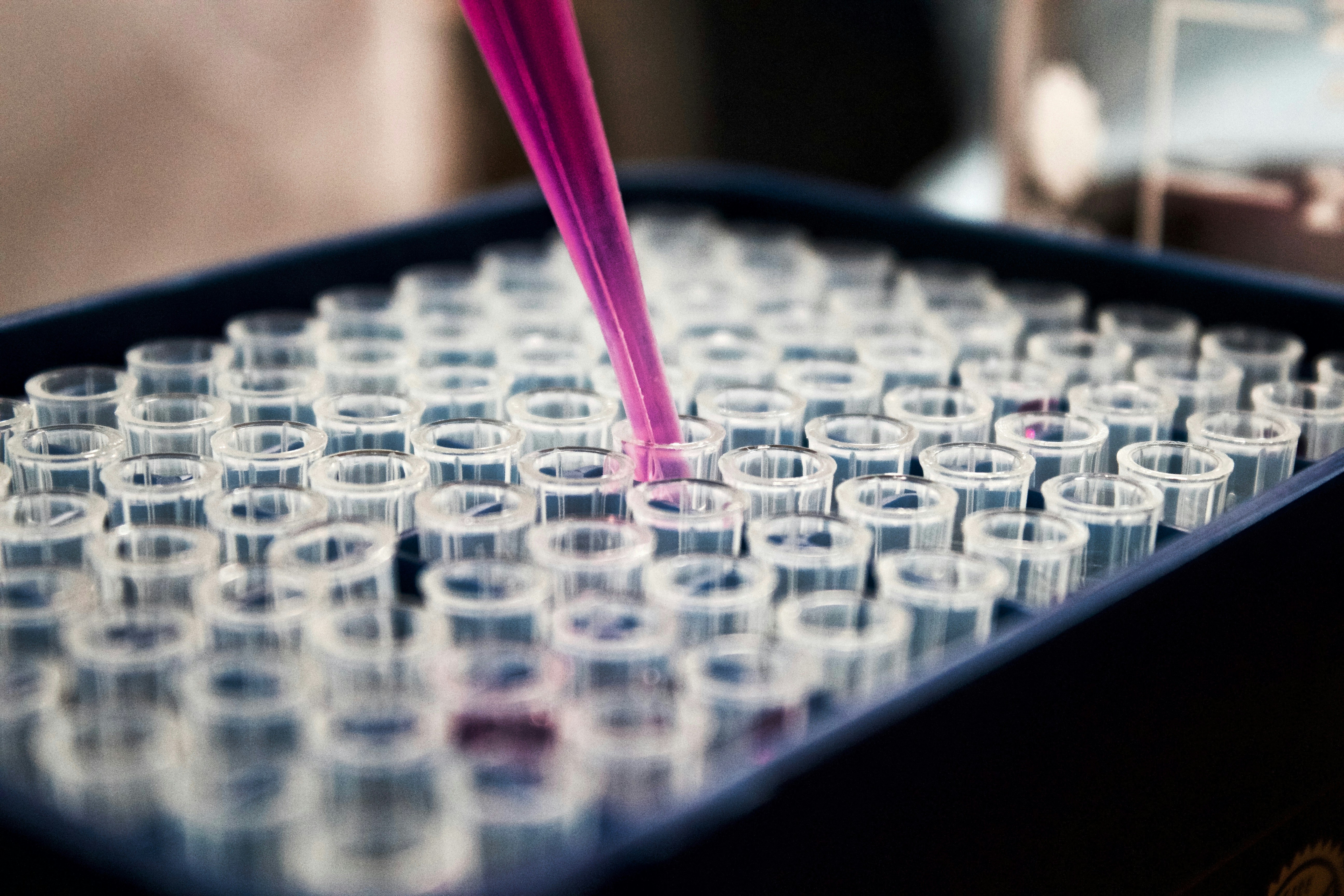
The Scientist's Toolkit: Building and Interrogating 3D Worlds
Creating and analyzing these complex models requires specialized tools:
| Reagent/Material | Function in Experiment | Why it's Crucial |
|---|---|---|
| Basement Membrane Extract | Provides a biologically relevant 3D scaffold mimicking the extracellular matrix (ECM) | Allows cells to self-organize into structures resembling real tissue architecture |
| Type I Collagen | Another key ECM component; forms fibrillar networks | Provides structural support and biochemical cues influencing cell behavior |
| Fluorescent Live-Cell Dyes | Stains live cells (green) and dead cells (red) without killing them | Enables real-time visualization and quantification of viability |
- Confocal microscope
- Light-sheet microscope
- High-content screening systems
- Imaris
- CellProfiler
- Custom AI solutions
The Future is Dynamic, 3D, and Quantified
The ability to precisely quantify how drugs dynamically reshape living 3D tissues marks a paradigm shift in drug discovery.
Key Advances:
- Moving beyond static snapshots to dynamic monitoring
- Revealing hidden mechanisms of drug efficacy and resistance
- Identifying subtle effects like early anti-metastatic activity
Future Directions:
- More sophisticated AI algorithms
- Advanced imaging technologies
- Integration with other omics data
- Personalized medicine applications